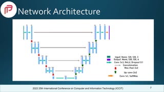
The document presents a study on brain tumor segmentation using an enhanced 2D U-Net model at the 2022 ICCIT conference, highlighting the challenges of segmenting MRI images due to overlapping modalities. It reviews related works achieving various dice scores and establishes research questions aimed at improving model performance. The findings indicate that the proposed model outperforms traditional machine learning models, with recommendations for future work including the exploration of 3D U-Net models to minimize information loss.

![2022 25th International Conference on Computer and Information Technology (ICCIT)
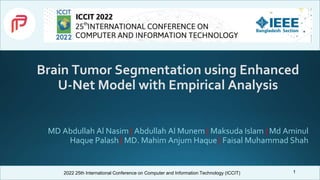
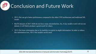
Related Work
4
• Agravat et al. [1] achieved 88.1% dice similarity on BraTS 2020 dataset using 3D encoder-decoder
FCNN model.
• Xu et al. [2] considered the correlations between the modalities of the BraTS dataset. They used
LSTM multi-modal 2D U-Net on BraTS 2015 dataset and achieved dice scores of 0.7309, 0.6235,
and 0.4254 for whole, core and enhancing tumor, respectively.
• Dong et al. [3] utilized 2D U-Net architecture on the BraTS 2015 dataset and gained dice scores of
0.86, 0.86, and 0.65 for whole, core, and enhancing tumors, respectively.
• Singh et al. [4] trained RCNN model using the BraTS 2020 dataset and got a precision of 0.79,
recall of 0.72, and dice coefficient of 0.75.
• Mobarakol et al. [5] integrated a 3D attention module of the decoder paths of simple U-Net
architecture and achieved mean dice scores of 0.704, 0.898, and 0.792, for enhancing tumor, whole,
and core respectively on BraTS 2019 Dataset.](https://image.slidesharecdn.com/393presentationslide-221219073445-63625143/85/Brain-Tumor-Segmentation-using-Enhanced-U-Net-Model-with-Empirical-Analysis-4-320.jpg)
![2022 25th International Conference on Computer and Information Technology (ICCIT)
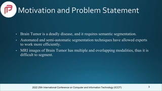
Comparison With Previous Works
Model
Dataset
(BraTS)
Dice Score
Core Whole Enhancing
SVM [6] 2012 0.61 0.71 0.73
Random
Forest [7]
2012 0.73 0.59 -
Random
Forest [8]
2013 0.70 0.76 0.67
2D CNN [8] 2013 0.73 0.83 0.69
3D CNN [9] 2015 0.73 0.87 0.77
3D encoder-
decoder
FCNN [1]
2020 0.83 0.88 0.78
3D FCNN
[10]
2020 0.74 0.88 0.71
Our
Proposed
Model
2019 0.871 0.95 0.94
11
Model Dataset
(BraTS)
Accuracy Sensitivity /
Recall
Specificity
R-CNN
[11]
2020 - 0.72 -
LeNET
[12]
2019 0.944 0.956 0.945
AlexNET
[12]
2019 0.961 0.952 0.951
R-CNN
[13]
2018 0.963 0.935 0.972
Hybrid
Fast
R-CNN
& SVM
[14]
2015 0.987 - -
Our
proposed
model
2019 0.998 0.997 0.999](https://image.slidesharecdn.com/393presentationslide-221219073445-63625143/85/Brain-Tumor-Segmentation-using-Enhanced-U-Net-Model-with-Empirical-Analysis-11-320.jpg)
![2022 25th International Conference on Computer and Information Technology (ICCIT)
Selected References
[1] R. R. Agravat and M. S. Raval, “3d semantic segmentation of brain tumor for overall survival prediction,” in International MICCAI Brainlesion
Workshop. Springer, 2020, pp. 215–227.
[2] F. Xu, H. Ma, J. Sun, R. Wu, X. Liu, and Y. Kong, “Lstm multi-modal unet for brain tumor segmentation,” in 2019 IEEE 4th international conference
on image, vision and computing (ICIVC). IEEE, 2019, pp. 236–240.
[3] H. Dong, G. Yang, F. Liu, Y. Mo, and Y. Guo, “Automatic brain tumor detection and segmentation using u-net based fully convolutional networks,” in
annual conference on medical image understanding and analysis. Springer, 2017, pp. 506–517.
[4] S. Singh, “A novel mask r-cnn model to segment heterogeneous brain tumors through image subtraction,” arXiv preprint arXiv:2204.01201, 2022.
[5] M. Islam, V. Vibashan, V. Jose, N. Wijethilake, U. Utkarsh, and H. Ren, “Brain tumor segmentation and survival prediction using 3d attention unet,” in
International MICCAI Brainlesion Workshop. Springer, 2019, pp. 262–272.
[6] S. Bauer, L.-P. Nolte, and M. Reyes, “Fully automatic segmentation of brain tumor images using support vector machine classification in combination
with hierarchical conditional random field regularization,” in International conference on medical image computing and computerassisted intervention.
Springer, 2011, pp. 354–361
[7] S. Bauer, T. Fejes, J. Slotboom, R. Wiest, L.-P. Nolte, and M. Reyes, “Segmentation of brain tumor images based on integrated hierarchical
classification and regularization,” in MICCAI BraTS Workshop. Nice: Miccai Society, vol. 11, 2012.
13](https://image.slidesharecdn.com/393presentationslide-221219073445-63625143/85/Brain-Tumor-Segmentation-using-Enhanced-U-Net-Model-with-Empirical-Analysis-13-320.jpg)
![2022 25th International Conference on Computer and Information Technology (ICCIT)
Selected References
[8] D. Zikic, Y. Ioannou, M. Brown, and A. Criminisi, “Segmentation of brain tumor tissues with convolutional neural networks,” Proceedings MICCAI-
BRATS, vol. 36, no. 2014, pp. 36–39, 2014.
[9] K. Kamnitsas, E. Ferrante, S. Parisot, C. Ledig, A. V. Nori, A. Criminisi, D. Rueckert, and B. Glocker, “Deepmedic for brain tumor segmentation,” in
International workshop on Brainlesion: Glioma, multiple sclerosis, stroke and traumatic brain injuries. Springer, 2016, pp. 138–149
[10] V. K. Anand, S. Grampurohit, P. Aurangabadkar, A. Kori, M. Khened, R. S. Bhat, and G. Krishnamurthi, “Brain tumor segmentation and survival
prediction using automatic hard mining in 3d cnn architecture,” in International MICCAI Brainlesion Workshop. Springer, 2020, pp.310–319.
[11] R. Raza, U. I. Bajwa, Y. Mehmood, M. W. Anwar, and M. H. Jamal, “dresu-net: 3d deep residual u-net based brain tumor segmentation from
multimodal mri,” Biomedical Signal Processing and Control, p. 103861, 2022.
[12] M. Gomathi and D. Dhanasekaran, “Glioma detection and segmentation using deep learning architectures,” Mathematical Statistician and Engineering
Applications, vol. 71, no. 4, pp. 452–461, 2022.
[13] Y. Zhuge, H. Ning, P. Mathen, J. Y. Cheng, A. V. Krauze, K. Camphausen, and R. W. Miller, “Automated glioma grading on conventional mri images
using deep convolutional neural networks,” Medical physics, vol. 47, no. 7, pp. 3044–3053, 2020.
[14] M. O. Khairandish, R. Gurta, and M. Sharma, “A hybrid model of faster r-cnn and svm for tumor detection and classification of mri brain images,” Int.
J. Mech. Prod. Eng. Res. Dev, vol. 10, no. 3, pp. 6863–6876, 2020.
14](https://image.slidesharecdn.com/393presentationslide-221219073445-63625143/85/Brain-Tumor-Segmentation-using-Enhanced-U-Net-Model-with-Empirical-Analysis-14-320.jpg)


![谷歌留痕技术 [ 𝙩𝙤𝙥 𝟮𝟯𝟯. 𝙘 𝙤𝙢 ]](https://cdn.slidesharecdn.com/ss_thumbnails/top233-260130174328-3833018c-thumbnail.jpg?width=640&height=640&fit=bounds)


