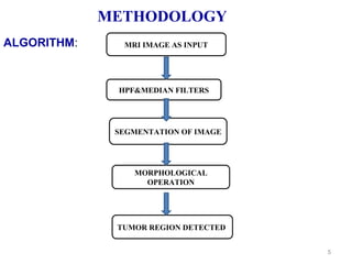
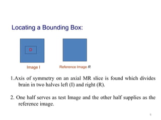
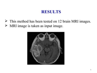
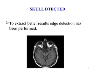
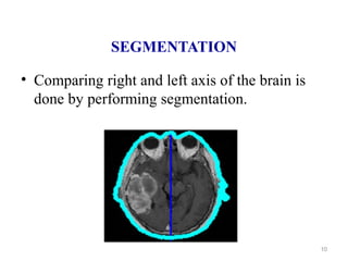
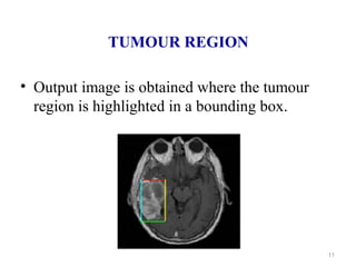
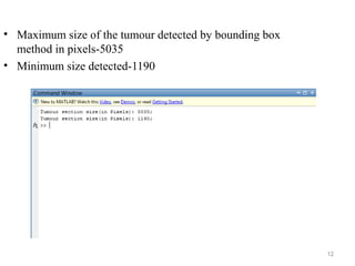
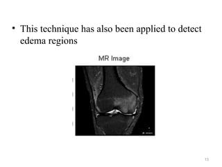
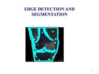
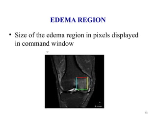
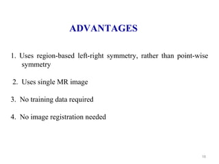
1
BRAIN TUMOUR DETECTION USING
BOUNDING BOX SYMMETRY
2
CONTENTS
 OBJECTIVE
INTRODUCTION
METHODOLOGY
 RESULTS
 ADVANTAGES
 CONCLUSION
 FUTURE SCOPE
3
OBJECTIVE
To detect the size and location of brain tumors and
edemas from the Magnetic Resonance Images.
4
INTRODUCTION
Brain tumor is an abnormal mass of tissue in which
cells grow and multiply uncontrollably seemingly
unchecked by the mechanisms that control normal
cells.
This change detection process uses a novel score
function based on Bhattacharya coefficient computed
with gray level intensity histograms.
 The score function admits a very fast search to
locate the bounding box.
5
METHODOLOGY
MRI IMAGE AS INPUT
HPF&MEDIAN FILTERS
SEGMENTATION OF IMAGE
MORPHOLOGICAL
OPERATION
TUMOR REGION DETECTED
ALGORITHM:
6
D
Locating a Bounding Box:
1.Axis of symmetry on an axial MR slice is found which divides
brain in two halves left (I) and right (R).
2. One half serves as test Image and the other half supplies as the
reference image.
Image I Reference Image R
7
3. Novel score function is used which identify the region of
change with two searches – one along the vertical direction and
other along the horizontal direction.
4. Novel score function uses Bhattacharya coefficient to
detects a rectangle D which represents the region of interest
between images I and R
8
RESULTS
 This method has been tested on 12 brain MRI images.
 MRI image is taken as input image.
SKULL DTECTED
To extract better results edge detection has
been performed.
9
SEGMENTATION
• Comparing right and left axis of the brain is
done by performing segmentation.
10
TUMOUR REGION
• Output image is obtained where the tumour
region is highlighted in a bounding box.
11
12
• Maximum size of the tumour detected by bounding box
method in pixels-5035
• Minimum size detected-1190
13
• This technique has also been applied to detect
edema regions
EDGE DETECTION AND
SEGMENTATION
14
EDEMA REGION
• Size of the edema region in pixels displayed
in command window
15
16
ADVANTAGES
1. Uses region-based left-right symmetry, rather than point-wise
symmetry
2. Uses single MR image
3. No training data required
4. No image registration needed
17
CONCLUSION
•The current method uses a computer aided system for brain MR
image segmentation for detection of tumour location using
bounding box symmetry.
•The resulting method is very fast, robust and reliable for indexing
tumour or edema images for both archival and retrieval purposes
and it can use as a vehicle for further clinical investigations.
18
FUTURE SCOPE
•In future, this technique can be developed to classify the
tumours based on feature extraction.
•This technique can be applied for ovarian, breast, lung, skin
tumours.
•Instead of rectangular boxes, can work with general boundaries:
level set based framework.
19

Tumour detection

  • 1.
    1 BRAIN TUMOUR DETECTIONUSING BOUNDING BOX SYMMETRY
  • 2.
  • 3.
    3 OBJECTIVE To detect thesize and location of brain tumors and edemas from the Magnetic Resonance Images.
  • 4.
    4 INTRODUCTION Brain tumor isan abnormal mass of tissue in which cells grow and multiply uncontrollably seemingly unchecked by the mechanisms that control normal cells. This change detection process uses a novel score function based on Bhattacharya coefficient computed with gray level intensity histograms.  The score function admits a very fast search to locate the bounding box.
  • 5.
    5 METHODOLOGY MRI IMAGE ASINPUT HPF&MEDIAN FILTERS SEGMENTATION OF IMAGE MORPHOLOGICAL OPERATION TUMOR REGION DETECTED ALGORITHM:
  • 6.
    6 D Locating a BoundingBox: 1.Axis of symmetry on an axial MR slice is found which divides brain in two halves left (I) and right (R). 2. One half serves as test Image and the other half supplies as the reference image. Image I Reference Image R
  • 7.
    7 3. Novel scorefunction is used which identify the region of change with two searches – one along the vertical direction and other along the horizontal direction. 4. Novel score function uses Bhattacharya coefficient to detects a rectangle D which represents the region of interest between images I and R
  • 8.
    8 RESULTS  This methodhas been tested on 12 brain MRI images.  MRI image is taken as input image.
  • 9.
    SKULL DTECTED To extractbetter results edge detection has been performed. 9
  • 10.
    SEGMENTATION • Comparing rightand left axis of the brain is done by performing segmentation. 10
  • 11.
    TUMOUR REGION • Outputimage is obtained where the tumour region is highlighted in a bounding box. 11
  • 12.
    12 • Maximum sizeof the tumour detected by bounding box method in pixels-5035 • Minimum size detected-1190
  • 13.
    13 • This techniquehas also been applied to detect edema regions
  • 14.
  • 15.
    EDEMA REGION • Sizeof the edema region in pixels displayed in command window 15
  • 16.
    16 ADVANTAGES 1. Uses region-basedleft-right symmetry, rather than point-wise symmetry 2. Uses single MR image 3. No training data required 4. No image registration needed
  • 17.
    17 CONCLUSION •The current methoduses a computer aided system for brain MR image segmentation for detection of tumour location using bounding box symmetry. •The resulting method is very fast, robust and reliable for indexing tumour or edema images for both archival and retrieval purposes and it can use as a vehicle for further clinical investigations.
  • 18.
    18 FUTURE SCOPE •In future,this technique can be developed to classify the tumours based on feature extraction. •This technique can be applied for ovarian, breast, lung, skin tumours. •Instead of rectangular boxes, can work with general boundaries: level set based framework.
  • 19.