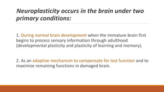
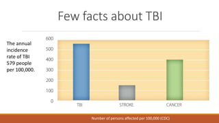
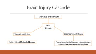
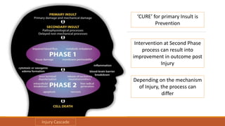
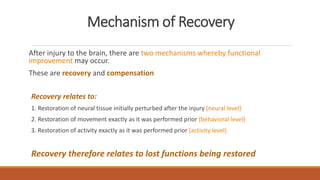
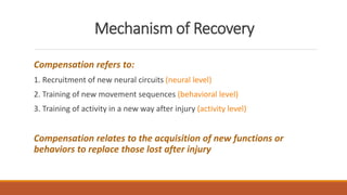
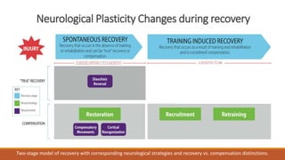
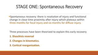
Brain Plasticity after TBI
DR RAVI SONI
DM GERIATRIC PSYCHIATRY
ASSISTANT PROFESSOR
HOSPITAL FOR MENTAL HEALTH
AHMEDABAD
What is Brain Plasticity?
Answer: Brain reacts and adapts in response to
challenge.
Brain Plasticity the capability of the brain to alter its
functional organization as a result of experience.
Brain Plasticity
Plasticity in physics : propensity of a material to undergo
permanent deformation under load.
Entire brain structure can change to better cope with the
environment.
Specifically when an area of the brain is damaged and
nonfunctional another area may take over some of the
function
Plasticity includes the brain's capacity to be
shaped or molded by experience, the capacity to
learn and remember, and the ability to
reorganize and recover after injury.
Plasticity: Brain’s ability to change: two
processes
Neurogenesis: new neurons migrate to different
areas
Synaptogenesis: development of connections with
neurons This means that the brain is actually
physically changing in response to information we
receive! “ creation of synapses”
Four types of Plasticity
Adaptive plasticity that enhances skill development or recovery
from brain injury
Impaired plasticity associated with cognitive impairment; as in
fragile X syndrome
Excessive plasticity leading to maladaptive brain circuits as in focal
dystonia
Plasticity that becomes the brain's `Achilles' Heel‘ because makes it
vulnerable to injury.
How does the brain change?
The brain can change in many ways such as:
A change in the internal structure of the neurons,
particularly at the area of synapses.
An increase in the number of synapses between
neurons.
Neuroplasticity occurs in the brain under two
primary conditions:
1. During normal brain development when the immature brain first
begins to process sensory information through adulthood
(developmental plasticity and plasticity of learning and memory).
2. As an adaptive mechanism to compensate for lost function and to
maximize remaining functions in damaged brain.
One rule that is more important in Brain
plasticity: Hebb Rule
1. Neurons that fire together wire together!
2. When cells are active together synapses are
strengthened and preserved.
3. The neurons & synapses that are activated
repeatedly are preserved while those who aren’t,
are pruned.
Few facts about TBI
The annual
incidence
rate of TBI
579 people
per 100,000.
Number of persons affected per 100,000 (CDC)
Brain Injury Cascade
Traumatic Brain Injury
Primary insult-Injury Secondary Insult-Injury
Two
Phases
Etiology: Direct Mechanical Damage Following mechanical damage, etiology being a
cascade of pathophysiological processes.
‘CURE’ for primary Insult is
Prevention
Intervention at Second Phase
process can result into
improvement in outcome post
Injury
Depending on the mechanism
of Injury, the process can
differ
Injury Cascade
Phase One:
Injuries typically include direct tissue damage, impaired cerebral blood flow, and
impaired metabolic activity, leading to edema formation and cyto-architecture
changes like membrane permeability.
Contact Forces: contusion, hemorrhage and lacerations throughout
Inertial forces: shearing and/or compression of brain tissue
These forces cause multifocal injuries (usually termed diffuse axonal injury)
affecting axons, blood vessels, junctions between white and gray matter, and
other select focal areas like the corpus callosum and junctions between the
frontal and parietal lobes.
Phase Two: Initial Injury
Disruption of Neurons (Neuronal
Depolarization)
Release of Excitatory
Neurotransmitters (Ca++ and Na+)
lead to intracellular breakdowns
Release of Caspases and Calpains
Cell Death
Release of
Caspases initiates
the process of
APOPTOSIS
(Programmed cell
death)
Release of Calpains quickly
leads to necrosis where cells
die as a response to
mechanical or hypoxic damage
and metabolic failure. This
leads to an inflammatory
response with the cells being
removed.
Neuroplasticity and Brain function after TBI
Easiest way to conceptualize neuroplasticity after injury to
the brain is to view it simply as re-learning
“the brain will rely on the same fundamental
neurobiological process it used to acquire those behaviors
initially. The basic rules governing how neural circuits adapt
to encode new behaviors do not change after injury” (Kleim
2011)
Neuroplasticity and Brain function after TBI
We can view re-establishing function as a re-learning process but
there are two conceptual differences when it occurs after a brain
injury.
First, because neural circuits for a particular function were
previously established during the brain’s neurodevelopmental
process, it may be possible to take advantage of those learned
behaviors if they persist in residual areas of the brain during the
rehabilitation (Kleim, 2011).
This presents as a potentially adaptive circumstance.
Neuroplasticity and Brain function after TBI
Second, a more maladaptive consequence which occurs post injury
relates to the concept of learned non-use.
Just as increasing dexterity of motor function leads to increased
motor cortex representation of neural circuitry (and therefore
improved function), non-use can lead to decreased motor cortex
representation, and therefore decreased function (Plowman and
Kleim, 2010).
Research indicates that learned non-use of a paretic limb,
combined with an increased reliance on the unaffected limb can
result in major brain reorganization.
Mechanism of Recovery
After injury to the brain, there are two mechanisms whereby functional
improvement may occur.
These are recovery and compensation
Recovery relates to:
1. Restoration of neural tissue initially perturbed after the injury (neural level)
2. Restoration of movement exactly as it was performed prior (behavioral level)
3. Restoration of activity exactly as it was performed prior (activity level)
Recovery therefore relates to lost functions being restored
Mechanism of Recovery
Compensation refers to:
1. Recruitment of new neural circuits (neural level)
2. Training of new movement sequences (behavioral level)
3. Training of activity in a new way after injury (activity level)
Compensation relates to the acquisition of new functions or
behaviors to replace those lost after injury
Neurological Plasticity Changes during recovery
Two-stage model of recovery with corresponding neurological strategies and recovery vs. compensation distinctions.
STAGE ONE: Spontaneous Recovery
Spontaneous recovery: there is resolution of injury and functional
change in close time proximity after injury which plateaus within
three months for focal injury and six months for diffuse injury.
Three processes have been theorized to explain this early recovery
1. Diaschisis reversal
2. Changes in kinematics.
3. Cortical reorganization.
Diaschisis Reversal
Diaschisis is a disturbance or loss of function in one part of the brain
due to a localized injury in another part of the brain, and these areas
can be of considerable distance from the lesion area including the
opposite hemisphere (Stein, 2012).
Reversal of Diaschisis: due to resolution of the inflammatory
process, blood flow changes, metabolic changes, edema, and
neuronal excitability
The result of diaschisis reversal is improved function due to intact
brain areas that were previously disrupted now being restored.
Changes in Kinematics
The second aspect of early recovery relates to changes in kinematic
(movement) patterns where compensatory patterns are utilized.
The individual begins to complete motor movements in a different
manner, resulting in improved function, sometimes in drastically
different ways than prior to injury.
While these new movements likely contribute to functional
improvement, these compensatory strategies have the potential to
be maladaptive.
Cortical Reorganization
The third strategy identified as spontaneous recovery is that the nervous
system undergoes within-area and between-area reorganization or
rewiring.
Cortical reorganization during spontaneous recovery is thought to be
compensatory as different circuits or networks of neurons are utilized post
injury than those utilized pre injury.
While spontaneous recovery occurs in the absence of rehabilitation, there
is certainly the opportunity for overlap of training induced recovery while
spontaneous recovery takes its course.
STAGE TWO: Training-induced recovery
Recovery in this stage involves compensation, in that either new
brain areas or neural networks are enlisted to complete previous
functions.
Through the process of training, neuroplasticity is induced.
Adaptive changes after injury are the outcome of new patterns of
activation which include plasticity in areas surrounding the damaged
cortex, reorganization of existing networks or recruitment of new
cortical areas or networks.
STAGE TWO: Training-induced recovery
1. Recruitment:
During training-induced recovery, areas which did not make a
significant contribution to that particular function pre-injury now
contribute to function post-injury.
Due to recruitment of neural areas from the undamaged
hemisphere.
Ultimate result is change in motor maps or cognitive functions in the
non-injured hemisphere, allows to take over the motor/cognitive
function of damaged Hemisphere.
STAGE TWO: Training-induced recovery
2. Retraining:
Retraining involves the training of residual brain areas, resulting in
reorganization within the cortex and compensation for lost function
(Kleim, 2007).
This often comes in the forms of reorganization within the damaged
hemisphere.
Ultimately, recruitment and retraining involve rewiring or reorganization
of neural networks.
Neurobiological Changes after Acquired Brain Injury
After injury to the brain, the processes of neuroplasticity are thought
to be the underpinnings of Recovery.
1. Increases or changes to synapses:
This includes synaptogenesis and synaptic plasticity
Dendrite changes including increased arborization, dendritic growth
and spine growth
Axonal changes including axonal sprouting
Neurobiological Changes after Acquired Brain Injury
2. Increased neuron growth:
Neurogenesis in specific brain areas like the hippocampus
subgranular zone of the dentate gyrus and subventricular zone in
some areas, substantia nigra and perinfarcted areas.
3. Angiogenesis:
Angiogenesis is the process through which new blood vessels form
from pre-existing vessels.
Neurobiological Changes after Acquired Brain Injury
4. Excitability changes:
Excitability refers to the ability of a neuron to generate action
potentials, which is a short-term change in the electrical potential on
the surface of a cell.
It is an all or nothing proposition as it either fires or does not fire
depending on the strength of the potential.
Neurobiological Changes after Acquired Brain Injury
The first two items (increase or changes to synapses and increased
neuronal growth) on the list above relate to increases in either the
number of neurons (this occurs in a very limited sense) or the numbers of
synapses or increased strength of existing synapses (this far more
prevalent).
These changes seen post injury are similar to changes seen in the intact
brain in the form of experience dependent learning.
But instead of it being a learning process, it is a relearning process, aided
substantially by rehabilitation.
Neurobiological Changes after Acquired Brain Injury
Experience Dependent Learning leads to:
◦ New synapses formation (synaptogenesis)
◦ Strengthening of synapses through changes in dendrites (new dendritic
spine formation), axonal sprouting and long term potentiation (synaptic
plasticity)
Synaptogenesis and synaptic plasticity are the main underpinnings
of cortical reorganization, recruitment and retraining as identified
in Mechanisms of recovery.
Findings Related to Neurobiological Changes
Synaptic, Dendritic and Axonal Related Changes:
Loss of synapses with other neurons in affected areas of brain
Dendritic arbors increased in non-affected areas of brain
Axonal sprouting and reorganization occurs
 This sprouting has adaptive consequences in that increased axonal growth leads to greater
levels of synapses allowing reinnervation
Restoration of motor/cognitive function through Synaptic Change
 This includes synaptogenesis where new synapses form through dendritic growth and axonal
sprouting, and synaptic plasticity which strengthens existing synapses through the process of
long-term potentiation
Angiogenesis
Angiogenesis is the process through which new blood vessels form from pre-
existing vessels.
The benefit is return of blood flow to previously damaged areas, which is assists
in establishing metabolic support
An area around the infarct affected by vascular compromise is more than just
dying cells and it is called penumbra – it may be a precursor of neuroplasticity.
Vascular endothelial growth factor (VEGF) is an important factor in post-injury
recovery vascular remodeling which ultimately promotes synaptogenesis and
neurogenesis.
Activation and Excitatory Changes
After injury, changes in the excitability of the damaged and intact hemispheres can impact
cortical functioning.
Interhemispheric rivalry model: where there are distinct differences in the excitability of
analogous areas between hemispheres (e.g., motor areas). For example in the damaged
hemisphere there is hyperpolarization (inhibition of neurons) and in the intact hemisphere there
is depolarization (excitation of neurons)
Better recovery is found if activation of the affected-side is more predominant than the
unaffected hemisphere over time. This shift of activation to the unaffected side is “the sign of a
distressed system”
 As a long term perspective, if the damaged side was more involved in function, that related to
better outcomes. However, if the patient had to rely on the unaffected side more for
function, that related to poorer outcomes.
The maladaptive side of neuroplasticity
Neuroplasticity has also its dark side:
Few examples include
oAddictions to alcohol, elicit substances or prescription drugs,
oPornography addictions,
oSeizure disorders post injury
oPhantom limb pain
oHand dystonia in musicians
oLearning and memory interference and
oChronic pain
References
Heidi Reyst, Neuroplasticity After Acquired Brain Injury. Rainbow Rehabilitation Centers.
YouRong Sophie Su, Anand Veeravagu, and Gerald Grant. Chapter 8-Neuroplasticity after
Traumatic Brain Injury. Translational Research in Traumatic Brain Injury.
Kleim JA, Jones TA. Principles of Experience-Dependent Neural Plasticity: Implications for
Rehabilitation After Brain Damage. Journal of Speech, Language, and Hearing Research,
February 2008, Vol. 51, S225-S239. doi:10.1044/1092-4388(2008/018)
Brain plasticity after Traumatic brain Injury

Brain plasticity after Traumatic brain Injury

  • 1.
    Brain Plasticity afterTBI DR RAVI SONI DM GERIATRIC PSYCHIATRY ASSISTANT PROFESSOR HOSPITAL FOR MENTAL HEALTH AHMEDABAD
  • 2.
    What is BrainPlasticity? Answer: Brain reacts and adapts in response to challenge. Brain Plasticity the capability of the brain to alter its functional organization as a result of experience.
  • 3.
    Brain Plasticity Plasticity inphysics : propensity of a material to undergo permanent deformation under load. Entire brain structure can change to better cope with the environment. Specifically when an area of the brain is damaged and nonfunctional another area may take over some of the function
  • 4.
    Plasticity includes thebrain's capacity to be shaped or molded by experience, the capacity to learn and remember, and the ability to reorganize and recover after injury.
  • 5.
    Plasticity: Brain’s abilityto change: two processes Neurogenesis: new neurons migrate to different areas Synaptogenesis: development of connections with neurons This means that the brain is actually physically changing in response to information we receive! “ creation of synapses”
  • 6.
    Four types ofPlasticity Adaptive plasticity that enhances skill development or recovery from brain injury Impaired plasticity associated with cognitive impairment; as in fragile X syndrome Excessive plasticity leading to maladaptive brain circuits as in focal dystonia Plasticity that becomes the brain's `Achilles' Heel‘ because makes it vulnerable to injury.
  • 7.
    How does thebrain change? The brain can change in many ways such as: A change in the internal structure of the neurons, particularly at the area of synapses. An increase in the number of synapses between neurons.
  • 8.
    Neuroplasticity occurs inthe brain under two primary conditions: 1. During normal brain development when the immature brain first begins to process sensory information through adulthood (developmental plasticity and plasticity of learning and memory). 2. As an adaptive mechanism to compensate for lost function and to maximize remaining functions in damaged brain.
  • 9.
    One rule thatis more important in Brain plasticity: Hebb Rule 1. Neurons that fire together wire together! 2. When cells are active together synapses are strengthened and preserved. 3. The neurons & synapses that are activated repeatedly are preserved while those who aren’t, are pruned.
  • 10.
    Few facts aboutTBI The annual incidence rate of TBI 579 people per 100,000. Number of persons affected per 100,000 (CDC)
  • 11.
    Brain Injury Cascade TraumaticBrain Injury Primary insult-Injury Secondary Insult-Injury Two Phases Etiology: Direct Mechanical Damage Following mechanical damage, etiology being a cascade of pathophysiological processes.
  • 12.
    ‘CURE’ for primaryInsult is Prevention Intervention at Second Phase process can result into improvement in outcome post Injury Depending on the mechanism of Injury, the process can differ Injury Cascade
  • 13.
    Phase One: Injuries typicallyinclude direct tissue damage, impaired cerebral blood flow, and impaired metabolic activity, leading to edema formation and cyto-architecture changes like membrane permeability. Contact Forces: contusion, hemorrhage and lacerations throughout Inertial forces: shearing and/or compression of brain tissue These forces cause multifocal injuries (usually termed diffuse axonal injury) affecting axons, blood vessels, junctions between white and gray matter, and other select focal areas like the corpus callosum and junctions between the frontal and parietal lobes.
  • 14.
    Phase Two: InitialInjury Disruption of Neurons (Neuronal Depolarization) Release of Excitatory Neurotransmitters (Ca++ and Na+) lead to intracellular breakdowns Release of Caspases and Calpains Cell Death Release of Caspases initiates the process of APOPTOSIS (Programmed cell death) Release of Calpains quickly leads to necrosis where cells die as a response to mechanical or hypoxic damage and metabolic failure. This leads to an inflammatory response with the cells being removed.
  • 15.
    Neuroplasticity and Brainfunction after TBI Easiest way to conceptualize neuroplasticity after injury to the brain is to view it simply as re-learning “the brain will rely on the same fundamental neurobiological process it used to acquire those behaviors initially. The basic rules governing how neural circuits adapt to encode new behaviors do not change after injury” (Kleim 2011)
  • 16.
    Neuroplasticity and Brainfunction after TBI We can view re-establishing function as a re-learning process but there are two conceptual differences when it occurs after a brain injury. First, because neural circuits for a particular function were previously established during the brain’s neurodevelopmental process, it may be possible to take advantage of those learned behaviors if they persist in residual areas of the brain during the rehabilitation (Kleim, 2011). This presents as a potentially adaptive circumstance.
  • 17.
    Neuroplasticity and Brainfunction after TBI Second, a more maladaptive consequence which occurs post injury relates to the concept of learned non-use. Just as increasing dexterity of motor function leads to increased motor cortex representation of neural circuitry (and therefore improved function), non-use can lead to decreased motor cortex representation, and therefore decreased function (Plowman and Kleim, 2010). Research indicates that learned non-use of a paretic limb, combined with an increased reliance on the unaffected limb can result in major brain reorganization.
  • 18.
    Mechanism of Recovery Afterinjury to the brain, there are two mechanisms whereby functional improvement may occur. These are recovery and compensation Recovery relates to: 1. Restoration of neural tissue initially perturbed after the injury (neural level) 2. Restoration of movement exactly as it was performed prior (behavioral level) 3. Restoration of activity exactly as it was performed prior (activity level) Recovery therefore relates to lost functions being restored
  • 19.
    Mechanism of Recovery Compensationrefers to: 1. Recruitment of new neural circuits (neural level) 2. Training of new movement sequences (behavioral level) 3. Training of activity in a new way after injury (activity level) Compensation relates to the acquisition of new functions or behaviors to replace those lost after injury
  • 20.
    Neurological Plasticity Changesduring recovery Two-stage model of recovery with corresponding neurological strategies and recovery vs. compensation distinctions.
  • 21.
    STAGE ONE: SpontaneousRecovery Spontaneous recovery: there is resolution of injury and functional change in close time proximity after injury which plateaus within three months for focal injury and six months for diffuse injury. Three processes have been theorized to explain this early recovery 1. Diaschisis reversal 2. Changes in kinematics. 3. Cortical reorganization.
  • 22.
    Diaschisis Reversal Diaschisis isa disturbance or loss of function in one part of the brain due to a localized injury in another part of the brain, and these areas can be of considerable distance from the lesion area including the opposite hemisphere (Stein, 2012). Reversal of Diaschisis: due to resolution of the inflammatory process, blood flow changes, metabolic changes, edema, and neuronal excitability The result of diaschisis reversal is improved function due to intact brain areas that were previously disrupted now being restored.
  • 23.
    Changes in Kinematics Thesecond aspect of early recovery relates to changes in kinematic (movement) patterns where compensatory patterns are utilized. The individual begins to complete motor movements in a different manner, resulting in improved function, sometimes in drastically different ways than prior to injury. While these new movements likely contribute to functional improvement, these compensatory strategies have the potential to be maladaptive.
  • 24.
    Cortical Reorganization The thirdstrategy identified as spontaneous recovery is that the nervous system undergoes within-area and between-area reorganization or rewiring. Cortical reorganization during spontaneous recovery is thought to be compensatory as different circuits or networks of neurons are utilized post injury than those utilized pre injury. While spontaneous recovery occurs in the absence of rehabilitation, there is certainly the opportunity for overlap of training induced recovery while spontaneous recovery takes its course.
  • 25.
    STAGE TWO: Training-inducedrecovery Recovery in this stage involves compensation, in that either new brain areas or neural networks are enlisted to complete previous functions. Through the process of training, neuroplasticity is induced. Adaptive changes after injury are the outcome of new patterns of activation which include plasticity in areas surrounding the damaged cortex, reorganization of existing networks or recruitment of new cortical areas or networks.
  • 26.
    STAGE TWO: Training-inducedrecovery 1. Recruitment: During training-induced recovery, areas which did not make a significant contribution to that particular function pre-injury now contribute to function post-injury. Due to recruitment of neural areas from the undamaged hemisphere. Ultimate result is change in motor maps or cognitive functions in the non-injured hemisphere, allows to take over the motor/cognitive function of damaged Hemisphere.
  • 27.
    STAGE TWO: Training-inducedrecovery 2. Retraining: Retraining involves the training of residual brain areas, resulting in reorganization within the cortex and compensation for lost function (Kleim, 2007). This often comes in the forms of reorganization within the damaged hemisphere. Ultimately, recruitment and retraining involve rewiring or reorganization of neural networks.
  • 28.
    Neurobiological Changes afterAcquired Brain Injury After injury to the brain, the processes of neuroplasticity are thought to be the underpinnings of Recovery. 1. Increases or changes to synapses: This includes synaptogenesis and synaptic plasticity Dendrite changes including increased arborization, dendritic growth and spine growth Axonal changes including axonal sprouting
  • 29.
    Neurobiological Changes afterAcquired Brain Injury 2. Increased neuron growth: Neurogenesis in specific brain areas like the hippocampus subgranular zone of the dentate gyrus and subventricular zone in some areas, substantia nigra and perinfarcted areas. 3. Angiogenesis: Angiogenesis is the process through which new blood vessels form from pre-existing vessels.
  • 30.
    Neurobiological Changes afterAcquired Brain Injury 4. Excitability changes: Excitability refers to the ability of a neuron to generate action potentials, which is a short-term change in the electrical potential on the surface of a cell. It is an all or nothing proposition as it either fires or does not fire depending on the strength of the potential.
  • 31.
    Neurobiological Changes afterAcquired Brain Injury The first two items (increase or changes to synapses and increased neuronal growth) on the list above relate to increases in either the number of neurons (this occurs in a very limited sense) or the numbers of synapses or increased strength of existing synapses (this far more prevalent). These changes seen post injury are similar to changes seen in the intact brain in the form of experience dependent learning. But instead of it being a learning process, it is a relearning process, aided substantially by rehabilitation.
  • 32.
    Neurobiological Changes afterAcquired Brain Injury Experience Dependent Learning leads to: ◦ New synapses formation (synaptogenesis) ◦ Strengthening of synapses through changes in dendrites (new dendritic spine formation), axonal sprouting and long term potentiation (synaptic plasticity) Synaptogenesis and synaptic plasticity are the main underpinnings of cortical reorganization, recruitment and retraining as identified in Mechanisms of recovery.
  • 33.
    Findings Related toNeurobiological Changes Synaptic, Dendritic and Axonal Related Changes: Loss of synapses with other neurons in affected areas of brain Dendritic arbors increased in non-affected areas of brain Axonal sprouting and reorganization occurs  This sprouting has adaptive consequences in that increased axonal growth leads to greater levels of synapses allowing reinnervation Restoration of motor/cognitive function through Synaptic Change  This includes synaptogenesis where new synapses form through dendritic growth and axonal sprouting, and synaptic plasticity which strengthens existing synapses through the process of long-term potentiation
  • 34.
    Angiogenesis Angiogenesis is theprocess through which new blood vessels form from pre- existing vessels. The benefit is return of blood flow to previously damaged areas, which is assists in establishing metabolic support An area around the infarct affected by vascular compromise is more than just dying cells and it is called penumbra – it may be a precursor of neuroplasticity. Vascular endothelial growth factor (VEGF) is an important factor in post-injury recovery vascular remodeling which ultimately promotes synaptogenesis and neurogenesis.
  • 35.
    Activation and ExcitatoryChanges After injury, changes in the excitability of the damaged and intact hemispheres can impact cortical functioning. Interhemispheric rivalry model: where there are distinct differences in the excitability of analogous areas between hemispheres (e.g., motor areas). For example in the damaged hemisphere there is hyperpolarization (inhibition of neurons) and in the intact hemisphere there is depolarization (excitation of neurons) Better recovery is found if activation of the affected-side is more predominant than the unaffected hemisphere over time. This shift of activation to the unaffected side is “the sign of a distressed system”  As a long term perspective, if the damaged side was more involved in function, that related to better outcomes. However, if the patient had to rely on the unaffected side more for function, that related to poorer outcomes.
  • 36.
    The maladaptive sideof neuroplasticity Neuroplasticity has also its dark side: Few examples include oAddictions to alcohol, elicit substances or prescription drugs, oPornography addictions, oSeizure disorders post injury oPhantom limb pain oHand dystonia in musicians oLearning and memory interference and oChronic pain
  • 37.
    References Heidi Reyst, NeuroplasticityAfter Acquired Brain Injury. Rainbow Rehabilitation Centers. YouRong Sophie Su, Anand Veeravagu, and Gerald Grant. Chapter 8-Neuroplasticity after Traumatic Brain Injury. Translational Research in Traumatic Brain Injury. Kleim JA, Jones TA. Principles of Experience-Dependent Neural Plasticity: Implications for Rehabilitation After Brain Damage. Journal of Speech, Language, and Hearing Research, February 2008, Vol. 51, S225-S239. doi:10.1044/1092-4388(2008/018)