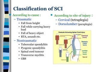
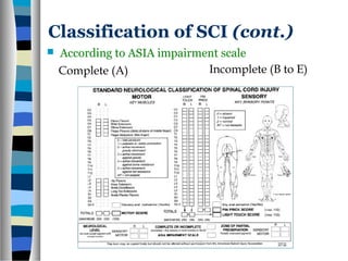
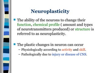
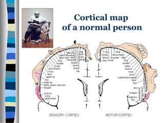
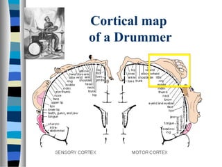
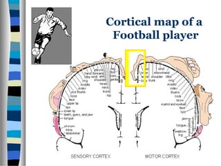
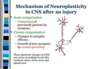
Neuroplasticity
Key to recovery after
spinal cord injury
Presented by : Dr. Shamim Khan
RMO, Medical Care Services
CRP, SAVAR
Classification of SCI
 According to cause :
– Traumatic
• Fall from height
• Fall while carrying heavy
load
• Fall of heavy object
• RTA, assault etc.
– Nontraumatic
• Tubercular spondylitis
• Pyogenic spondylitis
• Spinal cord tumour
• Transverse myelitis
• GBS
 According to site of injury :
– Cervical (tetraplegia)
– Dorsolumber (paraplegia)
Classification of SCI (cont.)
 According to ASIA impairment scale
Complete (A) Incomplete (B to E)
Spinal shock
 This is a time period after the transection of the
spinal cord during which all the spinal reflex
responses are profoundly depressed.
 Duration : Minimum 2 weeks
 Bulbocavernous reflex : First reflex to appear
following recovery of spinal shock.
Cellular mechanism of SCI
 Primary injury :
– Membrane dysruption
– Vascular damage
– Heamorrhage & edema.
– Ischemia (lack of O2)
 Secondary injury :
– Chemical mediators released
by activated macrophage and
glial cells
– Prolonged inflammation and
scarring.
– Neural cell death and
neurological damage.
Why SCI is an irreversible lesion?
 Once injured, CNS neurons
cannot regenerate their axons,
because :
– Lack of NGF.
– Inhibition of growth by
Oligodendrocytes.
– Clean up activities of
lymphocytes and Microglia.
– Increased GABAergic and
Glycinergic inhibition of spinal
networks.
Neuroplasticity
 The ability of the neurons to change their
function, chemical profile ( amount and types
of neurotransmitters produced) or structure is
referred to as neuroplasticity.
 The plastic changes in neuron can occur
– Physiologically according to activity and skill.
– Pathologically due to injury or disease of CNS.
Cortical map
of a normal person
Cortical map
of a Drummer
Cortical map of a
Football player
Mechanism of Neuroplasticity
in CNS after an injury
 Acute reorganization
– Unmasking of
previously present latent
synapses.
 Chronic reorganization
– Changes in synaptic
efficacy.
– Growth of new synapses
by axonal sprouting.
These plasticity changes in CNS
can occur at multiple levels like
cerebral cortex, brain stem and
spinal cord.
Cortical Plasticity
 Structural and functional reorganization of
cortical representation following injury is
known as cortical plasticity.
 Cortical plasticity can occur after :
– CNS injury (stroke, SCI)
– Loss of a body part (amputation of limb or digit).
 Changes in cortical map depends on :
– Spared connections available.
– Post injury survival time.
Cortical plasticity after
arm amputation
 In a person with a missing upper limb fMRI and TMS
study on somatosensory cortex shows the hand area
becomes reorganized for representation of the face.
Cortical plasticity in paraplegic patients
 In a complete paraplegic
patient after six months or
more, extensive use of hands
with least or no leg
movements results in plastic
invasion of cortical hand area
on the leg area.
 PET scan study demonstrated
extension of cortical hand
map into the cortical leg map.
Cortical plasticity in paraplegic
patients (cont.)
 By this way, the upper
limb gain strength and
lower limbs lose the
chance of functional
recovery.
 And the patient
becomes wheelchair
bound forever !!
Cortical plasticity
 It is desirable in a sense that, increased strength and
function of the upperlimbs of paraplegic pt can
compensate the weekness of lower limbs for
locomotion, bed transfer etc.
 It is degradable, because it weakens the chance of
lower limbs locomotor recovery.
Is it desirable or degradable ?
Plasticity in transected spinal cord
 Reorganization of severed descending pathways of
spinal cord can occur over time, and with the aid of
regenerative strategies.
1. Regeneration from the
severed fibre to the
original target.
2. Regeneration through a
haphazard pathway.
3. Sprouting from
neighbouring fibres onto
the denervated target
neuron.
4. Enhanced intrinsic
plasticity through
sensory feedback
training.
Plasticity in spinal pathways
Role of sensory feedback training
 Studies of spinal reflex
conditioning states that,
repeated cutaneous or
electrical stimulation on
paralysed lower limbs
can enhance motor
response by changing
synaptic efficacy along
the spinal reflex arc.
Motor tasks can be learned by
spinal cord after transection
 Can sensory feedback training help spinal cord to
acquire the ability to perform complex motor
activity, like walking or stepping?
 Several studies on complete thoracic spinal
transected cat trained on treadmill for
locomotion resulted full weight-bearing stepping.
 The spinal cord is able to integrate and adapt to
sensory information during locomotor training
and in response to sensory feedback, spinal
neurons learn to generate stepping in absence of
supraspinal input.
Can a complete spinal
transected human walk again ?
 Studies states that, if only 10% of descending
spinal tacts are spared, some voluntary control
of locomotion can be recovered.
 Task specific locomotor training triggers spinal
cord’s central pattern generator that can
sustain lower-limb repetitive movement
(walking), independent of direct brain control.
Strategies to enhance
recovery of locomotion
 Body weight supported treadmill training
(BWST).
 Pharmacological interventions.
 Biotechnology to regenerate spinal connectivity.
Body weight supported
treadmill training (BWST)
 About 50% of patients
body weight is suspended
in a harness.
 Therapists manually assist
his legs to step on a slowly
moving treadmill.
 The aim is to gradually
achieve full weight-
bearing at increasing
treadmill velocities.
BWST !! Light at the end of tunnel
 Of acutely injured paitents 92% who used wheelchairs became
independent walkers after treadmill training.
Researcher No. of
subjects
Durationof
injury
Training
period
Result
%improved Extent
Dr. Anton
Wernig(1995)
44 6 months
– 18 yrs
3 – 20 wks 36 indepen
dent
Dr. A. L.
Hicks(2005)
14 1.2 – 24
yrs
12 – 15
months
Dr. Marcus
Wirz (2005)
20 2-17 yrs 8 wks
Pharmacological intervention
to improve stepping after SCI
 Clonidine, a noradrenergic agonist.
 Bicuculline, a GABA antagonist.
 Strychnine, a glycinergic receptor
antagonist.
 Cyproheptadine, a serotonergic
antagonist.
Molecular Biology and Biotechnology
to regenerate spinal connectivity
 Peripheral nerve grafting.
 Transplantation of fetal nervous tissue.
 Administration of antibodies that block
growth inhibiting protein activity.
 Implantation of engineered cells.
Role of Surgical Decompression
and Stabilization
 Early decompression should be performed to
remove the tissue debris, bone and disc that
compress the spinal cord to alleviate pressure
and to improve the circulation of blood and
cerebrospinal fluid.
 Some Studies demonstrate that the longer
compression of the spinal cord exists, the worse
the prognosis for neurological recovery.
 Stabilization is obvious for discoligamentus
unstable spinal fractures.
 Early stabilization allows early mobilization and
locomotor training.
 Reduce chance of developing pressure sore,
postural hypotension and local pain.
 Reduce hospital staying period, so reduced
chance of acquired infections.

Neuroplasticity

  • 1.
    Neuroplasticity Key to recoveryafter spinal cord injury Presented by : Dr. Shamim Khan RMO, Medical Care Services CRP, SAVAR
  • 2.
    Classification of SCI According to cause : – Traumatic • Fall from height • Fall while carrying heavy load • Fall of heavy object • RTA, assault etc. – Nontraumatic • Tubercular spondylitis • Pyogenic spondylitis • Spinal cord tumour • Transverse myelitis • GBS  According to site of injury : – Cervical (tetraplegia) – Dorsolumber (paraplegia)
  • 3.
    Classification of SCI(cont.)  According to ASIA impairment scale Complete (A) Incomplete (B to E)
  • 4.
    Spinal shock  Thisis a time period after the transection of the spinal cord during which all the spinal reflex responses are profoundly depressed.  Duration : Minimum 2 weeks  Bulbocavernous reflex : First reflex to appear following recovery of spinal shock.
  • 5.
    Cellular mechanism ofSCI  Primary injury : – Membrane dysruption – Vascular damage – Heamorrhage & edema. – Ischemia (lack of O2)  Secondary injury : – Chemical mediators released by activated macrophage and glial cells – Prolonged inflammation and scarring. – Neural cell death and neurological damage.
  • 6.
    Why SCI isan irreversible lesion?  Once injured, CNS neurons cannot regenerate their axons, because : – Lack of NGF. – Inhibition of growth by Oligodendrocytes. – Clean up activities of lymphocytes and Microglia. – Increased GABAergic and Glycinergic inhibition of spinal networks.
  • 7.
    Neuroplasticity  The abilityof the neurons to change their function, chemical profile ( amount and types of neurotransmitters produced) or structure is referred to as neuroplasticity.  The plastic changes in neuron can occur – Physiologically according to activity and skill. – Pathologically due to injury or disease of CNS.
  • 8.
    Cortical map of anormal person
  • 9.
  • 10.
    Cortical map ofa Football player
  • 11.
    Mechanism of Neuroplasticity inCNS after an injury  Acute reorganization – Unmasking of previously present latent synapses.  Chronic reorganization – Changes in synaptic efficacy. – Growth of new synapses by axonal sprouting. These plasticity changes in CNS can occur at multiple levels like cerebral cortex, brain stem and spinal cord.
  • 12.
    Cortical Plasticity  Structuraland functional reorganization of cortical representation following injury is known as cortical plasticity.  Cortical plasticity can occur after : – CNS injury (stroke, SCI) – Loss of a body part (amputation of limb or digit).  Changes in cortical map depends on : – Spared connections available. – Post injury survival time.
  • 13.
    Cortical plasticity after armamputation  In a person with a missing upper limb fMRI and TMS study on somatosensory cortex shows the hand area becomes reorganized for representation of the face.
  • 14.
    Cortical plasticity inparaplegic patients  In a complete paraplegic patient after six months or more, extensive use of hands with least or no leg movements results in plastic invasion of cortical hand area on the leg area.  PET scan study demonstrated extension of cortical hand map into the cortical leg map.
  • 15.
    Cortical plasticity inparaplegic patients (cont.)  By this way, the upper limb gain strength and lower limbs lose the chance of functional recovery.  And the patient becomes wheelchair bound forever !!
  • 16.
    Cortical plasticity  Itis desirable in a sense that, increased strength and function of the upperlimbs of paraplegic pt can compensate the weekness of lower limbs for locomotion, bed transfer etc.  It is degradable, because it weakens the chance of lower limbs locomotor recovery. Is it desirable or degradable ?
  • 17.
    Plasticity in transectedspinal cord  Reorganization of severed descending pathways of spinal cord can occur over time, and with the aid of regenerative strategies. 1. Regeneration from the severed fibre to the original target. 2. Regeneration through a haphazard pathway. 3. Sprouting from neighbouring fibres onto the denervated target neuron. 4. Enhanced intrinsic plasticity through sensory feedback training.
  • 18.
    Plasticity in spinalpathways Role of sensory feedback training  Studies of spinal reflex conditioning states that, repeated cutaneous or electrical stimulation on paralysed lower limbs can enhance motor response by changing synaptic efficacy along the spinal reflex arc.
  • 19.
    Motor tasks canbe learned by spinal cord after transection  Can sensory feedback training help spinal cord to acquire the ability to perform complex motor activity, like walking or stepping?  Several studies on complete thoracic spinal transected cat trained on treadmill for locomotion resulted full weight-bearing stepping.  The spinal cord is able to integrate and adapt to sensory information during locomotor training and in response to sensory feedback, spinal neurons learn to generate stepping in absence of supraspinal input.
  • 20.
    Can a completespinal transected human walk again ?  Studies states that, if only 10% of descending spinal tacts are spared, some voluntary control of locomotion can be recovered.  Task specific locomotor training triggers spinal cord’s central pattern generator that can sustain lower-limb repetitive movement (walking), independent of direct brain control.
  • 21.
    Strategies to enhance recoveryof locomotion  Body weight supported treadmill training (BWST).  Pharmacological interventions.  Biotechnology to regenerate spinal connectivity.
  • 22.
    Body weight supported treadmilltraining (BWST)  About 50% of patients body weight is suspended in a harness.  Therapists manually assist his legs to step on a slowly moving treadmill.  The aim is to gradually achieve full weight- bearing at increasing treadmill velocities.
  • 23.
    BWST !! Lightat the end of tunnel  Of acutely injured paitents 92% who used wheelchairs became independent walkers after treadmill training. Researcher No. of subjects Durationof injury Training period Result %improved Extent Dr. Anton Wernig(1995) 44 6 months – 18 yrs 3 – 20 wks 36 indepen dent Dr. A. L. Hicks(2005) 14 1.2 – 24 yrs 12 – 15 months Dr. Marcus Wirz (2005) 20 2-17 yrs 8 wks
  • 24.
    Pharmacological intervention to improvestepping after SCI  Clonidine, a noradrenergic agonist.  Bicuculline, a GABA antagonist.  Strychnine, a glycinergic receptor antagonist.  Cyproheptadine, a serotonergic antagonist.
  • 25.
    Molecular Biology andBiotechnology to regenerate spinal connectivity  Peripheral nerve grafting.  Transplantation of fetal nervous tissue.  Administration of antibodies that block growth inhibiting protein activity.  Implantation of engineered cells.
  • 26.
    Role of SurgicalDecompression and Stabilization  Early decompression should be performed to remove the tissue debris, bone and disc that compress the spinal cord to alleviate pressure and to improve the circulation of blood and cerebrospinal fluid.  Some Studies demonstrate that the longer compression of the spinal cord exists, the worse the prognosis for neurological recovery.  Stabilization is obvious for discoligamentus unstable spinal fractures.  Early stabilization allows early mobilization and locomotor training.  Reduce chance of developing pressure sore, postural hypotension and local pain.  Reduce hospital staying period, so reduced chance of acquired infections.