Downloaded 1,066 times
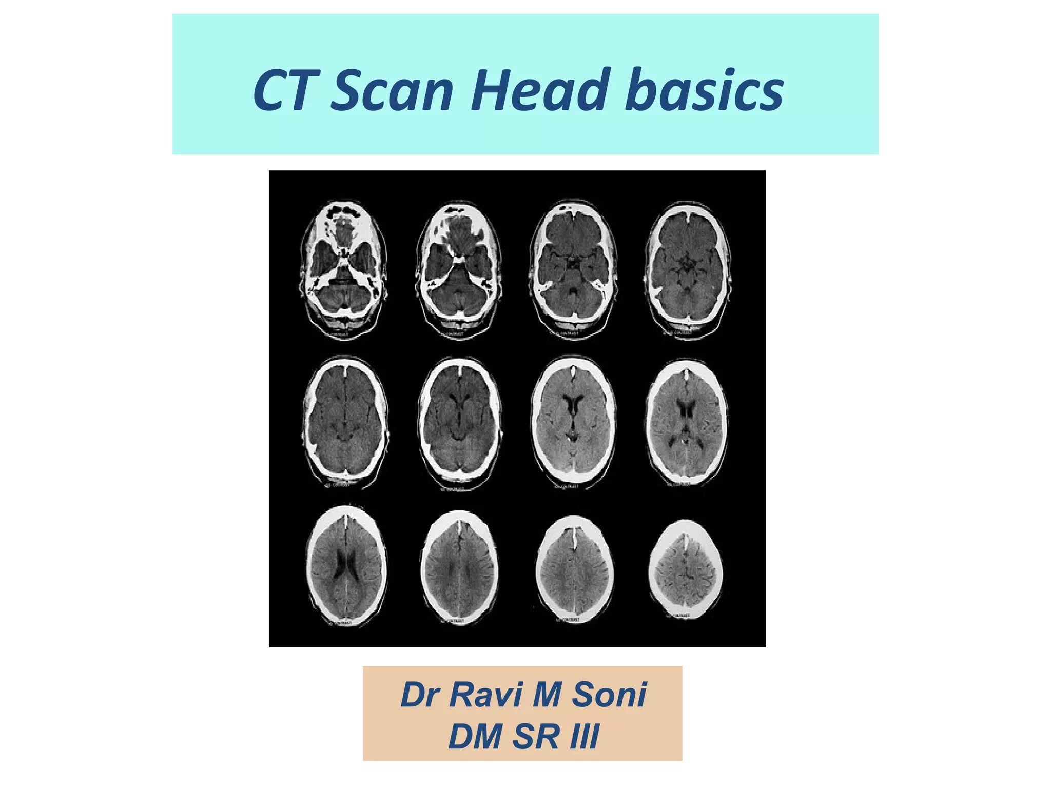


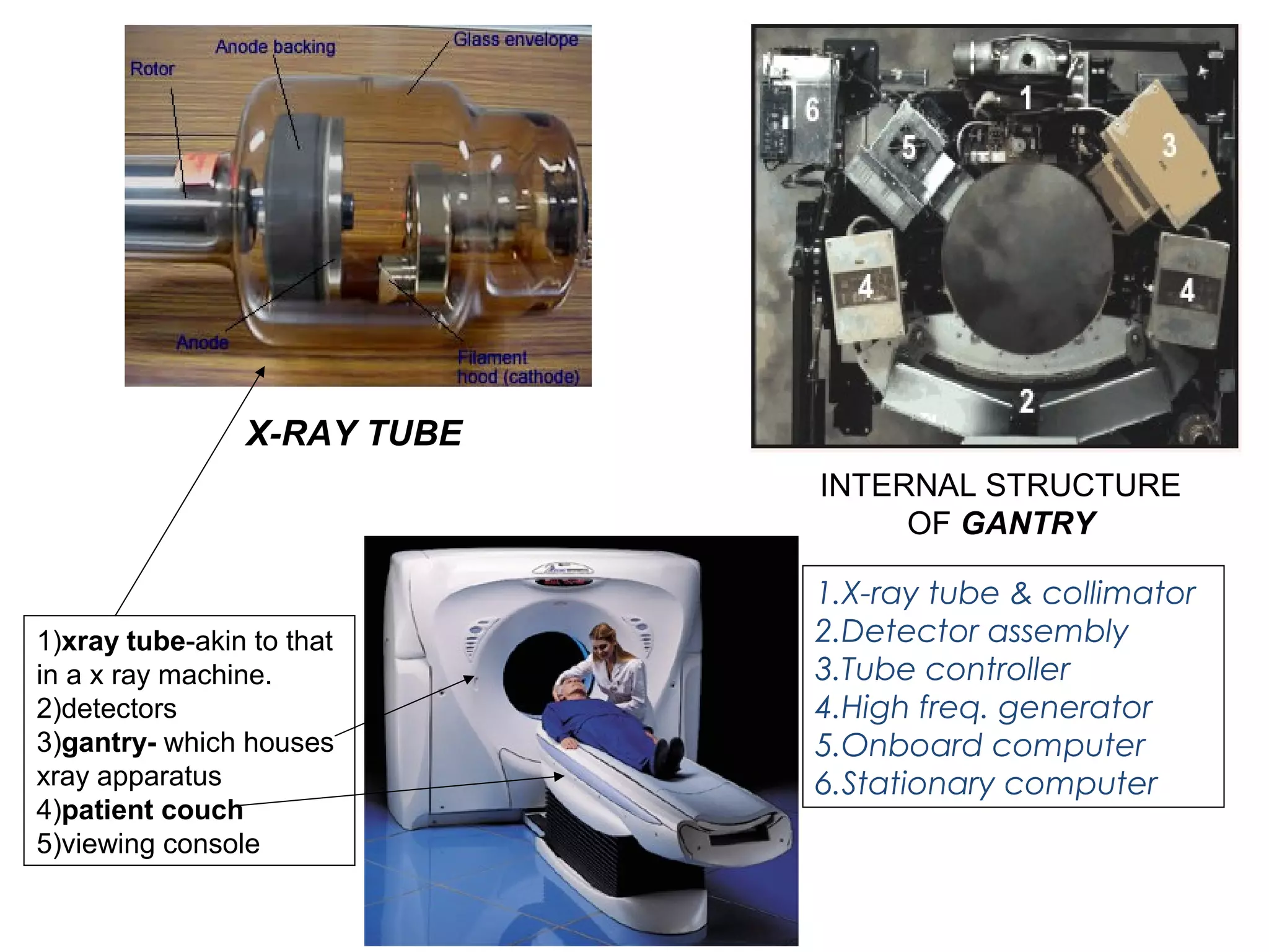

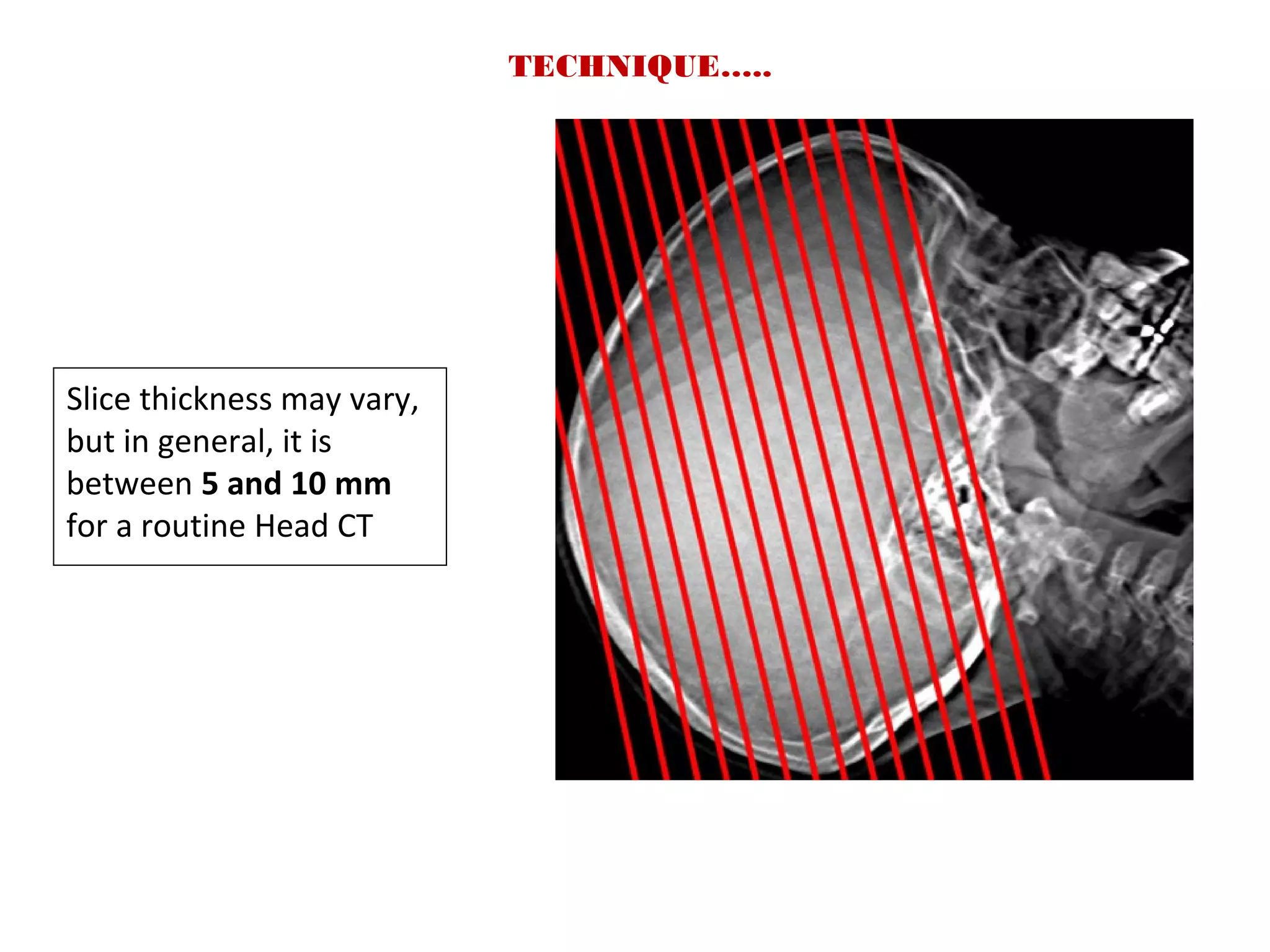


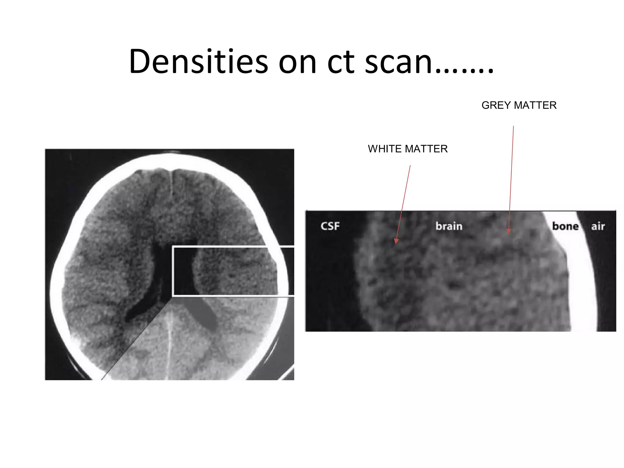


The document outlines the basics of CT scans of the head, including principles, neuroanatomy, and relevant pathologies. It details the components of CT scanners, the construction of images from x-ray data, and how various tissues appear on scans in terms of density. Additionally, it discusses the indications for CT scans in diagnosing neurological conditions, including trauma, infections, and tumors.


