Introduction
Ostracoderms (shell-skinned) are of several groups of extinct, primitive, jawless fishes that were covered in an armour of bony plates.
They appeared in the Cambrian, about 510 million years ago, and became extinct towards the end of the Devonian, about 377 million years ago. They were quite abundant during the upper Silurian and Devonian periods. Most of fossils of Ostracodermi were preserved in the bottom sediments of freshwater streams.
However, the opinion is sharply divided as to whether their habitat was freshwater or marine.
The first fossil fishes that were discovered were ostracoderms.
The Swiss anatomist Louis Agassiz received some fossils of bony armored fish from Scotland in the 1830s.
The ostracoderms resembled the present day cyclostomes (lampreys and hagfishes) in many respects and together with them constitute a special group of jawless vertebrates, the Agnatha.
Characteristics: They use gills exclusively for respiration but not for feeding . Earlier chordates with gills used them for both respiration and feeding. Ostracoderms had separate pharyngeal gill pouches along the side of the head, which were permanently open with no protective operculum. mostly small to medium-sized fishes, protected by a heavy, bony dermal (derived from skin) armor. bottom-dwellers; filter-feeders or grazers. no paired fins, but many with stabilizing paired flaps on either side of head.
(1) Ostracoderms were the first vertebrates.
(2) They were popularly called armoured fishes.
(4) They lived in freshwater.
(5) They were bottom dwellers.
(6) Their body was fish-like and did not exceed 30 cm in size.
(7) Paired fins were absent.
(8) Median and caudal fins were present.
(9) The caudal fin was of heterocercal type.
(10) The head and thorax were covered by heavy armour of bones. It protected ostracoderms from the giant scorpion like arthropods, eurypterids.
(11) Bony skull was well developed.
(12) Mouth was mostly present on the ventral side.
(13) They were having large number of gill slits.
(14) The nervous system had 10 pairs of cranial nerves.
(15) The head had a pair of lateral eyes, and a median pineal eye.
(16) They were filter feeders, feeding like a vacuum cleaner.
(17) The endoskeleton was either bony or cartilaginous.
ORIGIN OF CHORDATES
Animal kingdom is basically divided into two sub kingdoms:
Non-chordata- including animals without notochord.
Chordata- This comprising animals having notochord or chorda dorsalis.
Chordates were evolved sometime 500 million years ago during Cambrian period (invertebrates were also began to evolve in this period) .
Chamberlain (1900) pointed out that all modern chordates possess glomerular kidneys that are designed to remove excess water from body.
It is believed that Chordates have originated from invertebrates.
It is difficult to determine from which invertebrate group the chordates were developed.
Chordate ancestors were soft bodied animals. Hence they were not preserved as Fossils.
However, early fossils of chordates have all been recovered from marine sediments and even modern protochordates are all marine forms.
Also glomerular kidneys are also found in some marine forms such as myxinoids and sharks. That makes the marine origin of chordates more believable.
Chordates evolved from some deuterostome ancestor (echinoderms, hemichordates, pogonophorans etc.) as they have similarities in embryonic development, type of coelom and larval stages.
Many theories infers origin of chordates, hemichordates and echinoderms from a common ancestor.
It discusses basic information regarding a hemichordate animal called Balanoglossus or Acorn worm, which is also a good connecting link between the non-chordates and chordates.
Phylum Mollusca, Class Gastropoda, Torsion, Locomotion, Digestion,Reproductio...Dr. Muhammad Moosa
In this presentation, Phylum Mollusca Is described. After watching this you will learn Evolutionary Perspective of Mollusca and Relationships to Other Animals, Molluscan Characteristics, Class Gastropoda, Torsion, Shell Coiling, Locomotion, Feeding and Digestion, Other Maintenance Functions, Reproduction and Development, Gastropod Diversity, Class Bivalvia, Shell and Associated Structures Gas Exchange, Filter Feeding, and Digestion, Other Maintenance Functions Reproduction and Development, Bivalve Diversity, Class Cephalopoda, Shell, Locomotion, Feeding and Digestion, Other Maintenance Functions, Learning, Reproduction and Development, Class Polyplacophora, Class Scaphopoda, Class Monoplacophora, Class Solenogastres, Class Caudofoveata, Further Phylogenetic Considerations. It is part of BS Zoology Course, Animal diversity.
The alimentary canal of Scoliodon comprises:
the mouth,
buccal cavity,
pharynx,
oesophagus,
stomach,
intestine and
rectum opening in the cloaca through anus.
DENTITION IN MAMMALS
The study of arrangement structure and number of types of teeth collectively is called as dentition. Teeth are present in the foetal as well as in adults of mammals, based on the presence of teeth Mammals are two types.
Edentata : In some animals teeth are absent hence called as edentate. e.g., Echidna or spiny ant-eater (Tachyglossus) the teeth are absent in all stages of life.
Dentata : Teeth are present in all mammals though a secon¬dary toothless condition is found in some mammals. Modern turtles and birds lack teeth. The adult platypus (Ornithorhynchus) bears epidermal teeth but no true teeth are present. In platypus embryonic teeth are replaced by horny epidermal teeth in adult.
Classification According to the Shape and Size of the Teeth:
Homodont:
Homodont or Isodont type of teeth is a condition where the teeth are all alike in their shape and size in the toothed whales e.g., Pinnipedians. Fishes, amphibians, reptiles and in the extinct toothed birds.
Heterodont
Heterodont condition is the usual feature in mammals, i.e. the teeth are distinguished according to their shape, size and function. The function is also different at different parts of the tooth row.
According to the Mode of Attachment of Teeth:
Thecodont : The teeth are lodged in bony sockets or alveoli of the jaw bone and capillaries and nerves enter the pulp cavity through the open tips of the hollow roots e.g., mammals, crocodiles and in some fishes.
Acrodont: The teeth are fused to the surface of the underlying jawbone. They have no roots and are attached to the edge of the jawbone by fibrous membrane e.g., fishes, amphibians and some reptiles.
Pleurodont:
The teeth are attached to the inner-side of the jawbone. The tooth touches the bone only with the outer surface of its root. In acrodont and pleurodont types of dentition, there are no roots, and nerves and blood vessels do not enter the pulp cavity at the base, e.g., Necturus (Amphibia) and some reptiles.
According to the Succession or Replace¬ment of Teeth:
They are five pair in numbers
They includes
paired Antennules
Antennae
Mandibles
First maxillae
second maxillae
They are five pair in numbers
They includes
paired Antennules
Antennae
Mandibles
First maxillae
second maxillae
THIS PRESENTATION IS UPLOADED TO HELP THE EDUCATOR OF MEDICAL, NURSING & ALLIE HEALTH SCIENCES TO TEACH THEIR STUDENTS ABOUT THE NERVOUS SYSTEM. IT WILL ALSO CREATE AWARENESS AMONG THE COMMON PEOPLE REGARDING NERVOUS SYSTEM.
this slide will help undergraduate student to study the difference between Poisonous and non poisonous snakes with examples, Poison apparatus, venom and its uses.
(May 29th, 2024) Advancements in Intravital Microscopy- Insights for Preclini...Scintica Instrumentation
Intravital microscopy (IVM) is a powerful tool utilized to study cellular behavior over time and space in vivo. Much of our understanding of cell biology has been accomplished using various in vitro and ex vivo methods; however, these studies do not necessarily reflect the natural dynamics of biological processes. Unlike traditional cell culture or fixed tissue imaging, IVM allows for the ultra-fast high-resolution imaging of cellular processes over time and space and were studied in its natural environment. Real-time visualization of biological processes in the context of an intact organism helps maintain physiological relevance and provide insights into the progression of disease, response to treatments or developmental processes.
In this webinar we give an overview of advanced applications of the IVM system in preclinical research. IVIM technology is a provider of all-in-one intravital microscopy systems and solutions optimized for in vivo imaging of live animal models at sub-micron resolution. The system’s unique features and user-friendly software enables researchers to probe fast dynamic biological processes such as immune cell tracking, cell-cell interaction as well as vascularization and tumor metastasis with exceptional detail. This webinar will also give an overview of IVM being utilized in drug development, offering a view into the intricate interaction between drugs/nanoparticles and tissues in vivo and allows for the evaluation of therapeutic intervention in a variety of tissues and organs. This interdisciplinary collaboration continues to drive the advancements of novel therapeutic strategies.
Introduction:
RNA interference (RNAi) or Post-Transcriptional Gene Silencing (PTGS) is an important biological process for modulating eukaryotic gene expression.
It is highly conserved process of posttranscriptional gene silencing by which double stranded RNA (dsRNA) causes sequence-specific degradation of mRNA sequences.
dsRNA-induced gene silencing (RNAi) is reported in a wide range of eukaryotes ranging from worms, insects, mammals and plants.
This process mediates resistance to both endogenous parasitic and exogenous pathogenic nucleic acids, and regulates the expression of protein-coding genes.
What are small ncRNAs?
micro RNA (miRNA)
short interfering RNA (siRNA)
Properties of small non-coding RNA:
Involved in silencing mRNA transcripts.
Called “small” because they are usually only about 21-24 nucleotides long.
Synthesized by first cutting up longer precursor sequences (like the 61nt one that Lee discovered).
Silence an mRNA by base pairing with some sequence on the mRNA.
Discovery of siRNA?
The first small RNA:
In 1993 Rosalind Lee (Victor Ambros lab) was studying a non- coding gene in C. elegans, lin-4, that was involved in silencing of another gene, lin-14, at the appropriate time in the
development of the worm C. elegans.
Two small transcripts of lin-4 (22nt and 61nt) were found to be complementary to a sequence in the 3' UTR of lin-14.
Because lin-4 encoded no protein, she deduced that it must be these transcripts that are causing the silencing by RNA-RNA interactions.
Types of RNAi ( non coding RNA)
MiRNA
Length (23-25 nt)
Trans acting
Binds with target MRNA in mismatch
Translation inhibition
Si RNA
Length 21 nt.
Cis acting
Bind with target Mrna in perfect complementary sequence
Piwi-RNA
Length ; 25 to 36 nt.
Expressed in Germ Cells
Regulates trnasposomes activity
MECHANISM OF RNAI:
First the double-stranded RNA teams up with a protein complex named Dicer, which cuts the long RNA into short pieces.
Then another protein complex called RISC (RNA-induced silencing complex) discards one of the two RNA strands.
The RISC-docked, single-stranded RNA then pairs with the homologous mRNA and destroys it.
THE RISC COMPLEX:
RISC is large(>500kD) RNA multi- protein Binding complex which triggers MRNA degradation in response to MRNA
Unwinding of double stranded Si RNA by ATP independent Helicase
Active component of RISC is Ago proteins( ENDONUCLEASE) which cleave target MRNA.
DICER: endonuclease (RNase Family III)
Argonaute: Central Component of the RNA-Induced Silencing Complex (RISC)
One strand of the dsRNA produced by Dicer is retained in the RISC complex in association with Argonaute
ARGONAUTE PROTEIN :
1.PAZ(PIWI/Argonaute/ Zwille)- Recognition of target MRNA
2.PIWI (p-element induced wimpy Testis)- breaks Phosphodiester bond of mRNA.)RNAse H activity.
MiRNA:
The Double-stranded RNAs are naturally produced in eukaryotic cells during development, and they have a key role in regulating gene expression .
Slide 1: Title Slide
Extrachromosomal Inheritance
Slide 2: Introduction to Extrachromosomal Inheritance
Definition: Extrachromosomal inheritance refers to the transmission of genetic material that is not found within the nucleus.
Key Components: Involves genes located in mitochondria, chloroplasts, and plasmids.
Slide 3: Mitochondrial Inheritance
Mitochondria: Organelles responsible for energy production.
Mitochondrial DNA (mtDNA): Circular DNA molecule found in mitochondria.
Inheritance Pattern: Maternally inherited, meaning it is passed from mothers to all their offspring.
Diseases: Examples include Leber’s hereditary optic neuropathy (LHON) and mitochondrial myopathy.
Slide 4: Chloroplast Inheritance
Chloroplasts: Organelles responsible for photosynthesis in plants.
Chloroplast DNA (cpDNA): Circular DNA molecule found in chloroplasts.
Inheritance Pattern: Often maternally inherited in most plants, but can vary in some species.
Examples: Variegation in plants, where leaf color patterns are determined by chloroplast DNA.
Slide 5: Plasmid Inheritance
Plasmids: Small, circular DNA molecules found in bacteria and some eukaryotes.
Features: Can carry antibiotic resistance genes and can be transferred between cells through processes like conjugation.
Significance: Important in biotechnology for gene cloning and genetic engineering.
Slide 6: Mechanisms of Extrachromosomal Inheritance
Non-Mendelian Patterns: Do not follow Mendel’s laws of inheritance.
Cytoplasmic Segregation: During cell division, organelles like mitochondria and chloroplasts are randomly distributed to daughter cells.
Heteroplasmy: Presence of more than one type of organellar genome within a cell, leading to variation in expression.
Slide 7: Examples of Extrachromosomal Inheritance
Four O’clock Plant (Mirabilis jalapa): Shows variegated leaves due to different cpDNA in leaf cells.
Petite Mutants in Yeast: Result from mutations in mitochondrial DNA affecting respiration.
Slide 8: Importance of Extrachromosomal Inheritance
Evolution: Provides insight into the evolution of eukaryotic cells.
Medicine: Understanding mitochondrial inheritance helps in diagnosing and treating mitochondrial diseases.
Agriculture: Chloroplast inheritance can be used in plant breeding and genetic modification.
Slide 9: Recent Research and Advances
Gene Editing: Techniques like CRISPR-Cas9 are being used to edit mitochondrial and chloroplast DNA.
Therapies: Development of mitochondrial replacement therapy (MRT) for preventing mitochondrial diseases.
Slide 10: Conclusion
Summary: Extrachromosomal inheritance involves the transmission of genetic material outside the nucleus and plays a crucial role in genetics, medicine, and biotechnology.
Future Directions: Continued research and technological advancements hold promise for new treatments and applications.
Slide 11: Questions and Discussion
Invite Audience: Open the floor for any questions or further discussion on the topic.
Earliest Galaxies in the JADES Origins Field: Luminosity Function and Cosmic ...Sérgio Sacani
We characterize the earliest galaxy population in the JADES Origins Field (JOF), the deepest
imaging field observed with JWST. We make use of the ancillary Hubble optical images (5 filters
spanning 0.4−0.9µm) and novel JWST images with 14 filters spanning 0.8−5µm, including 7 mediumband filters, and reaching total exposure times of up to 46 hours per filter. We combine all our data
at > 2.3µm to construct an ultradeep image, reaching as deep as ≈ 31.4 AB mag in the stack and
30.3-31.0 AB mag (5σ, r = 0.1” circular aperture) in individual filters. We measure photometric
redshifts and use robust selection criteria to identify a sample of eight galaxy candidates at redshifts
z = 11.5 − 15. These objects show compact half-light radii of R1/2 ∼ 50 − 200pc, stellar masses of
M⋆ ∼ 107−108M⊙, and star-formation rates of SFR ∼ 0.1−1 M⊙ yr−1
. Our search finds no candidates
at 15 < z < 20, placing upper limits at these redshifts. We develop a forward modeling approach to
infer the properties of the evolving luminosity function without binning in redshift or luminosity that
marginalizes over the photometric redshift uncertainty of our candidate galaxies and incorporates the
impact of non-detections. We find a z = 12 luminosity function in good agreement with prior results,
and that the luminosity function normalization and UV luminosity density decline by a factor of ∼ 2.5
from z = 12 to z = 14. We discuss the possible implications of our results in the context of theoretical
models for evolution of the dark matter halo mass function.
2. The nervous system of Scoliodon includes:
(i) The central nervous system (CNS)
(ii) The peripheral nervous system (PNS)
(iii) The autonomous nervous system (ANS)
Nusrat Perween, AISC Pune 2
3. (i) Central nervous system:
(a) Brain:
(b) Spinal cord:
(ii) Peripheral nervous system:
• cranial nerves and spinal nerves.
(iii) Autonomous nervous system:
• series of paired ganglia (sympathetic and parasympathetic)
Nusrat Perween, AISC Pune 3
4. Introduction
• Brain of shark is heavy and large
• Externally covered by cranium, made up of cartilage and two
connective tissue membranes called meninges
• thick membrane dura- mater and thin membrane close to brain
pia- mater
• Brain and spinal cord is hallow from inside and cavities called
ventricles
• Space between meninges and ventricle of brain are filled with
cerebro-spinal fluid
• Special feature- Olfactory lobes and cerebellum well developed
Nusrat Perween, AISC Pune 4
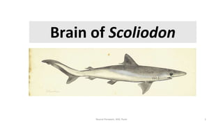
5. Brain of Scoliodon
The brain is divided into three
primary parts:
(a) Forebrain or Prosencephalon
(b) Midbrain or Mesencephalon
(c) Hindbrain or Rhombencephalon
5
6. a) Forebrain or Prosencephalon
Two part
1) Telencephalon-
i. Olfactory lobes
ii. Cerebrum
2) Diencephalon
6Dorsal view Ventral view
7. i) Olfactory lobes
• Anterior end of cerebral hemisphere
arise two stout olfactory peduncles;
each terminates into a large bilobed
olfactory lobe
1. Olfactory bulb
2. Olfactory sac
3. Olfactory peduncle
• Cavity of olfactory lobe- Rhinocoels
(1st ventricle)
• Function- sense of smell
7
Nusrat Perween, AISC Pune
8. ii) Cerebrum
• 2nd part of Telencephalon
• massive undivided mass of nervous tissue
• larger than other fishes.
• Neuropore- ventrally on the cerebrum, through which terminal or pre-
olfactory nerve comes out
• Cavities – paracoels or lateral ventricles (2nd ventricle)
• Function- all voluntary activities
Nusrat Perween, AISC Pune
8
9. 2. Diencephalon
• Present behind cerebrum (The
posterior part of forebrain)
• Completely covered by cerebellum
on dorsal side
i. Epithalamus (Pineal body)
ii. Hypothalamus
iii. Infundibulum
iv. Optic chiasma
v. Pituitary gland (hypophysis)
9
Nusrat Perween, AISC Pune
10. i. Anterior choroid plexus (telachoroidea):
• The roof of the diencephalon is thin, non-nervous and
highly vascular known as the anterior choroid plexus.
• Secretes cerebrospinal fluid and supplies nutrition to
cavities of brain
ii. Epithalamus:
• It comprises of epiphyris or pineal organ and habenular
gangalia
• Pineal body arises from posterior mid-dorsal end of
diencephalon
• Long and tubular
• The lateral walls of the diencephalon form two thickened
bodies called thalami.
• Function- not known, may be involved in reception of light
10
11. iii. Hypothalamus
• Ventral part of diencephalon
• Very well developed in fishes
• Contains neuro-secretory cells
• Functions- secretes neuro-secretary material
iv. Infundibulum
• Arises from floor of diencephalon
• Along the sides there are thick walled sacs called
inferior lobes
• Near distal end of each inferior lobe is wrinkled
glandular sac called saccus-vasculosus
• Saccus-vasculosus is highly pigmented ,vascular and
folded
• Saccus-vasculosus function- depth reception,
secretion of ventricular fluid
11
12. v. Optic chiasma
• On ventral side of brain in front of infundibulum
the two optic nerves arising from each eye ball
cross and form optic chiamsa (X)
vi. Pituitary gland
• Large in elasmobranchs
• Present on ventral side behind the infundibulum
(hidden)
• Divided into 3 parts- Anterior, dorsal and ventral
lobes
• Ventral lobe –prominent secretes TSH and FSH
• Cavity of diencephalon is called as diocoel (3rd
ventricle)
• Both cavities are join through foramen of Monro
12
13. • Dorsally and ventrally covered by
cerebellum and infundibulum
• large and consists of two round optic
lobes.
• Ventricle –optocoel connected to diocoel
by iter or aqueduct of sylvius
• Function –sense of vision
13
Midbrain or mesencephalon
15. • Highly developed cerebellum and a medulla
oblongata.
• Rhomboidal in shape
• The dorsal surface of the cerebellum shows
irregular folds
• Median longitudinal furrow – right and left
halves
• Two transverse furrow- three lobes
• Cavity – epicoel
• Function- center of co-ordination of body
movement
15
i. Cerebellum
16. • Posterior most part of brain
• Triangular in shape
• anterior end gives a pair of hollow
corpora restiformia
• The roof of the medulla oblongata is
non-nervous and highly vascular and
form posterior choroid plexus that
projects into ventricle and provide
nourishment
• The hind- brain controls swimming
movements.
• Cavity – metacoel or 4th ventricle
• Function- controls involuntary activities
16
ii. medulla oblongata or myelencephalon
18. 18
1. Olfactory lobes- Rhinocoel or 1st
ventricle
2. Cerebrum- paracoels or lateral
ventricles (2nd ventricle)
3. Diencephalon- diocoel (3rd ventricle)
4. Both diocoel are join through foramen
of Monro
5. Optic lobes- optocoel
6. Optocoel is connected to diocoel by iter
or aqueduct of sylvius
7. Cerebellum- epicoel
8. Medulla oblongata- metacoel or 4th
ventricle
Cavities or ventricles of brain