Anatomy of boneand
its functions
Dr.Navaneesh
Moderator:Dr.Anna mohan
2.
Contents
Formationof bone
Classification of bones
Structure of bone
Blood supply
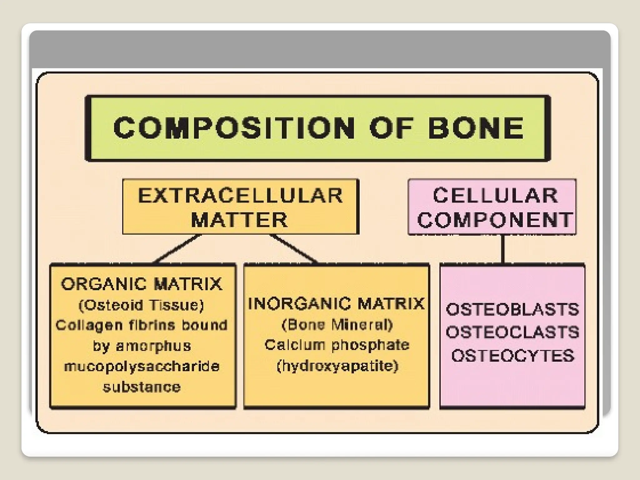
Composition of bone
3.
Bone(syn os-osteon)
Osseous tissue,a specialized form of
dense connective tissue consisting of bone
cells(osteocytes)
Embedded in a matrix of calcified
intercellular substance
Bone matrix contains collagen
fibers,minerals like calcium phosphate and
calcium carbonate
4.
Formation of bone
All bone is of mesodermal origin
Two types of ossification
Intramembranous ossification
Endochondral ossification
7.
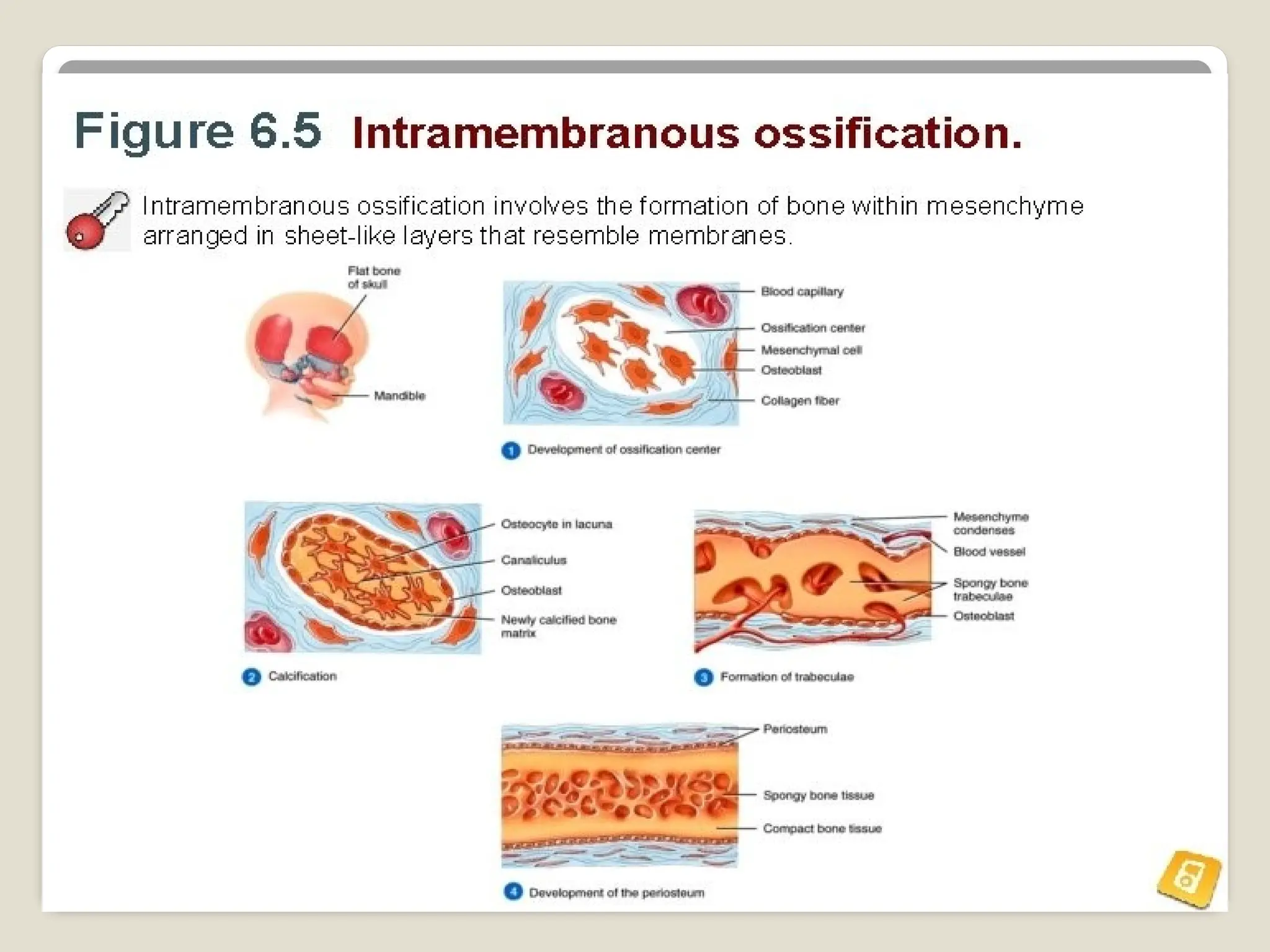
Intramembranous ossification
Mesenchymalcondensation
Highly vascular
Laying down of bundle of collagen fibers in
mesenchymal condensation
Osteoblast formation-osteiod
Calcium salts deposition-lamellus of bone
9.
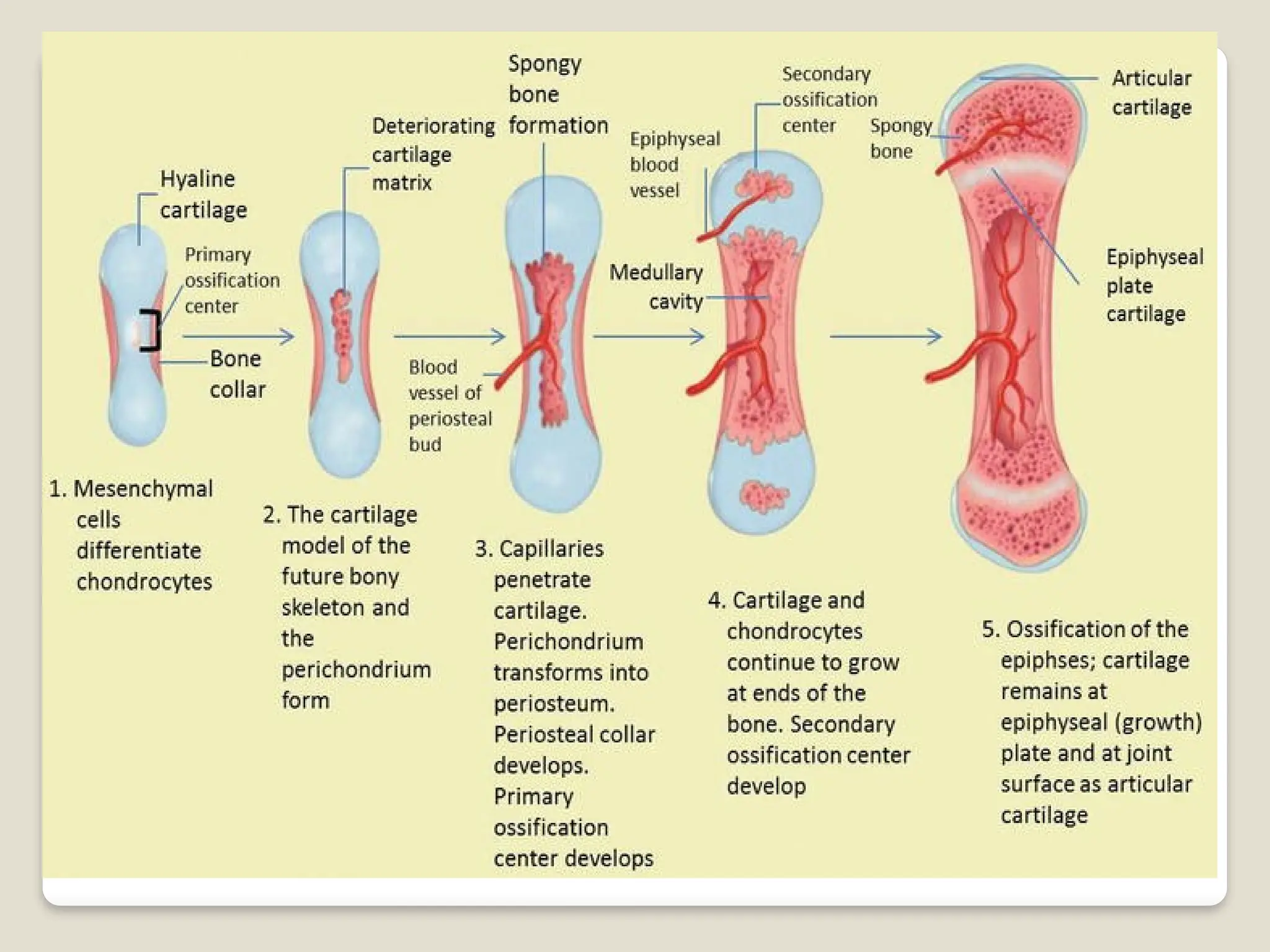
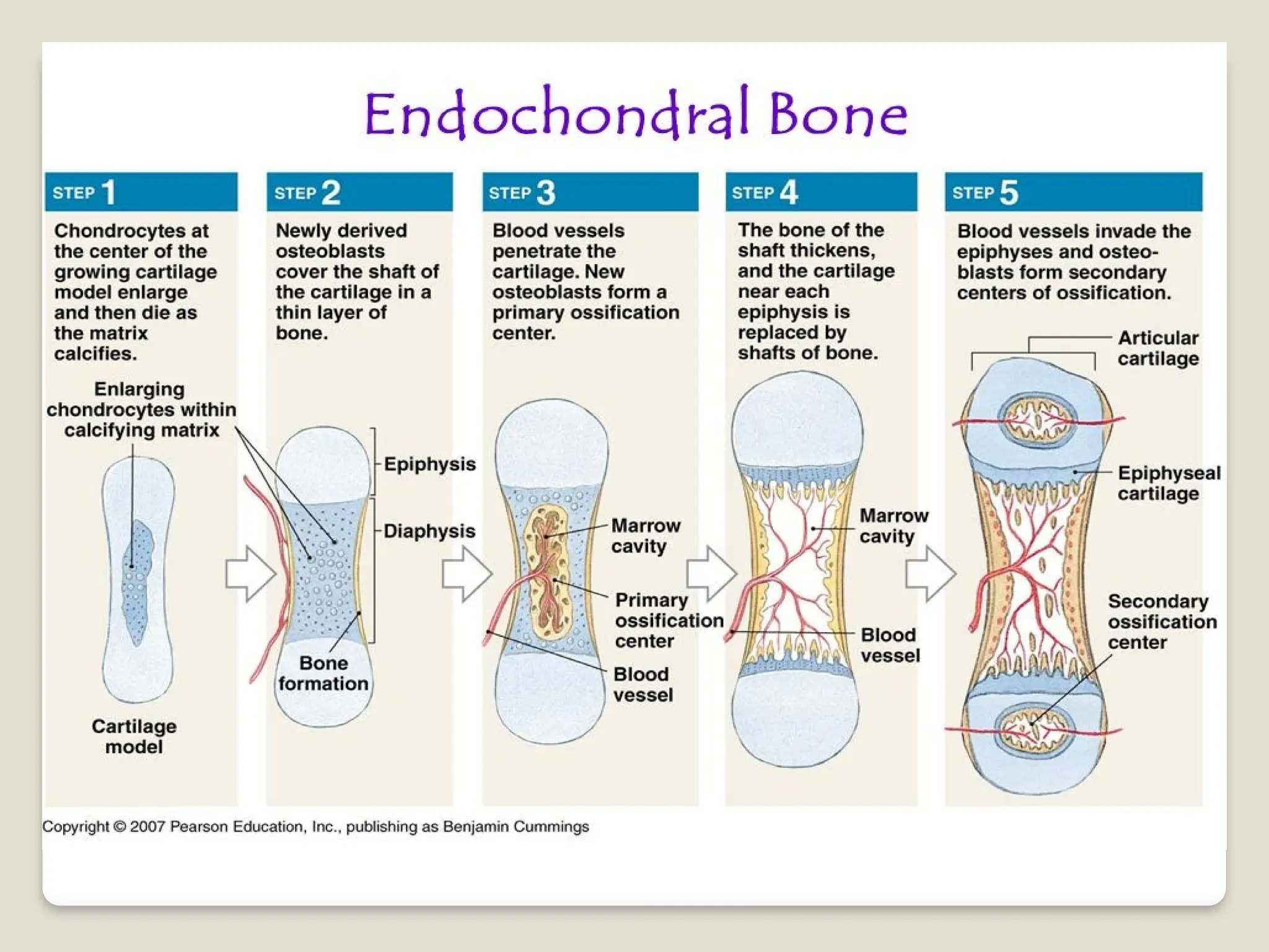
Enchondral ossification
Ossifiesbone that originate as hyaline
cartilage
Most bones originate as hyaline cartilage
Growth and ossification of long bones
occur in 6 steps
11.
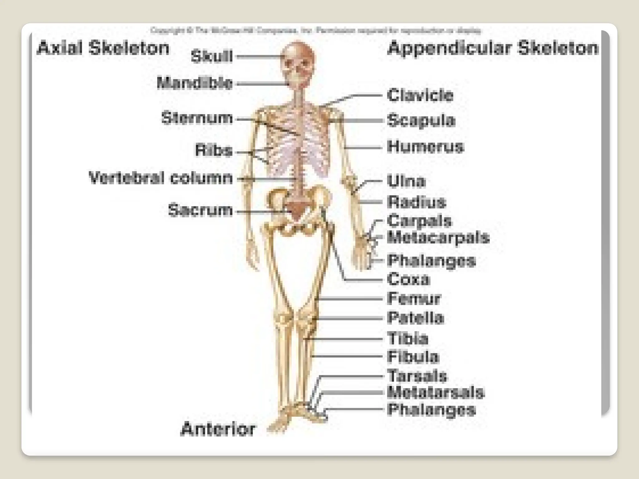
Skeletal organization
Theactual number of bones in human
skeleton varies from person to person
Typically there are about 206 bones
For convenience the skeleton is divided
into the
Axial skeleton
Appendicular skeleton
13.
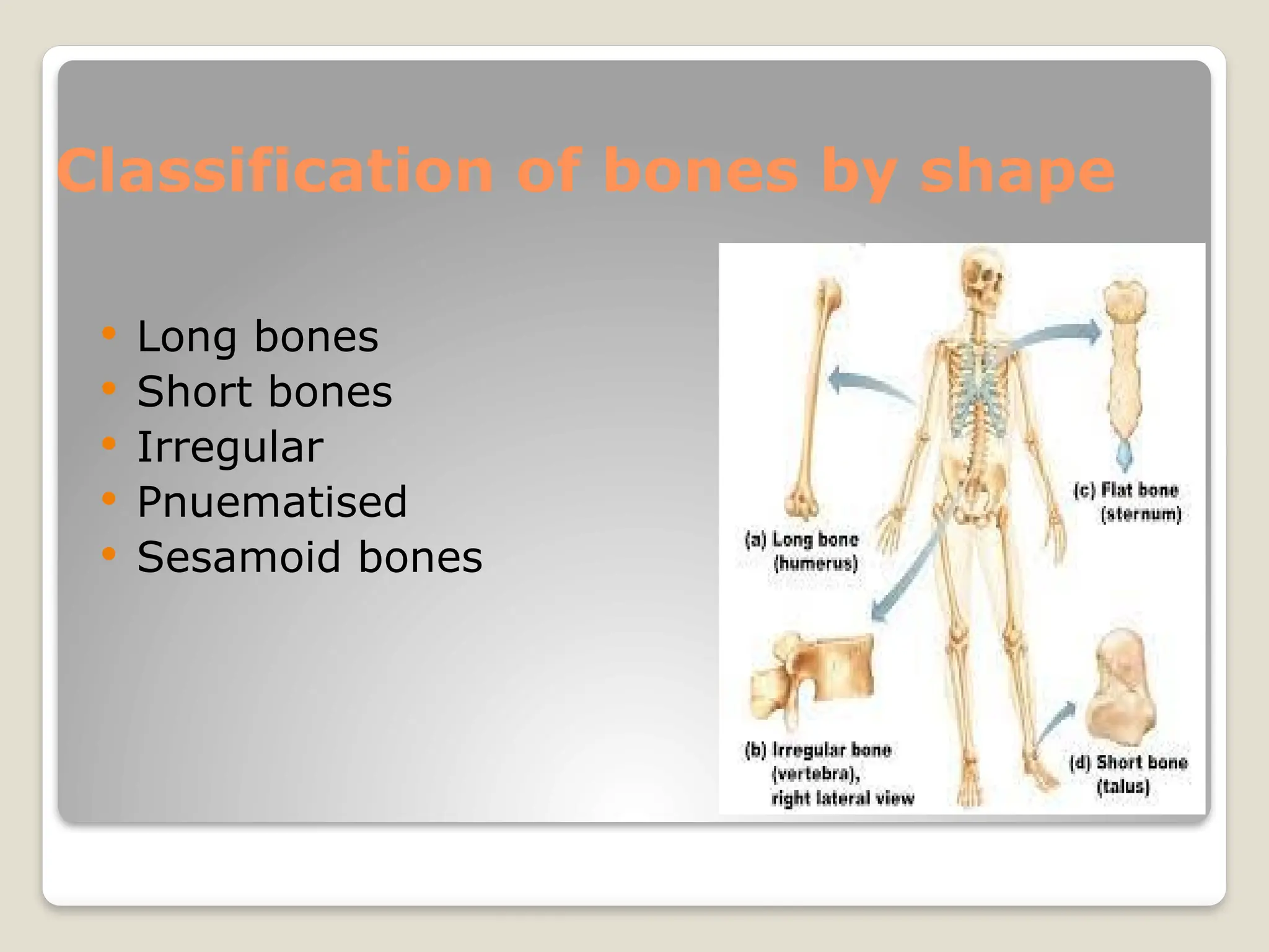
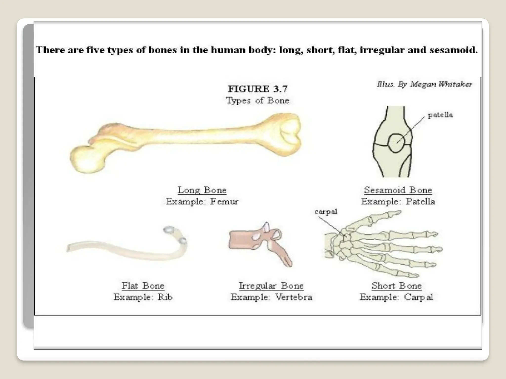
Classification of bonesby shape
Long bones
Short bones
Irregular
Pnuematised
Sesamoid bones
14.
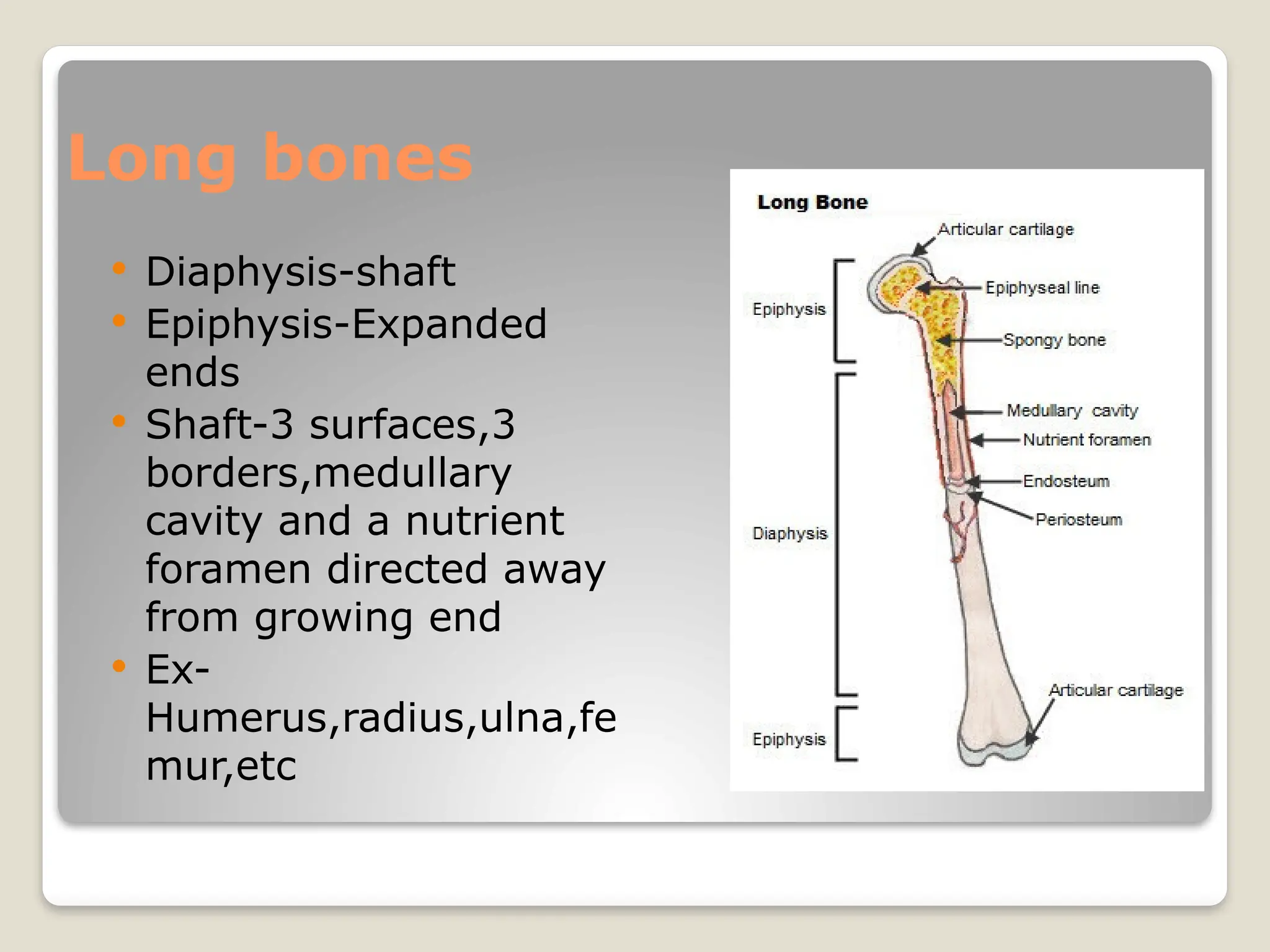
Long bones
Diaphysis-shaft
Epiphysis-Expanded
ends
Shaft-3 surfaces,3
borders,medullary
cavity and a nutrient
foramen directed away
from growing end
Ex-
Humerus,radius,ulna,fe
mur,etc
15.
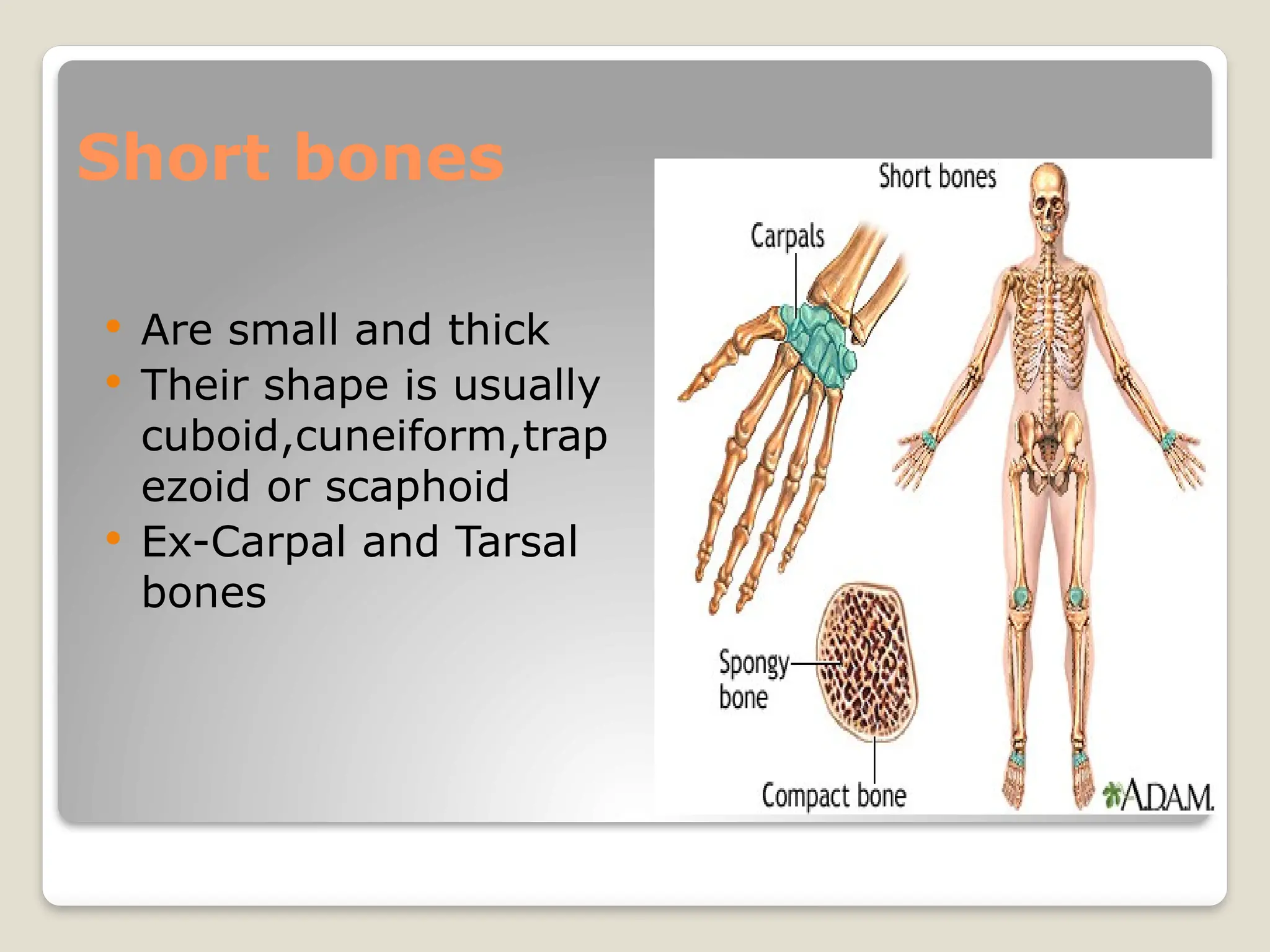
Short bones
Aresmall and thick
Their shape is usually
cuboid,cuneiform,trap
ezoid or scaphoid
Ex-Carpal and Tarsal
bones
16.
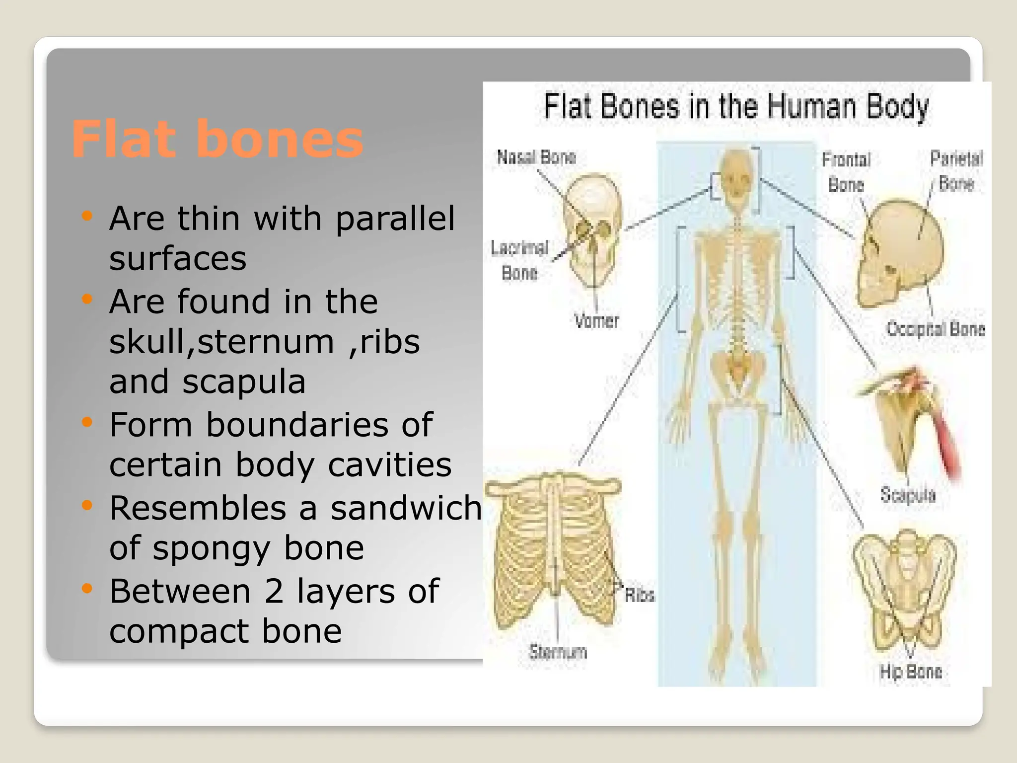
Flat bones
Arethin with parallel
surfaces
Are found in the
skull,sternum ,ribs
and scapula
Form boundaries of
certain body cavities
Resembles a sandwich
of spongy bone
Between 2 layers of
compact bone
17.
Pnuematic bones
Certainirregular bones contain large air
spaces lined by epithelium
Make the skull light in weight ,help in
resonance of voice and act as air
conditioning chambers for inspired air.
Ex –Maxilla,Sphenoid,Ethmoid,etc
18.
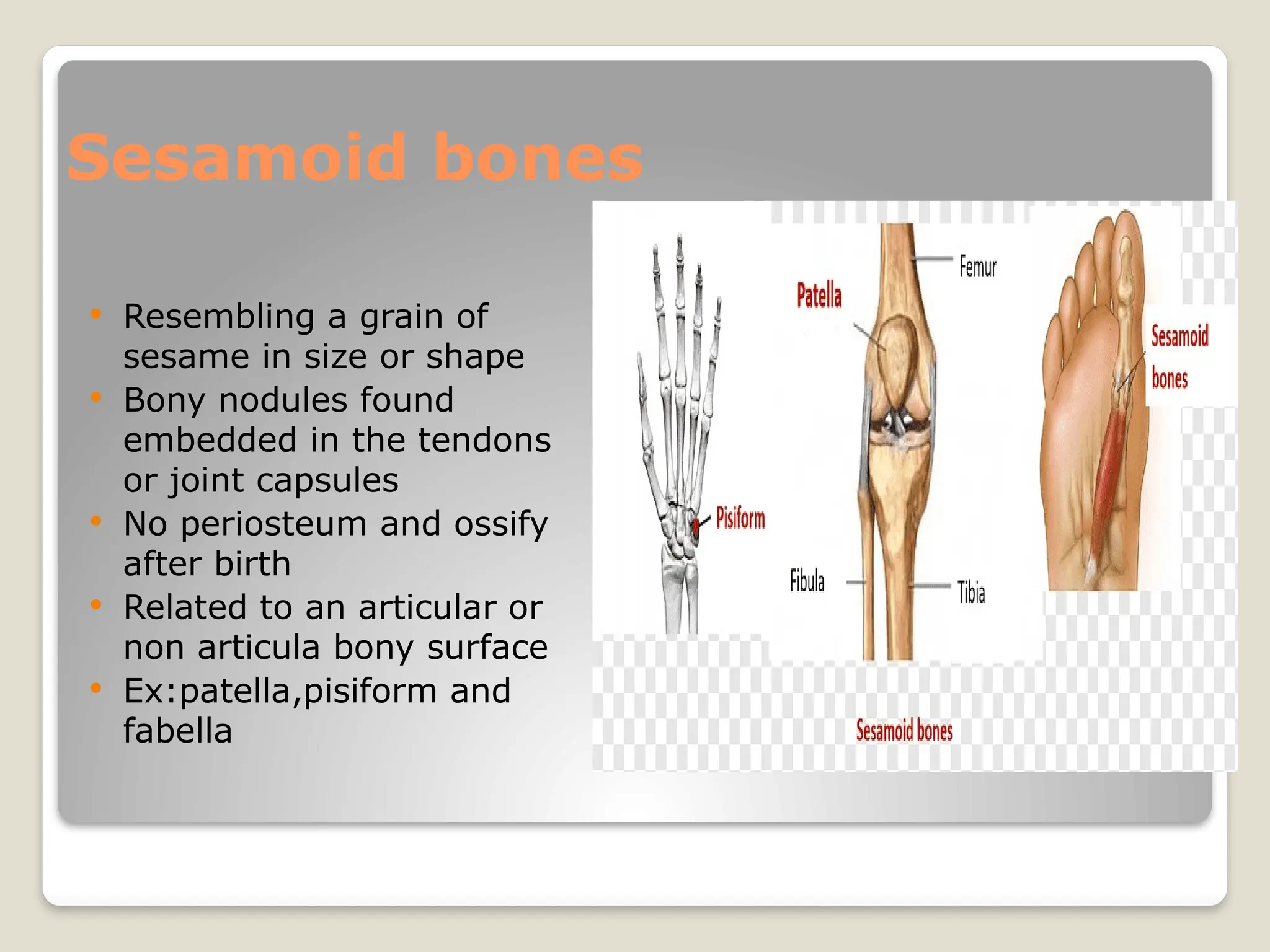
Sesamoid bones
Resemblinga grain of
sesame in size or shape
Bony nodules found
embedded in the tendons
or joint capsules
No periosteum and ossify
after birth
Related to an articular or
non articula bony surface
Ex:patella,pisiform and
fabella
Membrane(dermal )bones
Ossify in membrane(intramembranous of
mesenchymal.
Derived from mesenchymal condensations
Ex-bones of the vault of skull and facial
bones
Defect-Cleidocranial dysostosis
23.
Cartilaginous bones
Ossifyin cartilage(intracartilagenous or
endochondral)
Derived from preformed cartilaginous
models
Ex-Bone of limbs,Vertebral column and
thoracic cage
Defect-Common type of dwarfism called
achondroplasia.
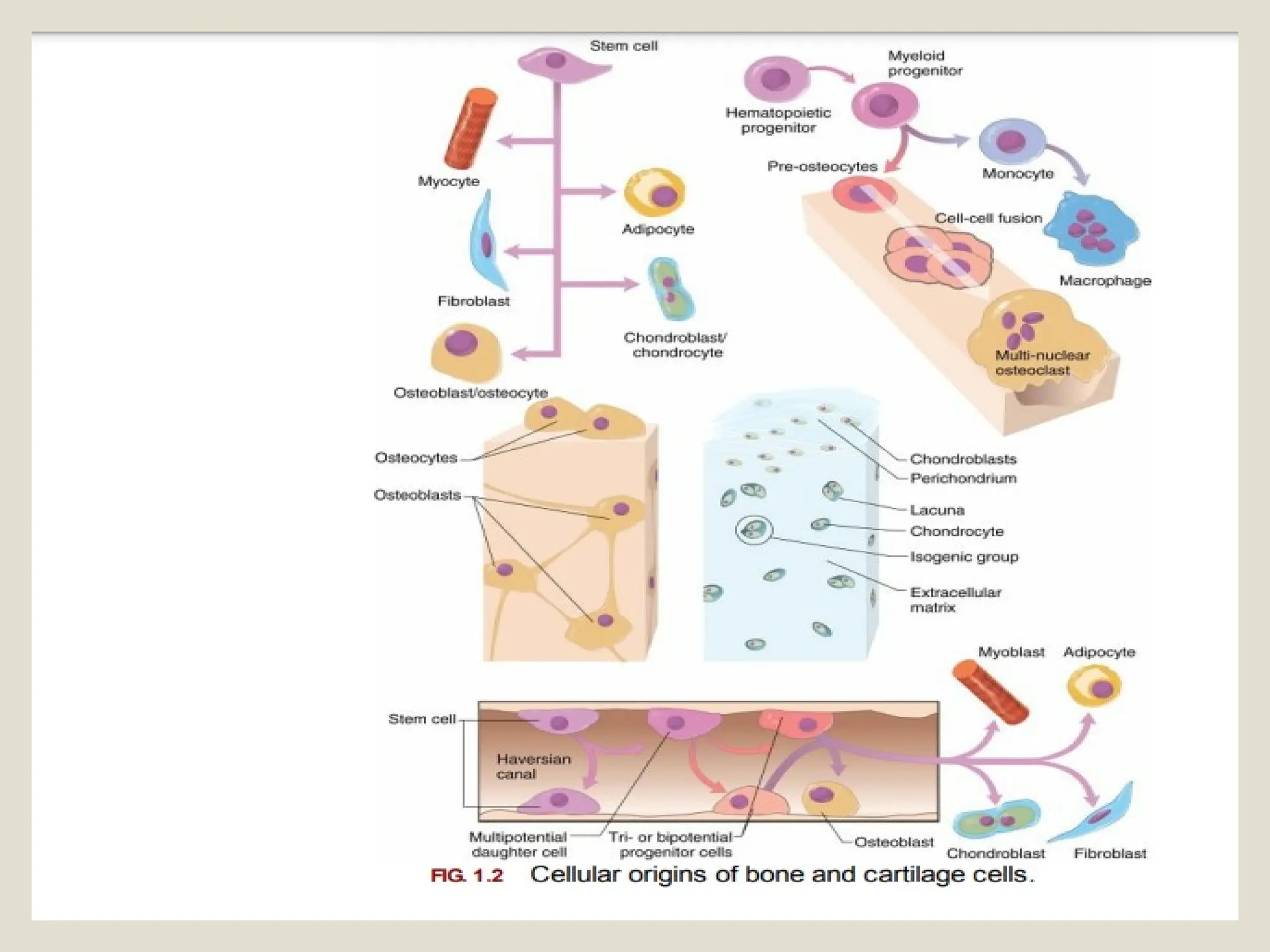
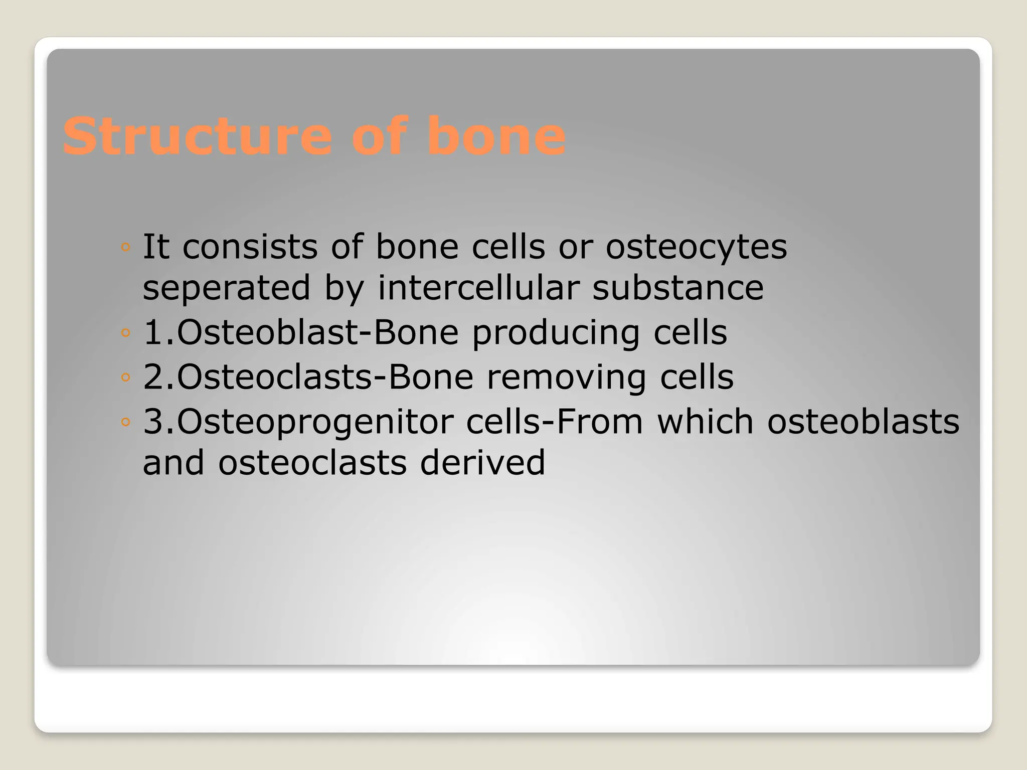
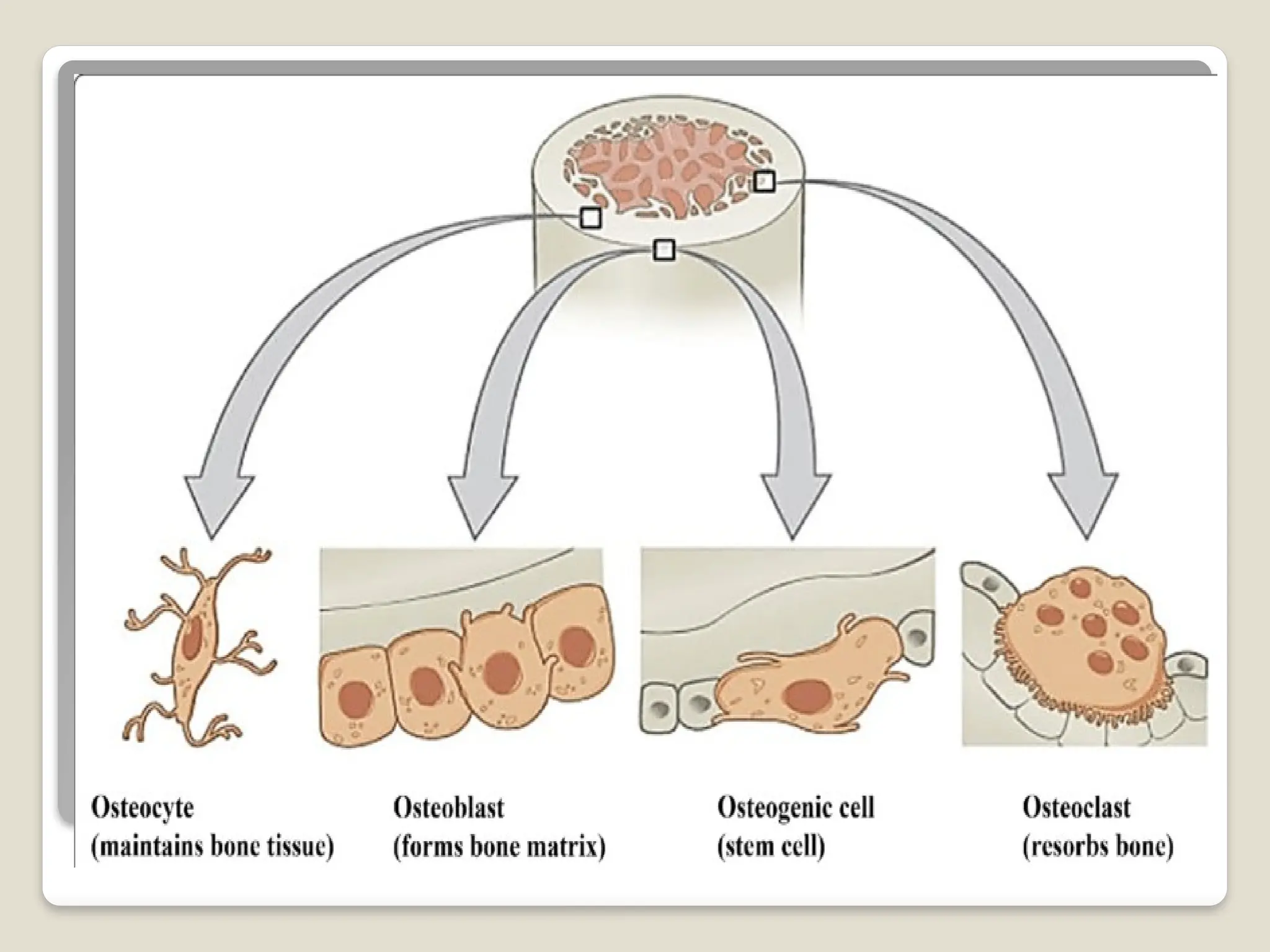
Structure of bone
◦It consists of bone cells or osteocytes
seperated by intercellular substance
◦ 1.Osteoblast-Bone producing cells
◦ 2.Osteoclasts-Bone removing cells
◦ 3.Osteoprogenitor cells-From which osteoblasts
and osteoclasts derived
27.
Osteoprogenitor cells
Mesenchymalstem cells that divide to
produce osteoblasts
Are located in inner,cellular layer of
periosteum(endosteum)
Assist in fracture repair
28.
Osteoblasts
Immature bonecells that secrete matrix
compounds(osteogenesis)
Matrix produced by osteoblasts,but not yet
calcified to form bone
Osteoblasts surrounded by bone become
osteocytes
29.
Osteocytes
Mature bonecells that maintain the bone
matrix
Live in lacunae
Are between layers (lamellae)of matrix
Connect by cytoplasmic extensions
through canaliculi in lamellae
Do not divide
30.
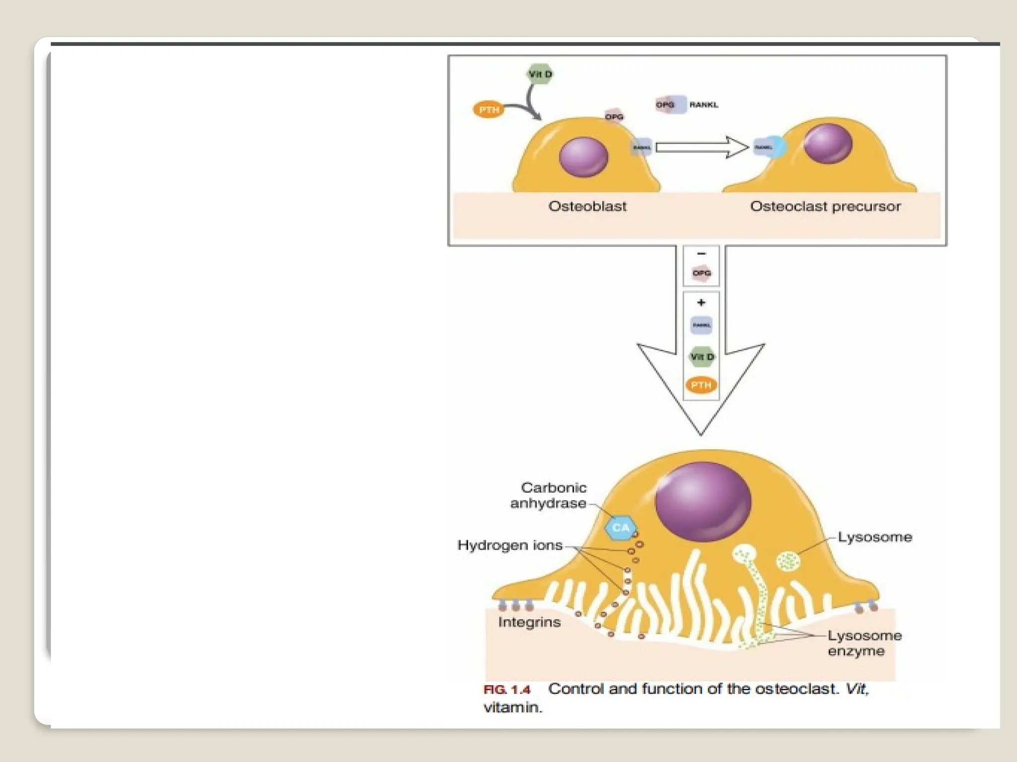
Osteoclast
Secrete acidsand protein digesting
enzymes
Giant,multinucleate cells
Dissolve bone matrix and release stored
minerals(osteolysis)
Are derived from stem cells that produce
macrophages
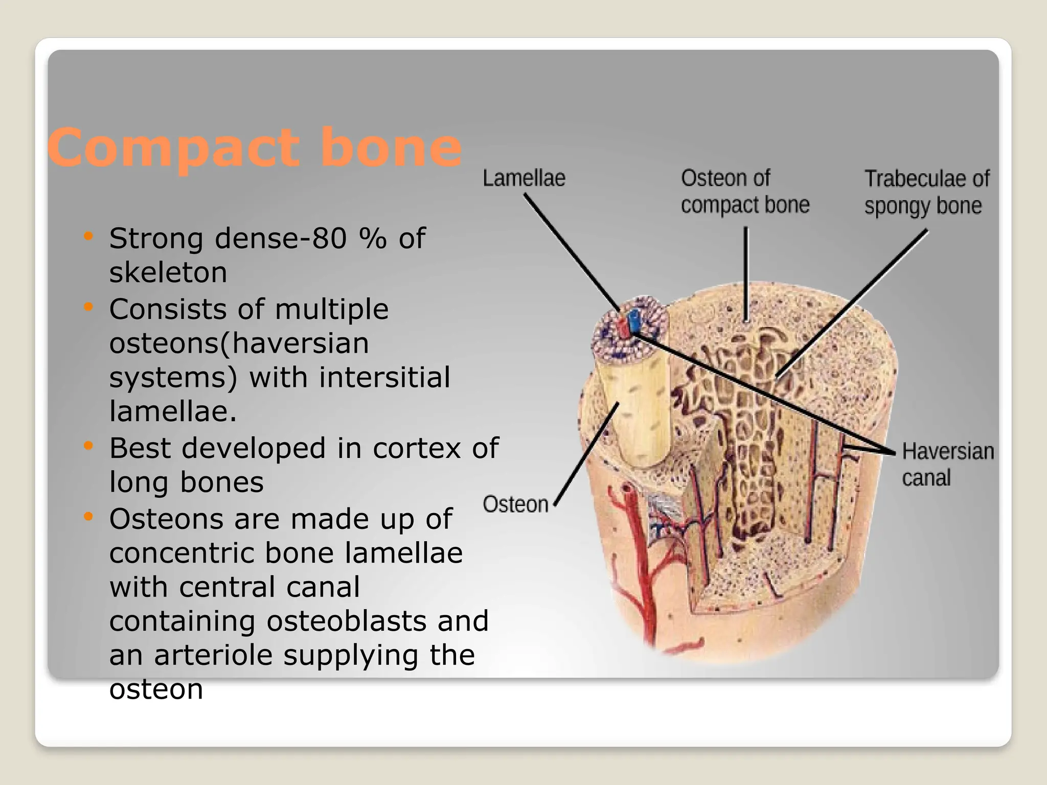
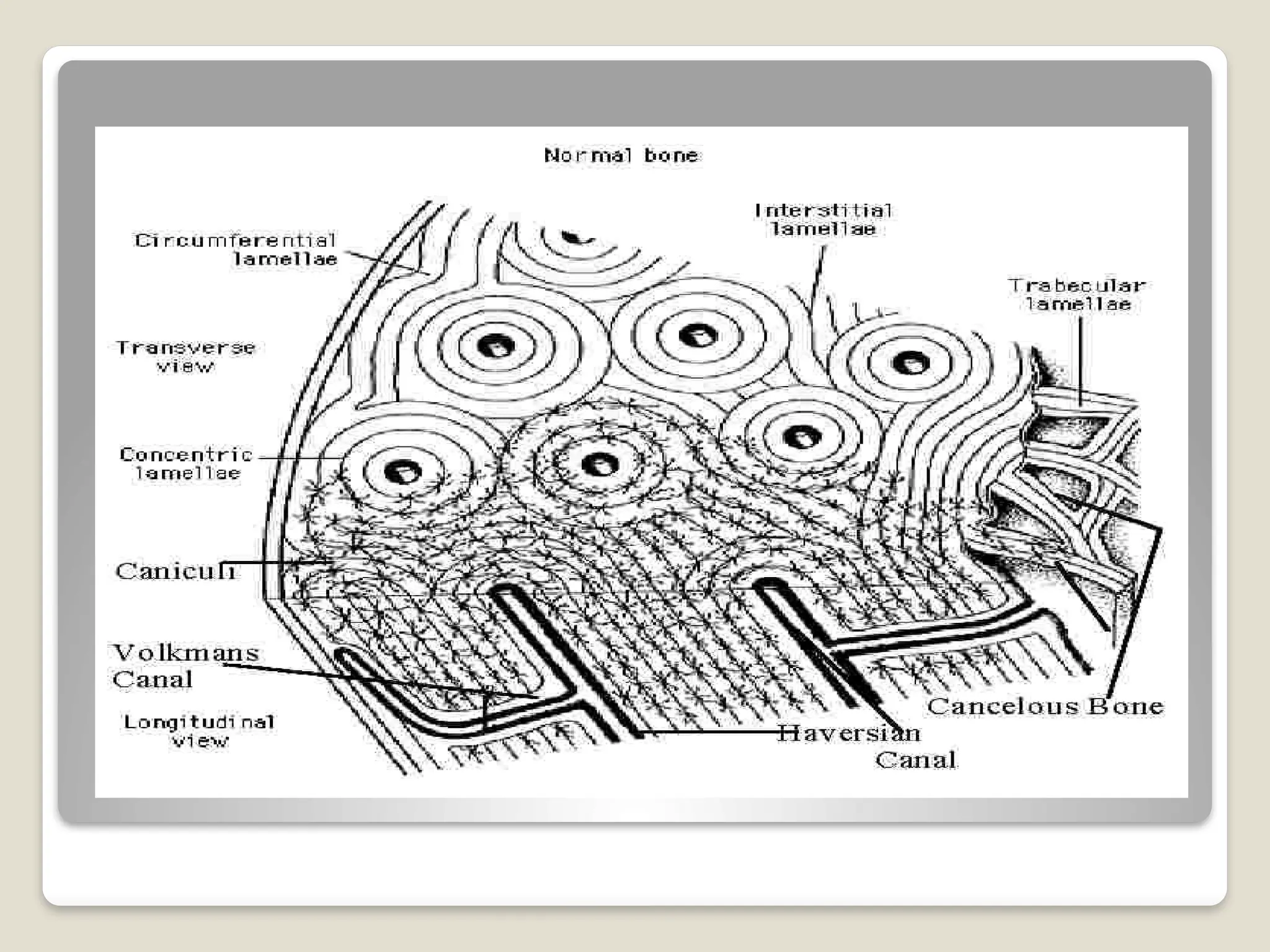
Compact bone
Strongdense-80 % of
skeleton
Consists of multiple
osteons(haversian
systems) with intersitial
lamellae.
Best developed in cortex of
long bones
Osteons are made up of
concentric bone lamellae
with central canal
containing osteoblasts and
an arteriole supplying the
osteon
34.
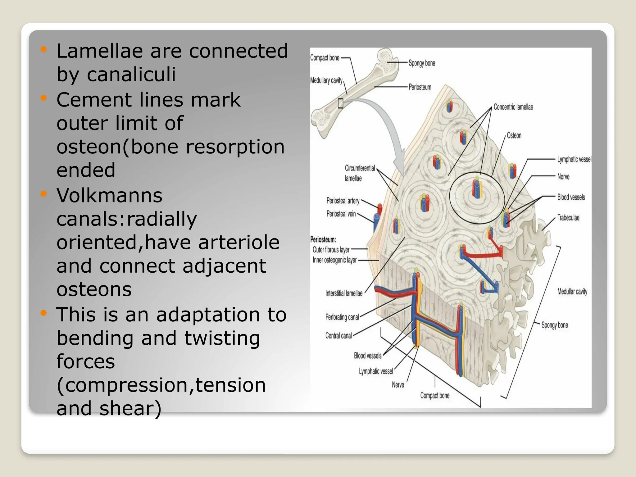
Lamellae areconnected
by canaliculi
Cement lines mark
outer limit of
osteon(bone resorption
ended
Volkmanns
canals:radially
oriented,have arteriole
and connect adjacent
osteons
This is an adaptation to
bending and twisting
forces
(compression,tension
and shear)
35.
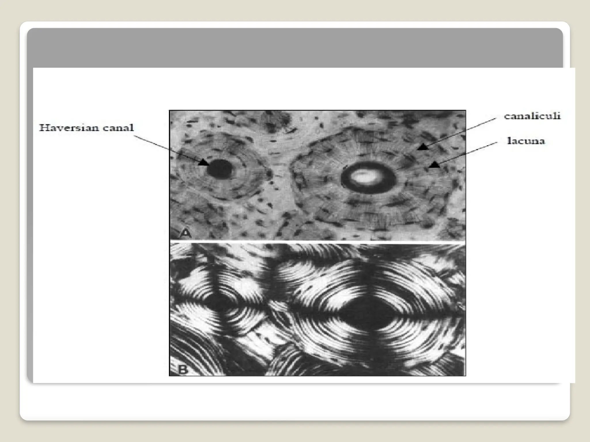
Osteon
The basicunit of mature compact bone
Osteocytes are arranged in concentric
lamellae
Around a central canal containing blood
vessels
38.
Cancellous bone(spongy or
trabecular)
Open in texture-Meshwork of trabeculae(rods
and plates)
Crossed lattice structure ,makes up 20% of
skeleton
High bone turnover rate
Bone is resorbed by osteoclasts in howship
lacunae and formed on the opposite side of
trabeculae by osteoblasts
Osteoporosis is common in cancellous
bones,making it susceptible to fracture.
39.
Commonly foundin the metaphysis and
epiphysis of long bones
Adaptation to compressive forces.
Does not have osteons
The matrix forms an open network of
trabeculae
Trabeculae have no blood vessels
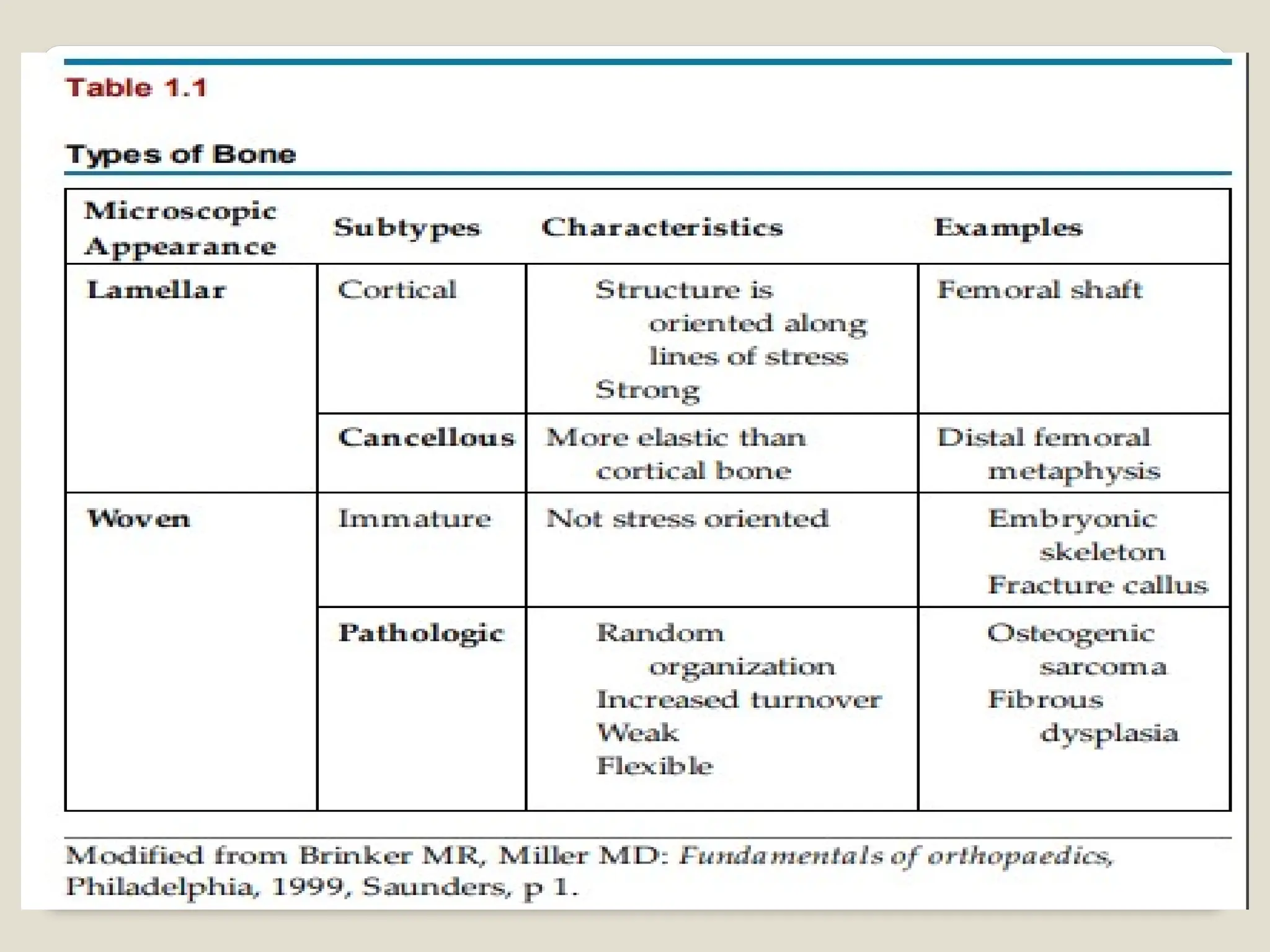
LAMELLAR BONE
Boneis made up of layers or lamellae
Lamellae is a thin plate of bone consisting
of collagen fibers and mineral
salts,deposited in gelatinous ground
substance
Between adjoining lamellae we see small
flattened spaces-lacunae
43.
Lacunae
containsone osteocyte
Have fine canals or canaliculi that
communicate with those from other
lacunae
Fibers of one lamellus run parallel to each
other ,but those of adjoining lamellae run
at varying angles to each other
44.
Woven bone
Foundin all newly formed bone –later
replaced by lamellar bone
Collagen fibers are present in bundles –
run randomly –interlacing with each other
Abnormal persistence-pagets disease
periosteum
External surfaceof any bone covered by a
membrane –periosteum
Two layer
Outer-fibrous membrane,inner –cellular
In young bones-inner layer-numerous
osteoblasts-osteogenitic layer
In adults-osteoblasts are not
conspicuous,but osteoprogenitor cells
present here can form osteoblasts when
need arises
47.
functions
Medium throughwhich muscles,tendons
and ligaments attached
Forms a nutritive function
Can form bone when required
Forms a limiting membrane that prevents
bone tissue from spilling out into
neighbouring tissues
48.
cortex
Is madeup of a compact bone which gives
the desired strength
Can withstand all possible mechanical
strains
49.
ENDOSTEUM
An incompletecellular layer
Lines the marrow cavity
Covers trabeculae of spongy bone and lines
central canals
Contains osteoblasts,osteoprogenitor cells
and osteoclasts
Is active in bone growth and repair
50.
Medullary cavity
Filledwith red or yellow bone marrow
Red-at birth-heamopoiesis
Yellow-as age advance-atrophies-fatty
Red marrow persists in the cancellous
ends of long bones
51.
Parts of youngbone
It ossifies in 3 parts
The two ends from the secondary centers
Intervening shaft from a primary center
52.
EPIPHYSIS
The endsof a bone which ossify from
secondary centers
Types
1.Pressure epiphysis-Transmission of
weight .Ex:head of femur
2.Traction epiphysis-Provide attachment
to one or more tendons which exerts a
traction on the epiphysis.Ex-Tronchanters
of femur
53.
Atavistic epiphysis-phylogeniticallyan
independent bone,which fuses to another
bone.Ex-corocoid process of scapula.
Aberrant epiphysis-not always
present .Ex-head of 1 st metacarpal and
base of other metacarpal
54.
DIAPHYSIS
It isthe elongated shaft of a long bone
which ossifies from a primary center
Made of thick cortical bone
Filled with bone marrow
55.
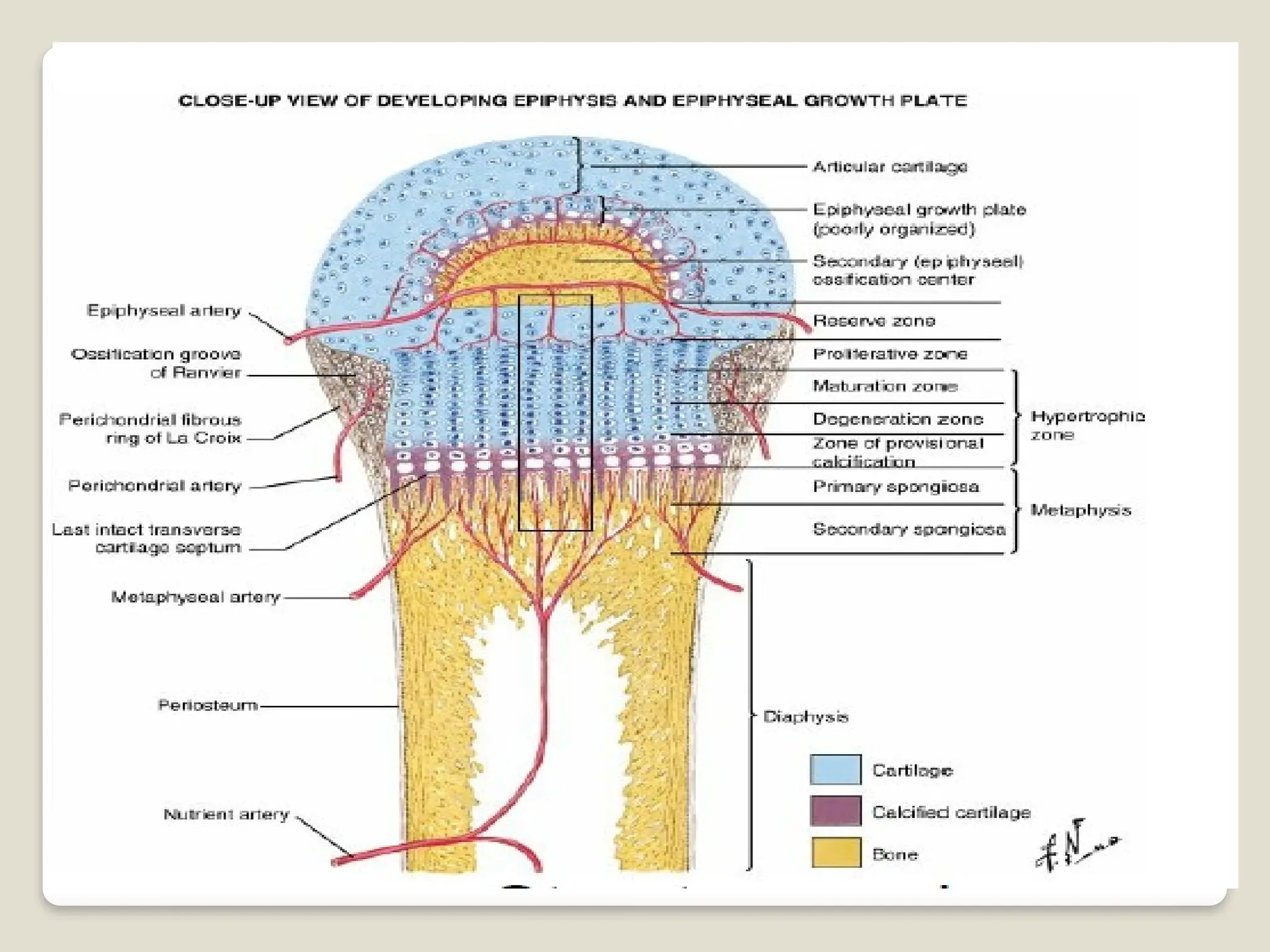
Metaphysis
Epiphysial endsof a diaphysis
Zone of active growth
Typically made up of cancellous bone
Hair pin bends of end arteries
56.
Epiphysial plate ofcartilage
It seperates epiphysis from metaphysis
Proliferation-Responsible for length wise
growth of long bone.
Epiphysial fusion-Can no longer grow
Nourished by both epiphysial and
metaphysial arteries.
58.
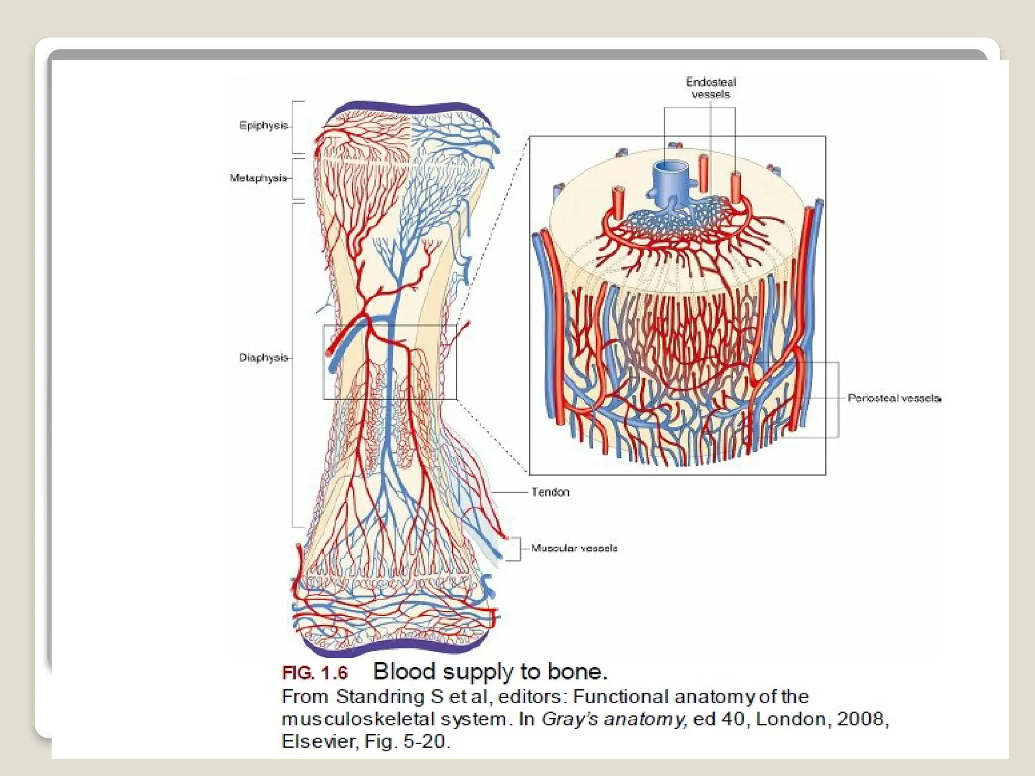
Blood supply ofbones
Long bones-derived from
1.Nutrient artery
2.Periosteal artery
3.Epiphysial artery
4.Metaphysial artery
60.
Nutrient artery
Entersthrough nutrient foramen
Divides into ascending and descending branches in
medullary cavity
Brach divides –small parallel channels-terminate in
adult metaphysis
Anatomosing with the epiphysial,metaphysial and
periosteal arteries
Supplies the medullary cavity,inner 2/3 rd of the
cortex and metaphysis
Nutrient foramen is directed away from the
growing end of the bone
61.
Periosteal artery
Numerousbeneath the muscular and
ligamentous attachments
Ramify beneath the periosteum and enter
the volkmanns canals to supply the outer
1/3 rd of the cortex
62.
Epiphyseal artery
Derivedfrom periarticular vascular
arcades(circulus vasculosus)
Out of the numerus vascular foramina in
this region –few admit arteries and rest
venous exits
Number size-idea of the relative
vascularity of two ends of the long bone.
63.
Metaphysial artery
Derivedfrom the neighbouring systemic
vessels
Pass directly into the metaphysis and
reinforce the metaphysial branches from
the primary nutrient artery
64.
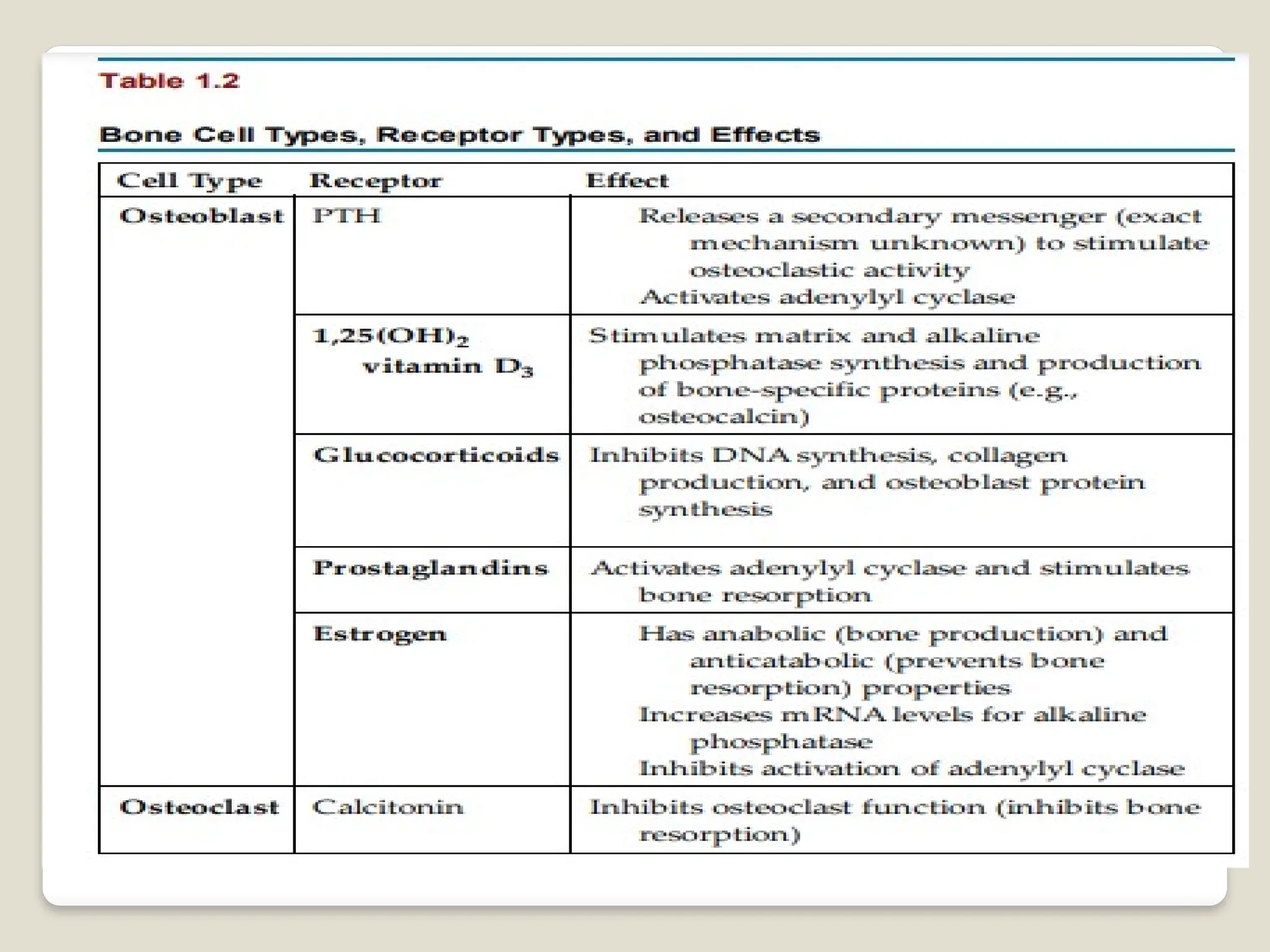
Homeostasis of bonetissue
Bone resorption –action of osteoclasts and
parathyroid hormone aka PTH
Bone deposition-Action of osteoblasts and
calcitonin
Occurs by direction of the thyroid and
parathyroid glands
67.
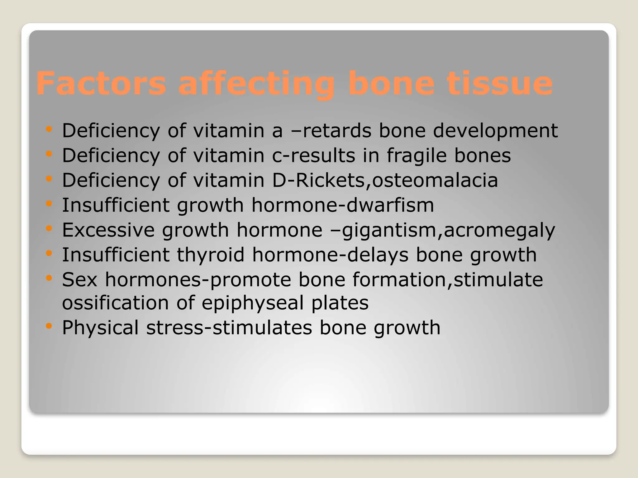
Factors affecting bonetissue
Deficiency of vitamin a –retards bone development
Deficiency of vitamin c-results in fragile bones
Deficiency of vitamin D-Rickets,osteomalacia
Insufficient growth hormone-dwarfism
Excessive growth hormone –gigantism,acromegaly
Insufficient thyroid hormone-delays bone growth
Sex hormones-promote bone formation,stimulate
ossification of epiphyseal plates
Physical stress-stimulates bone growth