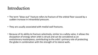
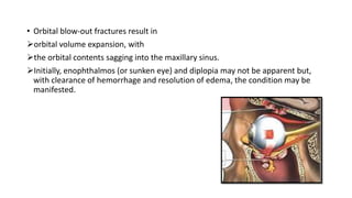
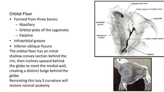
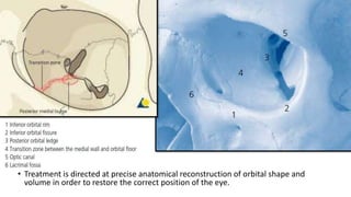
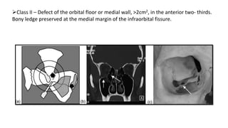
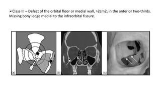
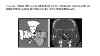
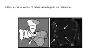
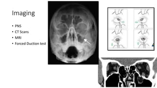
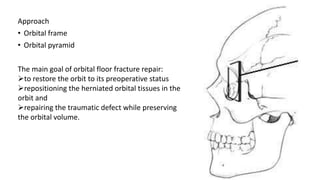
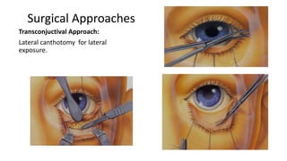
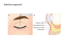
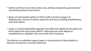
Blowout Fractures
Presented by
Dr Neha Umakant Chodankar
1st MDS OMFS
Introduction
• The term "blow-out" fracture refers to fracture of the orbital floor caused by a
sudden increase in intraorbital pressure.
• They are usually associated with medial wall fractures.
• Because of its ability to fracture selectively, similar to a safety valve, it allows the
dissipation of energy when orbit is struck and can be considered as an
evolutionary masterpiece, contributing to the orbit`s primary role of protecting
the globe in combination with the strength of its lateral walls.
• Orbital blow-out fractures result in
orbital volume expansion, with
the orbital contents sagging into the maxillary sinus.
Initially, enophthalmos (or sunken eye) and diplopia may not be apparent but,
with clearance of hemorrhage and resolution of edema, the condition may be
manifested.
Surgical Anatomy
• Pyramidal shape paired bony
cavities, housing and protecting
globe.
• Apex
• Base
• Four walls
Roof
Floor
Medial
Lateral
Orbital Floor
• Formed from three bones:
– Maxillary
– Orbital plate of the zygomatic
– Palatine
• Infraorbital groove
• Inferior oblique fissure
The orbital floor has an initial
shallow convex section behind the
rim, then inclines upward behind
the globe to meet the medial wall,
creating a distinct bulge behind the
globe.
Recreating this lazy S curvature will
restore normal anatomy
• Treatment is directed at precise anatomical reconstruction of orbital shape and
volume in order to restore the correct position of the eye.
Mechanisms
Buckling theory
• Lefort first described it
• If a force was to strike the orbital rim it
will cause the wall to undergo a rippling
effect and the weaker, paper thin walls
(especially the floor) will distort and
eventually fracture.
• limited to the anterior part of the
orbital floor
• 1.54 J
Hydraulic theory
• Given by Pfiffer
• The force of the blow received by the
eyeball was transmitted by it to the
walls of the orbit by increased
intraorbital pressure leading to fracture
of the delicate portions
• 1.22 J
Globe to wall theory
• direct injury to the globe forcing it into
the orbit
Pure Blowout Fractures: orbital floor fractures
Impure Blowout Fractures: orbital floor + rim fractures
1. Fractures limited to internal orbital skeleton.
• Orbital floor, medial wall, or roof can be involved. This type of fracture can be
further classified into:
o Trap door type of fracture – due to low velocity injuries
o Medial blow out fractures – due to intermediate velocity injuries
o Lateral blow out fractures – due to high velocity injuries
Classifications of Orbital Fractures
2. Fractures involving orbital rim / along with internal orbital skeleton. These
fractures may be subclassified into:
• Inferior rim fracture
• Superior rim fracture
• Lateral rim fracture
• Rim fracture in association with fractures involving internal orbital skeleton
3. Fractures of orbit associated with other fractures of facial skeleton.
• Zygomatico maxillary fracture
• Naso-orbito-ethmoid fracture
• Frontal sinus fracture
• Lefort II
• Lefort III
• 4. Orbital apex fractures : These fractures should be identified early because of
potential threat to neurovascular structures at superior orbital fissure and optic
canal. Optic canal injuries can lead to traumatic optic neuropathy
• According to classification by Jaquiery et al
Class I – Isolated defect of the orbital floor or the medial wall, 1-2cm2, in the
anterior two-thirds
Class II – Defect of the orbital floor or medial wall, >2cm2, in the anterior two- thirds.
Bony ledge preserved at the medial margin of the infraorbital fissure.
Class III – Defect of the orbital floor or medial wall, >2cm2, in the anterior two-thirds.
Missing bony ledge medial to the infraorbital fissure.
Class IV – Defect of the entire orbital floor and the medial wall, extending into the
posterior third. Missing bony ledge medial to the infraorbital fissure.
Class V – Same as class IV, defect extending into the orbital roof.
• Orbital Wall Fractures: Three Dimensional Computed Tomography Classification
Ahmed ElDegwi et al
type I, isolated medial wall or floor fracture
type II, medial wall and floor fractures
type III, medial wall floor, and zygomatic (trimalar) fractures
type IV, medial wall, floor, and complex fractures (i.e., maxillary, naso-orbital and
frontal fractures).
Clinical presentation Of a Blow-out fracture
The patient may complain of;
• Intraocular pain
• Numbness in certain regions of the face
• Blindness
• Unable to move eye
• Diplopia
There may also be signs of
• Enopthalmous
• Oedema
• Haematoma
• Globe displacement
• Minor or major eye injury
• Ocular mobility restrictions
• Infraorbital anaesthesia
• Proptosis if present indicates retrobulbar / peribulbar hemorrhage.
• Pupillary dysfunction associated with visual disturbances indicate injury to optic
nerve
A 3-mm downward displacement
of the entire floor
Orbital volume that is increased
by 1.5 cm3 a 5% increase
Producing 1-1.5 mm of
Enophthalmos
1957 by Smith & Regan
Imaging
• PNS
• CT Scans
• MRI
• Forced Duction test
Ophthalmologic assessment
• Complete ophthalmologic examination
• Hess charts (Lees screen)
Binocular Field of Vision
Hertel measurements
24
Initial Management
• Cold compresses over affected area for 48 hours
• Use of nasal decongestants
• Broad spectrum antibiotics
• Intravenous steroids
25
Absolute Indications for Surgical Repair
• Extraocular muscle entrapment
• Diplopia
• Enophthalmos >2 mm
26
Relative Indications for Surgery
• Substantial soft tissue herniation into maxillary sinus
• Intraoribital emphysema
• Hypoestheia in V2 distribution
27
Contraindications to surgery
• Hyphema
• Retinal detachment
• Globe perforation
• Only functional eye
• Medically unstable patient
28
Timing of Surgery
• Usually seven to ten days after trauma
Approach
• Orbital frame
• Orbital pyramid
The main goal of orbital floor fracture repair:
to restore the orbit to its preoperative status
repositioning the herniated orbital tissues in the
orbit and
repairing the traumatic defect while preserving
the orbital volume.
30
Surgical Approaches
Depend on the type of fracture, extent of fracture, aesthetic
considerations and an assessment of existing lacerations.
• Transconjunctival approach
• Subciliary approach
Surgical Approaches
Transconjuctival Approach:
Lateral canthotomy for lateral
exposure.
Subciliary approach
Complex orbital fractures require more than one material for reconstruction
Only hard tissue component corrected
• Defects of 25% or less of the surface area, without entrapment, generally heal
uneventfully without intervention.
• Repair of intermediate defects of 25% to 50% is based on degree of
displacement, amount of volume expansion and any co-existing enophthalmos,
even with edema.
• Larger or comminuted defects (greater than 50%) with significant disruption are
best treated with early repair (within 7 days) because some degree of
enophthalmos or diplopia is the norm when left unrepaired.
The decision to undertake surgical repair or reconstruction of these defects is
based on functional or cosmetic deformity.
37
Complications of Surgery
• Ectropion
• Lid retraction
• Persistent diplopia
• Malposition of eye
• hypoesthesia
• Extrusion of orbital floor implant
• Loss of vision
• Iatrogenic injuries
Conclusion
• The management of blow-out fractures takes considerable time and thought.
• No concrete guideline is available. However ,each fracture must be assessed on an
individual basis.
• The need of adequate assessment of the fracture sight for correct reduction and
evaluation of the defect is very important
• Diagnostic imaging , timing of treatment as well as evaluation of difficulty level of
reconstruction must be done prior and a treatment plan needs to be formulated
taking each of these into consideration.
Thank You

Blowout fractures.pptx

  • 1.
    Blowout Fractures Presented by DrNeha Umakant Chodankar 1st MDS OMFS
  • 2.
    Introduction • The term"blow-out" fracture refers to fracture of the orbital floor caused by a sudden increase in intraorbital pressure. • They are usually associated with medial wall fractures. • Because of its ability to fracture selectively, similar to a safety valve, it allows the dissipation of energy when orbit is struck and can be considered as an evolutionary masterpiece, contributing to the orbit`s primary role of protecting the globe in combination with the strength of its lateral walls.
  • 3.
    • Orbital blow-outfractures result in orbital volume expansion, with the orbital contents sagging into the maxillary sinus. Initially, enophthalmos (or sunken eye) and diplopia may not be apparent but, with clearance of hemorrhage and resolution of edema, the condition may be manifested.
  • 4.
    Surgical Anatomy • Pyramidalshape paired bony cavities, housing and protecting globe. • Apex • Base • Four walls Roof Floor Medial Lateral
  • 5.
    Orbital Floor • Formedfrom three bones: – Maxillary – Orbital plate of the zygomatic – Palatine • Infraorbital groove • Inferior oblique fissure The orbital floor has an initial shallow convex section behind the rim, then inclines upward behind the globe to meet the medial wall, creating a distinct bulge behind the globe. Recreating this lazy S curvature will restore normal anatomy
  • 6.
    • Treatment isdirected at precise anatomical reconstruction of orbital shape and volume in order to restore the correct position of the eye.
  • 7.
    Mechanisms Buckling theory • Lefortfirst described it • If a force was to strike the orbital rim it will cause the wall to undergo a rippling effect and the weaker, paper thin walls (especially the floor) will distort and eventually fracture. • limited to the anterior part of the orbital floor • 1.54 J
  • 8.
    Hydraulic theory • Givenby Pfiffer • The force of the blow received by the eyeball was transmitted by it to the walls of the orbit by increased intraorbital pressure leading to fracture of the delicate portions • 1.22 J Globe to wall theory • direct injury to the globe forcing it into the orbit
  • 9.
    Pure Blowout Fractures:orbital floor fractures Impure Blowout Fractures: orbital floor + rim fractures 1. Fractures limited to internal orbital skeleton. • Orbital floor, medial wall, or roof can be involved. This type of fracture can be further classified into: o Trap door type of fracture – due to low velocity injuries o Medial blow out fractures – due to intermediate velocity injuries o Lateral blow out fractures – due to high velocity injuries Classifications of Orbital Fractures
  • 10.
    2. Fractures involvingorbital rim / along with internal orbital skeleton. These fractures may be subclassified into: • Inferior rim fracture • Superior rim fracture • Lateral rim fracture • Rim fracture in association with fractures involving internal orbital skeleton 3. Fractures of orbit associated with other fractures of facial skeleton. • Zygomatico maxillary fracture • Naso-orbito-ethmoid fracture • Frontal sinus fracture • Lefort II • Lefort III
  • 11.
    • 4. Orbitalapex fractures : These fractures should be identified early because of potential threat to neurovascular structures at superior orbital fissure and optic canal. Optic canal injuries can lead to traumatic optic neuropathy
  • 12.
    • According toclassification by Jaquiery et al Class I – Isolated defect of the orbital floor or the medial wall, 1-2cm2, in the anterior two-thirds
  • 13.
    Class II –Defect of the orbital floor or medial wall, >2cm2, in the anterior two- thirds. Bony ledge preserved at the medial margin of the infraorbital fissure.
  • 14.
    Class III –Defect of the orbital floor or medial wall, >2cm2, in the anterior two-thirds. Missing bony ledge medial to the infraorbital fissure.
  • 15.
    Class IV –Defect of the entire orbital floor and the medial wall, extending into the posterior third. Missing bony ledge medial to the infraorbital fissure.
  • 16.
    Class V –Same as class IV, defect extending into the orbital roof.
  • 17.
    • Orbital WallFractures: Three Dimensional Computed Tomography Classification Ahmed ElDegwi et al type I, isolated medial wall or floor fracture type II, medial wall and floor fractures type III, medial wall floor, and zygomatic (trimalar) fractures type IV, medial wall, floor, and complex fractures (i.e., maxillary, naso-orbital and frontal fractures).
  • 18.
    Clinical presentation Ofa Blow-out fracture The patient may complain of; • Intraocular pain • Numbness in certain regions of the face • Blindness • Unable to move eye • Diplopia
  • 19.
    There may alsobe signs of • Enopthalmous • Oedema • Haematoma • Globe displacement • Minor or major eye injury • Ocular mobility restrictions • Infraorbital anaesthesia • Proptosis if present indicates retrobulbar / peribulbar hemorrhage. • Pupillary dysfunction associated with visual disturbances indicate injury to optic nerve
  • 20.
    A 3-mm downwarddisplacement of the entire floor Orbital volume that is increased by 1.5 cm3 a 5% increase Producing 1-1.5 mm of Enophthalmos 1957 by Smith & Regan
  • 21.
    Imaging • PNS • CTScans • MRI • Forced Duction test
  • 22.
    Ophthalmologic assessment • Completeophthalmologic examination • Hess charts (Lees screen)
  • 23.
    Binocular Field ofVision Hertel measurements
  • 24.
    24 Initial Management • Coldcompresses over affected area for 48 hours • Use of nasal decongestants • Broad spectrum antibiotics • Intravenous steroids
  • 25.
    25 Absolute Indications forSurgical Repair • Extraocular muscle entrapment • Diplopia • Enophthalmos >2 mm
  • 26.
    26 Relative Indications forSurgery • Substantial soft tissue herniation into maxillary sinus • Intraoribital emphysema • Hypoestheia in V2 distribution
  • 27.
    27 Contraindications to surgery •Hyphema • Retinal detachment • Globe perforation • Only functional eye • Medically unstable patient
  • 28.
    28 Timing of Surgery •Usually seven to ten days after trauma
  • 29.
    Approach • Orbital frame •Orbital pyramid The main goal of orbital floor fracture repair: to restore the orbit to its preoperative status repositioning the herniated orbital tissues in the orbit and repairing the traumatic defect while preserving the orbital volume.
  • 30.
    30 Surgical Approaches Depend onthe type of fracture, extent of fracture, aesthetic considerations and an assessment of existing lacerations. • Transconjunctival approach • Subciliary approach
  • 31.
  • 32.
  • 34.
    Complex orbital fracturesrequire more than one material for reconstruction
  • 35.
    Only hard tissuecomponent corrected
  • 36.
    • Defects of25% or less of the surface area, without entrapment, generally heal uneventfully without intervention. • Repair of intermediate defects of 25% to 50% is based on degree of displacement, amount of volume expansion and any co-existing enophthalmos, even with edema. • Larger or comminuted defects (greater than 50%) with significant disruption are best treated with early repair (within 7 days) because some degree of enophthalmos or diplopia is the norm when left unrepaired. The decision to undertake surgical repair or reconstruction of these defects is based on functional or cosmetic deformity.
  • 37.
    37 Complications of Surgery •Ectropion • Lid retraction • Persistent diplopia • Malposition of eye • hypoesthesia • Extrusion of orbital floor implant • Loss of vision • Iatrogenic injuries
  • 38.
    Conclusion • The managementof blow-out fractures takes considerable time and thought. • No concrete guideline is available. However ,each fracture must be assessed on an individual basis. • The need of adequate assessment of the fracture sight for correct reduction and evaluation of the defect is very important • Diagnostic imaging , timing of treatment as well as evaluation of difficulty level of reconstruction must be done prior and a treatment plan needs to be formulated taking each of these into consideration.
  • 39.