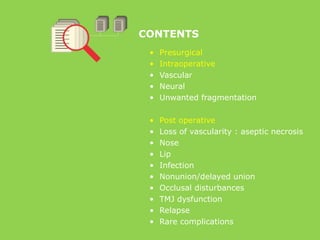
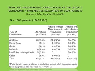
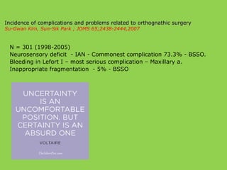
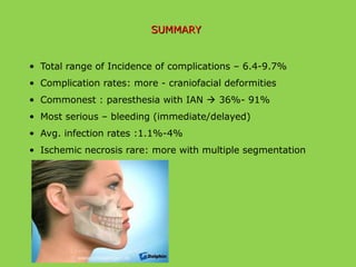
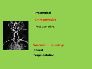
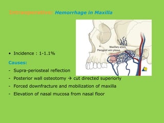
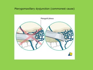
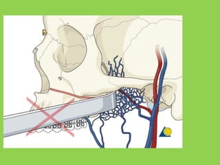
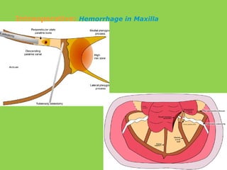
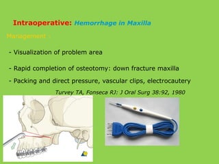
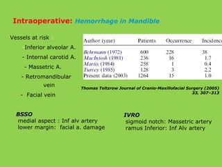
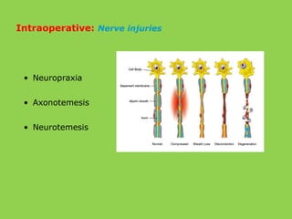
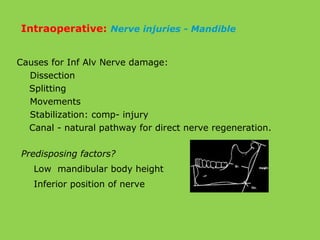
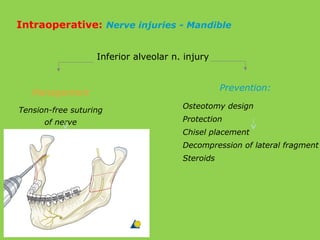
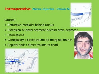
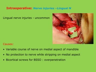
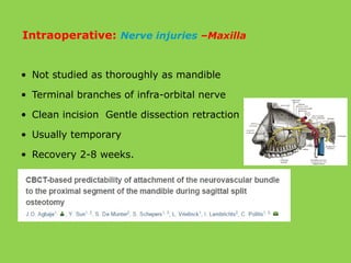
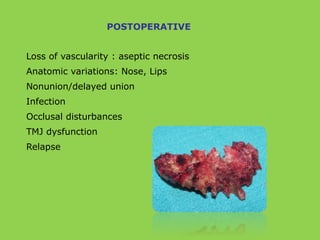
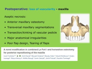
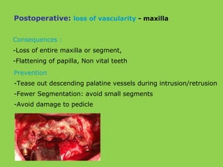
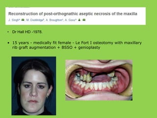
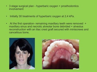
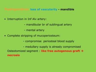
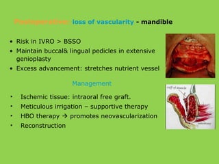
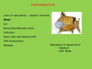
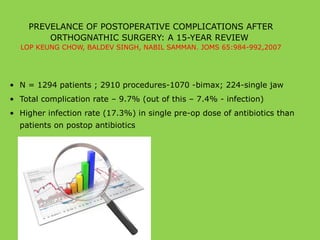
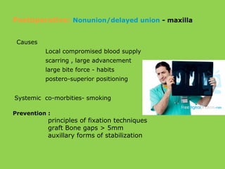
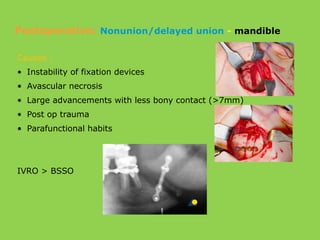
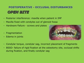
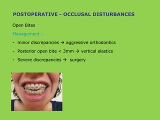
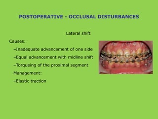
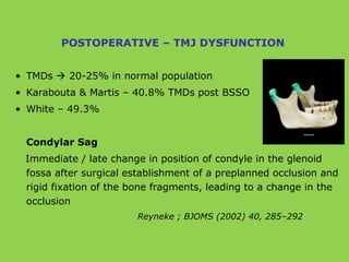
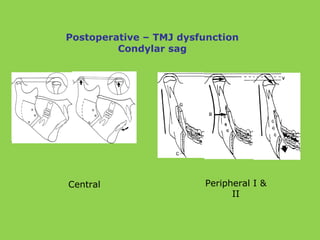
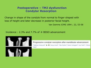
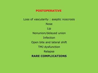
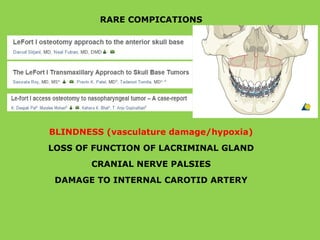
This document discusses various complications that can occur with orthognathic surgery, including:
- Common intraoperative complications are nerve injuries, most often to the inferior alveolar nerve during mandibular surgery, and hemorrhage, usually from the maxillary arteries.
- Frequent postoperative issues involve neurosensory deficits, nonunion or delayed bone healing, and infections, which have been reported in up to 9.7% of patients.
- Other risks include loss of vascularity leading to aseptic necrosis of the maxilla or mandible, nasal deformities, malocclusion, and TMJ dysfunction. Careful planning and technique aim to minimize complications while pursuing the benefits of orthognathic
The Indian Dental Academy is the Leader in continuing dental education , training dentists in all aspects of dentistry and
offering a wide range of dental certified courses in different formats.for more details please visit
www.indiandentalacademy.com
The Indian Dental Academy is the Leader in continuing dental education , training dentists in all aspects of dentistry and
offering a wide range of dental certified courses in different formats.for more details please visit
www.indiandentalacademy.com
Dr. Ahmed M. Adawy, Professor Emeritus, Dep. Oral & Maxillofacial Surgery. Former Dean, Faculty of Dental Medicine, Al-Azhar University. Mandibular angle fractures account for 23% to 42% of all facial fractures. Fracture of mandibular angle can be classified as (A) Vertical favorable or unfavorable, (B) Horizontally favorable of unfavorable. Traditionally, mandibular angle fractures have been treated with either closed reduction and inter-maxillary fixation or open reduction and internal fixation with or without inter-maxillary fixation. Patients treated with inter-maxillary fixation have a restricted airway and loose excess weight. Rigid internal fixation and early return to function have eliminated the use of wire osteosenthysis and prolonged use of inter-maxillary fixation. The principal of rigid fixation, however, have inherent set of disadvantages including damage to the inferior alveolar nerve and the marginal mandibular branch of facial nerve. Postoperative malocclusion rates are also high. With the introduction of semi-rigid technique fracture of the mandibular angle could be treated according to Champy’s Ideal lines of osteosenthysis. The technique involves placement of a single monocortial miniplate on the superior border of the mandible. However, some studies suggested using a second miniplate along the inferior border. Wether one or two miniplates should be used is still debatable. The application of 3D plates may provide additional stability in 3 dimension and good resistance against torque forces.
orthognathic surgery is very intresting and well knowing branch in oral surgery ....this presentation is dealing with jaw correction surgery in upper jaw.
Arthrocentesis of the temporomandibular jointAhmed Adawy
Dr. Ahmed M. Adawy, Professor Emeritus, Dep. Oral & Maxillofacial Surgery. Former Dean, Faculty of Dental Medicine, Al-Azhar University. Arthrocentesis of the temporomandibular joint refers to lavage of the upper joint space, hydraulic pressure and manipulation to release adhesions of the “anchored disc phenomenon” and improve motion. The technique of arthrocentesis is discussed together with the indications and contraindications of the procedure. Further, the presentation includes modifications of the standard technique.
Head and Neck Trauma by Dr. Kenneth DickieKenneth Dickie
Dr. Kenneth Dickie from Royal Centre of Plastic Surgery in Barrie, Ontario explained the care for Head & Neck Trauma.
f you have any questions, please contact Dr. Kenneth Dickie at http://royalcentreofplasticsurgery.com/
Lefort fractures /certified fixed orthodontic courses by Indian dental academy Indian dental academy
The Indian Dental Academy is the Leader in continuing dental education , training dentists in all aspects of dentistry and offering a wide range of dental certified courses in different formats.
Indian dental academy provides dental crown & Bridge,rotary endodontics,fixed orthodontics,
Dental implants courses.for details pls visit www.indiandentalacademy.com ,or call
0091-9248678078
The Indian Dental Academy is the Leader in continuing dental education , training dentists in all aspects of dentistry and offering a wide range of dental certified courses in different formats.
Indian dental academy provides dental crown & Bridge,rotary endodontics,fixed orthodontics,
Dental implants courses.for details pls visit www.indiandentalacademy.com ,or call
00919248678078
Complication of ortho gnathic surgery /certified fixed orthodontic courses by...Indian dental academy
The Indian Dental Academy is the Leader in continuing dental education , training dentists in all aspects of dentistry and offering a wide range of dental certified courses in different formats.
Indian dental academy provides dental crown & Bridge,rotary endodontics,fixed orthodontics,
Dental implants courses.for details pls visit www.indiandentalacademy.com ,or call
0091-9248678078
Dr. Ahmed M. Adawy
Professor Emeritus, Dep. Oral & Maxillofacial Surg.
Former Dean, Faculty of Dental Medicine
Al-Azhar UniversityOrthognathic surgery is the art and science of combining orthodontics and maxillofacial surgery to correct dento-facial deformities.The etiology, prevalence,diagnosis and preoperative planning,and Surgical procedures are presented.
The Indian Dental Academy is the Leader in continuing dental education , training dentists in all aspects of dentistry and
offering a wide range of dental certified courses in different formats.for more details please visit
www.indiandentalacademy.com
for online course please visit www.idalectures.com
for online interactive live courses/classes please visit
www.gotolectures.com.
HI THIS IS A NICE SEMINAR DESCRIBING ABOUT THE ORTHOGNATHIC SURGERY MAINLY RELATED TO ORTHODONTICS VIEWPOINT AND CEPH TRACING ITS INDICATION AND DIFFERENT TYPES OF SURGERIES. JUST HAVE A LOOK TO IT
Distraction osteogenesis versus bsso for advancement of the retrognathic mand...Indian dental academy
The Indian Dental Academy is the Leader in continuing dental education , training dentists in all aspects of dentistry and offering a wide range of dental certified courses in different formats.
Le fort fracture by Dr. Amit Suryawanshi .Dentist in Kolhapur (MDS). Oral &...All Good Things
Description:
Hi. This is Dr. Amit T. Suryawanshi. Dentist in Kolhapur (MDS) Oral & Maxillofacial surgeon from Kolhapur, India. I am here on slideshare.com to share some of my own presentations presented at various levels in the field of OMFS. Hope this would somehow be helpful to you making your presentations. All the best & your replies are welcomed!
This PowerPoint presentation delivers a technical analysis of the midface orthognathic procedure. Explore surgical techniques, anatomical considerations, and treatment objectives.
Pulmonary Thromboembolism - etilogy, types, medical- Surgical and nursing man...VarunMahajani
Disruption of blood supply to lung alveoli due to blockage of one or more pulmonary blood vessels is called as Pulmonary thromboembolism. In this presentation we will discuss its causes, types and its management in depth.
These simplified slides by Dr. Sidra Arshad present an overview of the non-respiratory functions of the respiratory tract.
Learning objectives:
1. Enlist the non-respiratory functions of the respiratory tract
2. Briefly explain how these functions are carried out
3. Discuss the significance of dead space
4. Differentiate between minute ventilation and alveolar ventilation
5. Describe the cough and sneeze reflexes
Study Resources:
1. Chapter 39, Guyton and Hall Textbook of Medical Physiology, 14th edition
2. Chapter 34, Ganong’s Review of Medical Physiology, 26th edition
3. Chapter 17, Human Physiology by Lauralee Sherwood, 9th edition
4. Non-respiratory functions of the lungs https://academic.oup.com/bjaed/article/13/3/98/278874
Factory Supply Best Quality Pmk Oil CAS 28578–16–7 PMK Powder in Stockrebeccabio
Factory Supply Best Quality Pmk Oil CAS 28578–16–7 PMK Powder in Stock
Telegram: bmksupplier
signal: +85264872720
threema: TUD4A6YC
You can contact me on Telegram or Threema
Communicate promptly and reply
Free of customs clearance, Double Clearance 100% pass delivery to USA, Canada, Spain, Germany, Netherland, Poland, Italy, Sweden, UK, Czech Republic, Australia, Mexico, Russia, Ukraine, Kazakhstan.Door to door service
Hot Selling Organic intermediates
Ozempic: Preoperative Management of Patients on GLP-1 Receptor Agonists Saeid Safari
Preoperative Management of Patients on GLP-1 Receptor Agonists like Ozempic and Semiglutide
ASA GUIDELINE
NYSORA Guideline
2 Case Reports of Gastric Ultrasound
micro teaching on communication m.sc nursing.pdfAnurag Sharma
Microteaching is a unique model of practice teaching. It is a viable instrument for the. desired change in the teaching behavior or the behavior potential which, in specified types of real. classroom situations, tends to facilitate the achievement of specified types of objectives.
The prostate is an exocrine gland of the male mammalian reproductive system
It is a walnut-sized gland that forms part of the male reproductive system and is located in front of the rectum and just below the urinary bladder
Function is to store and secrete a clear, slightly alkaline fluid that constitutes 10-30% of the volume of the seminal fluid that along with the spermatozoa, constitutes semen
A healthy human prostate measures (4cm-vertical, by 3cm-horizontal, 2cm ant-post ).
It surrounds the urethra just below the urinary bladder. It has anterior, median, posterior and two lateral lobes
It’s work is regulated by androgens which are responsible for male sex characteristics
Generalised disease of the prostate due to hormonal derangement which leads to non malignant enlargement of the gland (increase in the number of epithelial cells and stromal tissue)to cause compression of the urethra leading to symptoms (LUTS
Flu Vaccine Alert in Bangalore Karnatakaaddon Scans
As flu season approaches, health officials in Bangalore, Karnataka, are urging residents to get their flu vaccinations. The seasonal flu, while common, can lead to severe health complications, particularly for vulnerable populations such as young children, the elderly, and those with underlying health conditions.
Dr. Vidisha Kumari, a leading epidemiologist in Bangalore, emphasizes the importance of getting vaccinated. "The flu vaccine is our best defense against the influenza virus. It not only protects individuals but also helps prevent the spread of the virus in our communities," he says.
This year, the flu season is expected to coincide with a potential increase in other respiratory illnesses. The Karnataka Health Department has launched an awareness campaign highlighting the significance of flu vaccinations. They have set up multiple vaccination centers across Bangalore, making it convenient for residents to receive their shots.
To encourage widespread vaccination, the government is also collaborating with local schools, workplaces, and community centers to facilitate vaccination drives. Special attention is being given to ensuring that the vaccine is accessible to all, including marginalized communities who may have limited access to healthcare.
Residents are reminded that the flu vaccine is safe and effective. Common side effects are mild and may include soreness at the injection site, mild fever, or muscle aches. These side effects are generally short-lived and far less severe than the flu itself.
Healthcare providers are also stressing the importance of continuing COVID-19 precautions. Wearing masks, practicing good hand hygiene, and maintaining social distancing are still crucial, especially in crowded places.
Protect yourself and your loved ones by getting vaccinated. Together, we can help keep Bangalore healthy and safe this flu season. For more information on vaccination centers and schedules, residents can visit the Karnataka Health Department’s official website or follow their social media pages.
Stay informed, stay safe, and get your flu shot today!
TEST BANK for Operations Management, 14th Edition by William J. Stevenson, Ve...kevinkariuki227
TEST BANK for Operations Management, 14th Edition by William J. Stevenson, Verified Chapters 1 - 19, Complete Newest Version.pdf
Title: Sense of Smell
Presenter: Dr. Faiza, Assistant Professor of Physiology
Qualifications:
MBBS (Best Graduate, AIMC Lahore)
FCPS Physiology
ICMT, CHPE, DHPE (STMU)
MPH (GC University, Faisalabad)
MBA (Virtual University of Pakistan)
Learning Objectives:
Describe the primary categories of smells and the concept of odor blindness.
Explain the structure and location of the olfactory membrane and mucosa, including the types and roles of cells involved in olfaction.
Describe the pathway and mechanisms of olfactory signal transmission from the olfactory receptors to the brain.
Illustrate the biochemical cascade triggered by odorant binding to olfactory receptors, including the role of G-proteins and second messengers in generating an action potential.
Identify different types of olfactory disorders such as anosmia, hyposmia, hyperosmia, and dysosmia, including their potential causes.
Key Topics:
Olfactory Genes:
3% of the human genome accounts for olfactory genes.
400 genes for odorant receptors.
Olfactory Membrane:
Located in the superior part of the nasal cavity.
Medially: Folds downward along the superior septum.
Laterally: Folds over the superior turbinate and upper surface of the middle turbinate.
Total surface area: 5-10 square centimeters.
Olfactory Mucosa:
Olfactory Cells: Bipolar nerve cells derived from the CNS (100 million), with 4-25 olfactory cilia per cell.
Sustentacular Cells: Produce mucus and maintain ionic and molecular environment.
Basal Cells: Replace worn-out olfactory cells with an average lifespan of 1-2 months.
Bowman’s Gland: Secretes mucus.
Stimulation of Olfactory Cells:
Odorant dissolves in mucus and attaches to receptors on olfactory cilia.
Involves a cascade effect through G-proteins and second messengers, leading to depolarization and action potential generation in the olfactory nerve.
Quality of a Good Odorant:
Small (3-20 Carbon atoms), volatile, water-soluble, and lipid-soluble.
Facilitated by odorant-binding proteins in mucus.
Membrane Potential and Action Potential:
Resting membrane potential: -55mV.
Action potential frequency in the olfactory nerve increases with odorant strength.
Adaptation Towards the Sense of Smell:
Rapid adaptation within the first second, with further slow adaptation.
Psychological adaptation greater than receptor adaptation, involving feedback inhibition from the central nervous system.
Primary Sensations of Smell:
Camphoraceous, Musky, Floral, Pepperminty, Ethereal, Pungent, Putrid.
Odor Detection Threshold:
Examples: Hydrogen sulfide (0.0005 ppm), Methyl-mercaptan (0.002 ppm).
Some toxic substances are odorless at lethal concentrations.
Characteristics of Smell:
Odor blindness for single substances due to lack of appropriate receptor protein.
Behavioral and emotional influences of smell.
Transmission of Olfactory Signals:
From olfactory cells to glomeruli in the olfactory bulb, involving lateral inhibition.
Primitive, less old, and new olfactory systems with different path
2. CONTENTS
• Presurgical
• Intraoperative
• Vascular
• Neural
• Unwanted fragmentation
• Post operative
• Loss of vascularity : aseptic necrosis
• Nose
• Lip
• Infection
• Nonunion/delayed union
• Occlusal disturbances
• TMJ dysfunction
• Relapse
• Rare complications
3. “Unintended consequence of the surgery that causes harm
to the patient, occurring either intra-operatively or early
and late post-operatively.”
• A complication is so named because it complicates the
situation.
4. • “No matter what measures are taken, doctors will
sometimes falter, and it isn't reasonable to ask that we
achieve perfection. What is reasonable is to ask that we
never cease to aim for it.”
•
― Atul Gawande, Complications: A Surgeon's Notes on an Imperfect Science
5. N = 1000 patients (1983-2002)
INTRA AND PERIOPERATIVE COMPLICATIONS OF THE LEFORT IINTRA AND PERIOPERATIVE COMPLICATIONS OF THE LEFORT I
OSTEOTOMY: A PROSPECTIVE EVALUATION OF 1000 PATIENTSOSTEOTOMY: A PROSPECTIVE EVALUATION OF 1000 PATIENTS
Kramer; J CrFac Surg Vol 15,6 Nov’04
6. Incidence of complications and problems related to orthognathic surgeryIncidence of complications and problems related to orthognathic surgery
Su-Gwan Kim, Sun-Sik Park ; JOMS 65;2438-2444,2007
N = 301 (1998-2005)
Neurosensory deficit - IAN - Commonest complication 73.3% - BSSO.
Bleeding in Lefort I – most serious complication – Maxillary a.
Inappropriate fragmentation - 5% - BSSO
7. SUMMARYSUMMARY
• Total range of Incidence of complications – 6.4-9.7%
• Complication rates: more - craniofacial deformities
• Commonest : paresthesia with IAN 36%- 91%
• Most serious – bleeding (immediate/delayed)
• Avg. infection rates :1.1%-4%
• Ischemic necrosis rare: more with multiple segmentation
10. Pre-surgical
Lack of pre-treatment objectives
• Failure to recognize underlying skeletal abnormality
• Unexpected adverse growth
• Lack of patient co-operation
• Gross skeletal deformity correction:
mainly orthodontics & minimal surgery
11. Inability to perform the ideal procedure
Undesired esthetic and occlusal results
Creation of new problems and revision procedures
Presurgical : Lack of pre treatment objectives
20. Management :
- Visualization of problem area
- Rapid completion of osteotomy: down fracture maxilla
- Packing and direct pressure, vascular clips, electrocautery
Turvey TA, Fonseca RJ: J Oral Surg 38:92, 1980
Intraoperative: Hemorrhage in Maxilla
21. Thomas Teltzrow Journal of Cranio-Maxillofacial Surgery (2005)
33, 307–313
Vessels at risk :
-Inferior alveolar A.
- Internal carotid A.
- Massetric A.
- Retromandibular
vein
- Facial vein
BSSO
medial aspect : Inf alv artery
lower margin: facial a. damage
IVRO
sigmoid notch: Massetric artery
ramus Inferior: Inf Alv artery
Intraoperative: Hemorrhage in Mandible
24. Causes for Inf Alv Nerve damage:
Dissection
Splitting
Movements
Stabilization: comp- injury
Canal - natural pathway for direct nerve regeneration.
Intraoperative: Nerve injuries - Mandible
Predisposing factors?
Low mandibular body height
Inferior position of nerve
25. Inferior alveolar n. injury
Prevention:
Management
Tension-free suturing
of nerve
Osteotomy design
Protection
Chisel placement
Decompression of lateral fragment
Steroids
Intraoperative: Nerve injuries - Mandible
26. Causes:
• Retraction medially behind ramus
• Extension of distal segment beyond prox. segment
• Haematoma
• Genioplasty : direct trauma to marginal branch
• Sagittal split : direct trauma to trunk
Intraoperative: Nerve injuries –Facial N.
27. Lingual nerve injuries - uncommon
Causes:
• Variable course of nerve on medial aspect of mandible
• No protection to nerve while stripping on medial aspect
• Bicortical screws for BSSO : overpenetration
Intraoperative: Nerve injuries –Lingual N
28. • Not studied as thoroughly as mandible
• Terminal branches of infra-orbital nerve
• Clean incision Gentle dissection retraction
• Usually temporary
• Recovery 2-8 weeks.
Intraoperative: Nerve injuries –Maxilla
30. “Deviation from osteotomy line during osteotomy procedure,
resulting in osteotomy in area unrelated to surgery”
Maxilla Mandible
Intraoperative: Fragmentation
31. Factors:
• Bone architecture
• Bone density
• Unanticipated fractures
• Difficult fixation
• Impacted third molar
Intraoperative: Fragmentation
32. Sequalae :
• Infection
• Sequestration of the fragments
• Delayed bone healing
• Pseudoarthrosis
• Post operative instability & Relapse
• TMJ
Intraoperative: Fragmentation
34. POSTOPERATIVE
Loss of vascularity : aseptic necrosis
Anatomic variations: Nose, Lips
Nonunion/delayed union
Infection
Occlusal disturbances
TMJ dysfunction
Relapse
35. Aseptic necrosis:
• Anterior maxillary osteotomy
• Transversal maxillary segmentations
• Transection/kinking of vascular pedicle
• Major anatomical irregularities
• Poor flap design, Tearing of flaps
Postoperative: loss of vascularity - maxilla
36. Consequences :
-Loss of entire maxilla or segment,
-Flattening of papilla, Non vital teeth
Prevention
-Tease out descending palatine vessels during intrusion/retrusion
-Fewer Segmentation: avoid small segments
-Avoid damage to pedicle
Postoperative: loss of vascularity - maxilla
37. • Dr Hall HD -1978.
• 15 years - medically fit female - Le Fort I osteotomy with maxillary
rib graft augmentation + BSSO + genioplasty
38. • 3 stage surgical plan - hyperbaric oxygen + prosthodontics
involvement
• Initially 30 treatments of hyperbaric oxygen at 2.4 kPa.
• At the first operation- remaining maxillary teeth were removed +
maxillary sinus and necrotic alveolar bone debrided + alveolus
reconstruction with an iliac crest graft secured with miniscrews and
cancellous bone,
39. • Interruption in Inf Alv artery:
- mandibular br of sublingual artery
- mental artery
• Complete stripping of mucoperiosteum:
- compromise periosteal blood supply
- medullary supply is already compromised
Osteotomized segment : like free autogenous graft
necrosis
Postoperative: loss of vascularity - mandible
41. POSTOPERATIVE
Alteration in Nasal form
- Septum
- Alar Base
Loss of vascularity : aseptic necrosis
Nose
Lip
Nonunion/delayed union
Infection
Open bite and lateral shift
TMJ dysfunction
Relapse
42. Nasal Septum deviation:
- Maxillary impaction : encroachment on Presurgical
dimension of nasal septum
- Maxillary advancement buckling
Failure to reposition :
- Septal deviation – obstruction
- Abnormal position of columella/nasal tip
Postoperative: Nose
43. Intraop
- Resection of inferior aspect of septum
- Trim septal spurs if present
- Trim bone from nasal crest of maxilla
- Groove in superior aspect of maxilla
Septal deviation - How to avoid?
Management
-Reoperation
- Delayed septoplasty
Postoperative: Nose
44. Alteration in alar base and perioral structures
• Alar base widening
• Prominent alar groove
• Upturning of nasal tip – obtuse nasolabial angle
• Flattening and thinning of upper lip
• Downturning of labial commisures
Postoperative: Nose
46. POSTOPERATIVE
Loss of vascularity : aseptic necrosis
Nose
Lip
Nonunion/delayed union
Infection
Occlusal disturbances
TMJ dysfunction
Relapse
Rare Complications
48. POSTOPERATIVE
Loss of vascularity : aseptic necrosis
Nose
Lip
Infection
Nonunion/delayed union
Occlusal disturbances
TMJ dysfunction
Relapse
Rare complications
49. PREVELANCE OF POSTOPERATIVE COMPLICATIONS AFTER
ORTHOGNATHIC SURGERY: A 15-YEAR REVIEW
LOP KEUNG CHOW, BALDEV SINGH, NABIL SAMMAN. JOMS 65:984-992,2007
• N = 1294 patients ; 2910 procedures-1070 -bimax; 224-single jaw
• Total complication rate – 9.7% (out of this – 7.4% - infection)
• Higher infection rate (17.3%) in single pre-op dose of antibiotics than
patients on postop antibiotics
50. POSTOPERATIVE
Loss of vascularity : aseptic necrosis
Nose
Lip
Infection
Nonunion/delayed union
Occlusal disturbances
TMJ dysfunction
Relapse
Blindness
51. Causes
Local compromised blood supply
scarring , large advancement
large bite force - habits
postero-superior positioning
Systemic co-morbities- smoking
Prevention :
principles of fixation techniques
graft Bone gaps > 5mm
auxillary forms of stabilization
Postoperative: Nonunion/delayed union - maxilla
52. Causes :
• Instability of fixation devices
• Avascular necrosis
• Large advancements with less bony contact (>7mm)
• Post op trauma
• Parafunctional habits
IVRO > BSSO
Postoperative: Nonunion/delayed union - mandible
53. POSTOPERATIVE
Loss of vascularity : aseptic ncecrosis
Nose
Lip
Nonunion/delayed union
Infection
Occlusal disturbances
TMJ dysfunction
Relapse
Rare Complications
54. POSTOPERATIVE - OCCLUSAL DISTURBANCES
- Posterior interference: maxilla when patient in IMF
- Maxilla fixed with condyles out of glenoid fossa
- Hardware Failure - screws and plates
- Fragmentation
- Edema in joints
- Condylar torque, condylar sag, incorrect placement of fragments
- BSSO- failure of rigid fixation at the osteotomy site, occlusal shifts
during fixation, and finally condylar sag
55. Open Bites
Management :
- minor discrepancies aggressive orthodontics
- Posterior open bite < 3mm vertical elastics
- Severe discrepancies surgery
POSTOPERATIVE - OCCLUSAL DISTURBANCES
56. POSTOPERATIVE - OCCLUSAL DISTURBANCES
Lateral shift
Causes:
–Inadequate advancement of one side
–Equal advancement with midline shift
–Torqueing of the proximal segment
Management:
–Elastic traction
57. Postoperative
Loss of vascularity : aseptic ncecrosis
Nose
Lip
Nonunion/delayed union
Infection
Open bite and lateral shift
TMJ dysfunction
Relapse
Rare Complications
58. Intraoperative position of condyle influenced by:
• Incorrect vector during condylar positioning
• Incomplete or green-stick split prevents condylar seating
• Muscular, ligamentous or periosteal interference
• Intra-articular hemorrhage or edema
• Flexion in proximal segment while placing rigid fixation
POSTOPERATIVE – TMJ DYSFUNCTION
59. • TMDs 20-25% in normal population
• Karabouta & Martis – 40.8% TMDs post BSSO
• White – 49.3%
Condylar Sag
Immediate / late change in position of condyle in the glenoid
fossa after surgical establishment of a preplanned occlusion and
rigid fixation of the bone fragments, leading to a change in the
occlusion
Reyneke ; BJOMS (2002) 40, 285–292
POSTOPERATIVE – TMJ DYSFUNCTION
62. • The condyle is seated with the condylar seating tool + light digital
pressure at the angle
• resultant vector is anterosuperior
63.
64.
65.
66. Change in shape of the condyle from normal to finger shaped with
loss of height and later decrease in posterior facial height.
Van Damme JCMS 1994 ; 22, 53-58
Incidence : 2.3% and 7.7% of BSSO advancement
Postoperative – TMJ dysfunction
Condylar Resorption
67. POSTOPERATIVE
Loss of vascularity : aseptic ncecrosis
Nose
Lip
Nonunion/delayed union
Infection
Open bite and lateral shift
TMJ dysfunction
Relapse
Rare Complications
68. Stability depends on :
- Adequate presurgical orthodontics
- Long-term maxillomandibular fixation (MMF)
- Nonrigid fixation that allow muscular adaptation
- Minimal muscle alteration
- Good bony contact, and control of the proximal segment
POSTOPERATIVE - RELAPSE
69. Factors :
• Magnitude of mandibular advancement or setback,
• Stretch of surrounding soft tissue,
• Positioning of mandibular condyles
• Method of fixation
• Growth of mandible
• skeletal behavior among hyper/hypodivergent skeletal patterns
POSTOPERATIVE – RELAPSE
MANDIBLE
70. • Obligate relapse after mandibular advancements >7mm
• Mandibular setback >12 mm - less skeletal relapse
• Closure of anterior open bite with only mandibular osteotomies
POSTOPERATIVE – RELAPSE
MANDIBLE
How to reduce/avoid :
• Counterclockwise rotation of the mandible be avoided
• Mandibular advancement limited to < 7mm
• Bimaxillary surgery
71. Depends on :
• Degree of surgical advancement
• Degree of inferior repositioning of anterior maxilla
• Use of bone grafts in large advancements
POSTOPERATIVE – RELAPSE
MAXILLA
72. Other Causes :
- Increased soft tissue stretching results in drift of the
screws during bone healing
- Reduced area of bone contact at the lateral aspects of the
maxilla - compromised union
- Preoperative scarring - Cleft maxilla
Postoperative – Relapse
Maxilla
73. • Postoperative relapse was not considerable after total maxillary
setback surgery.
• Although the amount of maxillary setback was greater,
postoperative relapse did not increase significantly.
• Significant osseous regeneration at the pterygomaxillary region
occurred in the early phase of recovery.
74. • On average, 18% of the horizontal maxillary repositioning was lost.
• Most of the change (89%) occurred during the first 6 months
postoperatively.
• Relapse increased significantly with degree of surgical advancement
and degree of inferior repositioning of anterior maxilla.
75. Remedy for prevention:
• Advance the maxilla at least 2mm more than the ideal overjet
to compensate for relapse
• Provision of a period of MMF (3—4 weeks) in addition to rigid
fixation in large advancements –
Postoperative – Relapse
Maxilla - Management
Van Sickels BJOMS 1996;34:279—85.
76. POSTOPERATIVE
Loss of vascularity : aseptic ncecrosis
Nose
Lip
Nonunion/delayed union
Infection
Open bite and lateral shift
TMJ dysfunction
Relapse
RARE COMPLICATIONS
78. • Abnormalities of the pterygoid plates ranging from mild
hypoplasia to complete absence.
• Excessive thickness of the posterior maxillary wall, which is
normally hypoplastic,
79. Devastating complication – mechanism not clear
• Immediate swelling eyelids
• 1st
post-op unable to open eye
• Manual lift –no light perception
• Intense chemosis, loss of
abduction, pupillary dilatation
88
80. • MRI- NAD
• CT- Complex fractures of the pterygoid plates on both sides
greater wing sphenoid, sinus
• Bone fragments in inferior orbital fissure
81. PTERYGOMAXILLARY DYSJUNCTION
schuchardt 1942
Maxillary tuberosity
+
Pyramidal process of palatine bone
+
Pterygoid plates of sphenoid
Disarticulated easily during childood (melsen & ousterhout 1987)
Complexity of sutures increases with age
Cause: adverse transmission of forces to skull base via sphenoid bone
Precaution during Pterygomaxillary dysjunction
88. • Statistically significant reduction in intraoperative blood loss
• Statistically significant correlation between the surgeon's perception
of the quality of the surgical field and intraoperative blood pressure,
• No statistically significant decrease in operative time when
hypotensive anesthesia was used.
89. • 3rd
post-op day - CSF discharge - left nostril,
• confirmed by laboratory analysis- did not resolve
• CT cysternogram was performed.
• A lumbar drain was placed and the CSF leak resolved over several
days. There were no long-term sequelae.
90. • Nuerological condition of unknown orgin
• Anisocoria-inequality of pupils
• Damage to innervation of ciliary muscles / ciliary ganglion
• Complete recovery in 48 hours
91.
92.
93. Facial Dysmorphophobia
• Distorted perception of one’s self appearance
• Defect may be imagined
• Minor defect excessive concern
• No other mental disorder associated
• ‘Doctor shopping’ and frequent requests for surgery
• History taking – most important
• Psychiatric counselling
94. Cognitive behavior therapy (CBT) - effective treatment BDD.
A meta-analysis found CBT more effective than medication after 16
weeks of treatment.
CBT may improve connections between the orbitofrontal cortex and
the amygdala
95. CONVERSION DISORDER,
4-DAY BLUES, DEPRESSION
• Arises from the situation that has overwhelmed their usual
ability to cope - hysteria
• reassure them of recovery, minimize secondary gain that
may prolong recovery, honest disclosure about diagnosis,
and reinforce
96.
97. OTHERS
• Dysphagia- Constricted eosophageal sphincter hypoesthesia
due to change in anatomy of the hyoid region- reduced
tension in supra-hyoid musculature – reduced dilator effect
on sphincter
• Perforation of lateral nasal mucosa by fixation screws
• OAF, Eustachian tube malfunction- damage TVP
99. “A surgeon who has not come to cross paths with
complications,
is the one who has not operated enough ”
100. CONCLUSION
When a true complication occurs, early recognition, rapid
response and effective resolution is essential
101. REFERENCES
Contemporary Oral and Maxillofacial Surgery- Larry J. Peterson
Oral and Maxillofacial Surgery 2nd Edition- Raymond J. Fonseca
volume 3
Essentials of Orthognathic Surgery- Johan P. Reyneke
Online resource via Science-direct & Pub-Med.
Editor's Notes
SURGERY CAN GET MESSED UP EVEN BEFORE YOU OPERATE
ALTERNATELY BASED ON THE MAGNITUDE OF COMPLICATIONS OR LAWYER FEES
When hemodynamics of intramedullary and periosteal circulation are altered in orthognathic surgery, many cortical, medullary and soft tissue blood vessels become more functional
Superior reposition of maxilla increase in cross-sec area decrease airway resistance increase in breathing.
large bite force – parafunctional habits
postero-superior positioning of maxilla insufficient bony interface.
Central sag :
Condyle positioned inferiorly in the glenoid fossa no contact with fossa (Fig. 1A).
Removal of the IMF and in the absence of intracapsular edema or hemarthrosis, the condyle moves superiorly malocclusion
Peripheral sag:
Type 1 : condyle positioned inferior with peripheral fossa contact .
Type 2 : correct condyle position in fossa, , incorrect rigid fixation flexural stress in the proximal segment
To reduce the increased anterior facial height in patients with a hyperdivergent facial pattern, for example, surgeons might rotate the mandible counterclockwise. This movement is considered to be an unfavorable movement leading to relapse.