Mandibular fractures
•
26 likes•2,817 views

Dr. Bhavik Miyani, Resident Doctor in Department of Oral and Maxillofacial Surgery, Narsinhbhai Patel Dental College and Hospital, Visnagar.

Recommended
More Related Content
What's hot
What's hot (20)
Similar to Mandibular fractures
Similar to Mandibular fractures (20)
More from Dr Bhavik Miyani
More from Dr Bhavik Miyani (20)
Recently uploaded
Recently uploaded (20)
Mandibular fractures
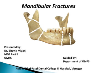
- 1. Presented by: Dr. Bhavik Miyani MDS Part ll OMFS Guided by: Department of OMFS Narsinhbhai Patel Dental College & Hospital, Visnagar Mandibular Fractures
- 2. Intoduction Etiology Epidemiology Classification Diagnosis- clinical & Radiological Management Complications References
- 3. INTRODUCTION
- 4. U-shaped bone Bilateral joint articulations Only mobile bone of the facial/cranial region Unique skeletal position of the mandible forms an inviting landmark as a recipient of an intentional or unintentional violence resulting in fracture
- 5. Thick cortical bone with single vessel for endosteal blood supply ◦ Varies with patient’s age and amount of dentition ◦ With atrophic mandibles, endosteal blood supply is decreased and periosteal blood supply is the dominant
- 6. Writings on mandible fractures appared as early as 1650 BC, when Egyptian described the examination, diagnosis and treatment of mandible fractures. Hippocrates described direct reapproximation of the fracture segments with the use of circumdental wiring. 1795, Chopart and Desault were the first to use dental prosthetic devices in an attempt to immobilize fracture segments. Chopart and Desault
- 7. John Barton described his Barton Bandage in order to immbilize and stabilize fracture fragment 1886, Thomas Brain Gunning was the first person to use a custom fitted intraoral dental splint for immobilization. He used the splint in conjunction with an external head appliance.
- 8. Glimer credited with being the first to use method of intermaxillary fixation. In 1881, Glimer described a method of mandibular fracture fixation that used two heavy rods placed on either side of the fracture and wired together. Luhr – 1960, developed mandibular compression plates Michelet and champy- 1970’s, placement of small bendable non- compression plates
- 9. Multiple studies have shown that greater than 75% of mandible fractures begin in areas of tension Exception to this is comminuted intracapsular condylar fractures which are totally compression in origin
- 10. Huelke(1961) has shown that anterior forces applied to the symphysis menti, over one mental foramen or over the mandibular body leads to strain at condylar necks and along the lingual plates in the opposite molar region.
- 11. Huelke(1961) & Hodgson(1967) stated that bone fracture at sites of tensile strain, since their resistance to compressive forces is greater. Once the mandible is loaded, the forces are distributed across the entire length of the mandible However, due to irregularities of the mandibular arch (foramen, concavities, convexities, ridges, and cross sectional thickness differences) load is distributed differently in areas
- 12. The weakness of the angle is produced by the abrupt change in direction between the body and ascending ramus in two planes. Between the junction of body & Ramus,there occurs in the vertical plane, a change of direction of almost 20 degrees, while in the horizontal plane, it is about 70 degrees at the upper border. Impacted third molars increases the risk of mandibular angle fractures.
- 14. FACTORS INFLUENCING DISPLACEMENT OF FRACTURE : Direction and intensity of the traumatic force Site of fracture Direction of fracture line muscle pull exerted on fractured fragments. Presence or absence of teeth. Extent of soft tissue wound.
- 15. Typical causes : A. Due to direct violence Interpersonal fights Blow due to stick, metal rods, bricks Fall Road traffic accident Occupational hazards-athletic injuries and industrial mishaps Iatrogenic causes-during dental treatment, fracture of a tooth, alveolus, maxillary tuberosity, fracture of mandible
- 16. B. Indirect violence Fall from height Counter coup fractures - due to excessive muscle contraction 2. Crush injuries Automobile accidents - RTA Aero plane crashes Mining accident
- 17. 3. High velocity missiles 4. Predisposing causes- Presence of cysts, osteomyelitis, tumors, presence of the third molars and systemic diseases affecting the formation of the structure of the bone Most of the severe injuries are caused by road traffic accident.
- 18. Mandible is second most frequently fractured bone of the face and tenth most frequently fractured bone of the body(Archer,1961). Males>Females Age: 16-30 years Assault>MVA>Falls>Sports for most common cause of fracture With concomitant facial injuries, 45% included at least 1 mandible fracture
- 19. Mandible fractures in conjunction with other injuries: ◦ Generally relevant to mode of injury Assault- 90% mandible only MVA- 46% with other injuries Spinal Cord injuries- varies according to studies : 3-49%
- 21. Multiple schemes exist to classify fractures Relate – ◦ fracture type, ◦ anatomic location, ◦ muscular relation, ◦ dentition relation, ◦ AO Classification etc.
- 22. Fracture types: Classification by kruger: ◦ Simple/closed- not opened to the external environment ◦ Compound/opened- fracture extends into external environment ◦ Comminuted- splintered or crushed ◦ Greenstick- only one cortex fractured ◦ Pathologic- pre-existing disease of bone lead to fracture
- 23. Fracture types: ◦ Multiple- two or more lines of fractures on the same bone that do not communicate ◦ Impacted- fracture which is driven into another portion of bone ◦ Indirect- a fracture at a point distant from the site of injury ◦ Complicated/complex- damage to adjacent soft tissue, can be simple or compound
- 24. Anatomic Classification: ◦ Developed by Dingman and Natvig Symphysis Parasymphyseal Body Angle Ramus Condyle process Coronoid process Alveolar process
- 25. Dentition Classification: ◦ Developed by Kazanjian and Converse ◦ Class I: teeth are present on both sides of the fracture line ◦ Class II: Teeth present only on one side of the fracture line ◦ Class III: Patient is edentulous
- 26. Muscle Action Classification: ◦ Vertically Favorable vs. Non Favorable Resistance to medial pull ◦ Horizontal Favorable vs. Non Favorable Resistance to upward movement Generally apply to angle and body fractures
- 27. AO Classification (Relevant to internal fixation) F : Number of fracture or fragments L : Location or site of the fracture O : Status of occlusion S : Soft tissue involvement A : Associated fractures of the facial skeleton
- 28. These criteria can be objectified clinically and radiographically : F : Number of fracture F0 : Incomplete fracture F1 : Single fracture F2 : Multiple fracture F3 : Comminuted fracture F4 : Fracture with a bone defect
- 29. 2. Categories of localization L1 : Precanine region L2 : Canine L3 : Postcanine L4 : Angle L5 : Supra – angular L6 : Condyle L7 : Coronoid L8 : Alveolar process
- 30. 3. Category of occlusion O0 : No malocclusion O1 : Malocclusion O2 : Nonexistent occlusion – edentulous mandible 4. Categories of soft tissue involvement S0 : Closed S1 : Open intraorally S2 : Open extraorally S3 : Open intra and extraorally S4 : Soft tissue defect
- 31. 5. Categories of associated fractures A0 : None A1 : Fracture and/or loss of teeth A2 : Nasal bone A3 : Zygoma A4 : Lefort I A5 : Lefort II A6 : Lefort III
- 32. Prior to examination, it is important to gain the following information ◦ Mechanism of injury ◦ Previous facial fractures ◦ Pre-existing TMJ disorders ◦ Pre-existing occlusion ◦ Past medical history (epilepsy, alcoholic, mental retardation, diabetes, psychiatric, immune status)
- 33. Physical examination: ◦ Tenderness to palpation ◦ Malocclusion- Anterior open bite- bilateral condylar or angle Unilateral open bite- ipsilateral angle and parasymphyseal fracture Posterior cross bite- symphyseal and condylar fractures with splaying of the posterior segments Prognathic bite- TMJ effusions Retrognathic bite- condylar or angle fractures
- 34. Physical examination: ◦ Loss of Normal form- bony contour change, soft tissue depressions, deformities ◦ Step deformity ◦ Loss of function- can be from pain, trismus Deviation on opening towards side of condylar fracture Inability to open due to impingement of coronoid or ramus on the zygomatic arch Premature contacts from alveolar, angle, ramus, or symphysis
- 35. Physical examination: ◦ Edema ◦ Abrasions/lacerations- potential for compound fracture ◦ Ecchymosis- especially floor of mouth Symphyseal or body fracture ◦ Crepitus with manipulation ◦ Altered sensation/parasthesia ◦ Dolor/Tumor/Rubor- signs of inflammation ◦ Fracture, luxation or subluxation of teeth
- 38. Radiographic Evaluation ◦ Panoramic radiograph: Most informative radiographic tool Shows entire mandible and direction of fracture (horizontal favorable, unfavorable) Disadvantages: Patient must sit up-right Difficult to determine buccal/lingual bone and medial condylar displacement Some detail is lost/blurred in the symphysis, TMJ and dentoalveolar regions
- 39. Radiographic Evaluation ◦ Reverse Towne’s radiograph: Ideal for showing lateral or medial condylar displacement
- 40. Radiographic Evaluation ◦ Lateral oblique radiograph: Used to visualize ramus, angle, and body fractures Easy to do Disadvantage: Limited visualization of the condylar region, symphysis, and body anterior to the premolars
- 41. Radiographic Evaluation ◦ Posteroanterior (PA) radiograph: Shows displacement of fractures in the ramus, angle, body, and symphysis region Disadvantage: Cannot visualize the condylar region
- 42. Radiographic Evaluation ◦ Occlusal views: Used to visualize fractures in the body in regards to medial or lateral displacement Used to visualize symphyseal fractures for anterior and posterior displacement
- 43. Radiographic Evaluation ◦ Computed tomography CT: Excellent for showing intracapsular condyle fractures Can get axial and coronal views, 3-D reconstructions Disadvantage: Expensive Larger dose of radiation exposure compared to plain film Difficult to evaluate direction of fracture from individual slices (reformatting to 3-D overcomes this)
- 46. Radiographic Evaluation ◦ Ideally need 2 radiographic views of the fracture that are oriented 90’ from one another to properly work up fractures Panorex and Towne’s CT axial and coronal cuts ◦ Single view can lead to misdiagnosis and complications with treatment
- 47. This Towne’s view show a body fracture that is displaced in a medial to lateral direction and a subcondylar fracture with lateral displacement
- 48. However, Panorex clearly shows the superior displacement of the right body fracture
- 50. 1. Patient’s general physical status 2. Diagnosis and treatment of mandibular fractures should not be approached with an “emergency-type” mentality
- 51. 3. Dental injuries should be evaluated and treated concurrently with the treatment of mandibular fractures 4. Re-establishment of occlusion is the primary goal in the treatment of mandibular fractures 5. With multiple facial fractures, mandibular fractures should be treated first
- 52. 6. Intermaxillary fixation time should vary according to the type, location, number, and severity of the mandibular fractures as well as the patient’s health and age, and the method used for reduction and immobilization
- 53. 7. Prophylactic antibiotics should be used for mandibular fractures 8. Nutritional needs should be monitored closely postoperatively 9. Most mandibular fractures can be treated with closed reduction
- 54. Bone healing is altered by types of fixation and mobility of the fracture site in relation to function Can be primary or secondary bone healing
- 55. Primary bone healing: No fracture callus forms Heals by a process of 1. Haversian remodeling directly across the fracture site if no gap exists (Contact healing), or 2. Deposition of lamellar bone if small gaps exist (Gap healing) Requires absolute rigid fixation with minimal gaps
- 56. Contact Healing Gap Healing
- 57. Secondary bone healing: ◦ Bony callus forms across fracture site to aid in stability and immobilization ◦ Occurs when there is mobility around the fracture site
- 58. Secondary bone healing involves the formation of a subperiosteal hematoma, granulation tissue, then a thin layer of bone forms by membranous ossification. Hyaline cartilage is deposited, replaced by woven bone and remodels into mature lamellar bone
- 60. Airway ◦ Tongue falling back ◦ Blood clots ◦ Fractured teeth segments ◦ Broken fillings ◦ Dentures Hemorrhage Soft tissue lacerations Support of bone fragments Pain control Infection control e.g. compound fractures Food and Fluid
- 61. Three type of management of fracture : 1. No treatment 2. Closed reduction 3. Open reduction
- 62. No treatment •Cases in which mandible appears stable •Favorable fracture pattern •No displacement of bony segments •No change in occlusion •Motivated patient •Management - Careful observation - Liquid diet, limited physical activity Remain prepared to intervene
- 63. Fracture reduction that involves techniques of not opening the skin or mucosa covering the fracture site Fracture site heals by secondary bone healing This is also a form of non-rigid fixation
- 64. Indications: ◦ “It is safe to say that the vast majority of fractures of the mandible may be treated satisfactorily by the method of closed reduction” ◦ “If the principle of using the simplest method to achieve optimal results is to be followed, the use of closed reduction for mandibular fractures should be widely used” Peterson’s Principle of Oral and Maxillofacial Surgery 2nd edition
- 65. Indications: ◦ Simply stated as all cases that open reduction is not indicated or is contraindicated ◦ Comminuted fractures- especially gunshot wounds ◦ Lack of soft tissue covering for avulsive type injuries
- 66. Indications: ◦ Nondisplaced favorable fractures ◦ Mandibular fractures in children with developing dentition ◦ Condylar fractures ◦ Edentulous fractures with use of prosthesis with circumandibular wires
- 67. Contraindications: ◦ Medical conditions that should avoid intermaxillary fixation Alcoholics Seizure disorder Mental retardation Nutritional concerns Respiratory diseases (COPD) ◦ Unfavorable fractures
- 68. Advantages: ◦ Low cost ◦ Short procedure time ◦ Can be done in clinical setting with local anesthesia or sedation ◦ Easy procedure ◦ No foreign body in patients
- 69. Disadvantages: ◦ Not absolute stability (secondary bone healing) ◦ Oral hygiene difficult ◦ Possible TMJ sequelae Muscular atrophy/stiffness Myofibrosis Possible affect on TMJ cartilage Decrease range of motion ◦ Non-compliance
- 70. Techniques: ◦ Direct interdental wiring ◦ Indirect interdental wiring ◦ Continuous or multiple loop wiring ◦ Arch bars – Erich arch bars ◦ Ivy loops ◦ Essig Wire ◦ Intermaxillary fixation screws ◦ Splints
- 71. ADVANTAGE: Simple and rapid method of immobilization METHOD: A 15cm length, prestretched 0.35mm soft s.s. wire is passed around a tooth emerging through the interdental spaces. With the wire placed around the neck of tooth the two ends are tightened by twisting together to produce a 3cm tail.
- 73. Adequate no. of teeth are wired. IMF is effected after reduction of the fracture by twisting the saperate tails together obtaining a crossbracing where possible. Bend the cut ends into interdental spaces or cover them with wax or gutta-percha. LIMITATIONS: 1) the wires tends to loosen, 2) a broken direct wire cannot be replaced without first removing and then replacing all of the others, 3) this technique is considered as a first aid method for temporary immobilization .
- 76. DRAWBACKS: ( Leonard 1977) ◦ Elastic traction is time consuming to apply ◦ Loops get loosen and break easily WILLIAM’S MODIFICATION BUTTON WIRING CLOVE HITCH WIRING
- 81. A 25cm long 26 guage wire Twisted around necks of second molar of each side. Both ends of wire twisted along its entire length Twisted wires from both ends are brought together in the midline, adapted according to the shape of arch on buccal side & final twisting is done. Forming the base wire. Additional interdental wires are used to secure this base wire.
- 83. ARCH BARSARCH BARS RISDON ARCH BARRISDON ARCH BAR JELENKO ARCH BARJELENKO ARCH BAR ERICH ARCH BARERICH ARCH BAR HAMILTON (1967)HAMILTON (1967)
- 84. ARCH BARS- custom madeARCH BARS- custom made schuchardt(1956)schuchardt(1956) schuchardt&metz(1966)schuchardt&metz(1966) stanhope(1969)stanhope(1969) clarke(1977)clarke(1977)
- 86. INDICATIONS: 1. For prolonged fixation on mandibular teeth in a patient with fracture of the tooth bearing segment of the mandible & bilateral displaced fractures of the condylar neck. 2. A portion of mandible is missing with considerable soft tissue loss. Here the cap splint maintains correct relationship of remaining tooth bearing segments till the reconstruction is completed. 3. In patients with severe periodontal disease. Here the cap splint helps to splint the loose teeth together and facilitates the application of IMF
- 90. Length of Intermaxillary fixation: ◦ Based on multiple factors Type and pattern of fracture Age of patient Involvement of intracapsular fractures ◦ Average adult: 3-4 weeks ◦ Children 15 years or younger- 2-3 weeks ◦ Elderly patients- 6-8 weeks ◦ Condylar fractures- 2-4 weeks
- 91. Intermaxillary fixation: ◦ Multiple studies show clinical bone union (no mobility, no pain, reduced on films) in 4 weeks in adults and 2 weeks in children ◦ Condylar process fractures tend to need only short periods of IMF to aid with pain and occlusion; usually 2 weeks
- 92. Technique of fracture repair by using transcutaneous pins threaded into the lateral surface of the mandible. The pin segments are then connected together with an acrylic bar, metal framework, or graphite rods. Synonymous with the Joe Hall Morris appliance
- 93. Indications: ◦ Comminuted mandible fractures with/without displacement ◦ Avulsive gunshot wounds ◦ Edentulous mandible fractures ◦ Can be used on patients that are poor candidate for open reduction and closed reduction (may increase likelihood of follow-up)
- 94. Joe Hall Morris appliance applied to mandibular defect
- 95. Different portions of the mandible will undergo different patterns of force in relation to loading
- 96. Mandibular Angle Region: ◦ Generally vertical pull due to masseter, medial pterygoid, and temporalis muscle ◦ Rarely is there any medial or lateral rotational forces ◦ Therefore, fixation/stabilization is to address the vertical component
- 97. Mandibular Body Region: ◦ Transitional zone ◦ Contains both vertical and horizontal movements ◦ Fixation/stabilization is directed towards countering both directions
- 98. Anterior Mandible: ◦ Direction of forces tends to alter with function ◦ Zones of compression and tension may actually alter with function ◦ Undergoes shearing and torsional forces
- 99. Implies the opening of skin or mucosa to visualize the fracture and reduction of the fracture Can be used for manipulation of fracture only Can be used for the non-rigid and rigid fixation of the fracture
- 100. Indications: ◦ Unfavorable/unstable mandibular fractures ◦ Patients with multiple facial fractures that require a stable mandible for basing reconstruction ◦ Fractures of an edentulous mandible fracture with severe displacement
- 101. Indications: ◦ Edentulous maxillary arch with opposing mandible fracture ◦ Delayed treatment with interposition of soft tissue that prevents closed reduction techniques to re-approximate the fragments
- 102. Indications: ◦ Medically compromised patients Gastrointestinal diseases Seizure disorders Compromised pulmonary health Mental retardation Nutritional disturbances Substance abuse patients
- 103. Contraindications: ◦ If a simpler method of repair is available, may be better to proceed with those options ◦ Severely comminuted fractures ◦ Patients with healing problems (radiation, chronic steroid use, transplant patients) ◦ Mandible fractures that are grossly infected
- 104. Advantage : ◦ Accurate reduction and stable immobilization-primary bone healing ◦ Normal nutrition in a matter of days ◦ Early return to normal jaw function ◦ Avoidance of airway problems ◦ Oral hygiene can be maintained ◦ No IMF – no occupational inconvenience ◦ Good patient compliance-decreased discomfort ◦ Less myoatrophy/TMJ disorders/MPDS ◦ Less trismus/derangement of jaw movements
- 105. Disadvantage : ◦ Need for surgically invasive techniques ◦ Risk of damage to anatomic structures ◦ Success depends on technical expertise ◦ Expensive equipment/materials required ◦ No leeway for occlusion to adjust ◦ Scarring ◦ No bridging of small bone defects ◦ Might lead to loss of multiple, small bone fragments ◦ Might require secondary procedure to remove hardware
- 106. Rigid fixation: ◦ Any form of fixation that counters any biomechanical forces that are acting upon the fracture site ◦ Prevents any inter-fragmentary motion across that fracture site ◦ Heals with primary (contact or gap) bone healing, produces no callus around fracture site
- 107. Lag screw technique: ◦ Utilizes screws that create a compression of the fracture segments by only engaging the screw threads in the remote segment and screw head in the near cortex ◦ Should be used to gain rigid fixation
- 108. Lag screw technique: ◦ Advantages: Low cost, less equipment Faster technique than plating Rigid fixation ◦ Disadvantages: Screw must be placed perpendicular to fracture Can be technique sensitive
- 109. Lag screw technique: ◦ Utilizes 2-3 screws to overcome rotational forces ◦ Smaller diameter drill used for portion of screw engaged in distant segment ◦ A single lag screw can be placed in the angle region to resist tension
- 111. Compression plate technique: ◦ Technique that creates rigid fixation ◦ When screws engage plate, they impart compression across the fracture segments ◦ Results in the fragments being brought together with compression and interfragmentary friction
- 113. Compression plate technique: ◦ Advantages: Rigid fixation Thicker hardware ◦ Disadvantages: Technique sensitive- plates must be adapted properly or mal- alignment can occur More expensive then miniplates Bicortical screws
- 114. Compression plate technique: ◦ With regards to the regional dynamic forces of the mandible, the ideal area to place the compression plate would be the alveolus (due to tension). However, due to the presence of the dentition, bicortical screws cannot be placed.
- 115. Compression plate technique: ◦ Therefore, compression plates are placed at the inferior border of the mandible with bicortical screws. ◦ Must utilize a tension band at the superior surface to counteract compressive spread of superior surface by the compression plate Arch bars Miniplates with monocortical screws (3 on each side ideal) ◦ Tension band placed prior to compression plate
- 116. Compression plate technique: ◦ Two types of compression plates exist Dynamic compression plates (DCP)- require tension band, can be placed intra-orally Eccentric dynamic compression plate (EDCP)- designed with the most lateral holes angled in a superior/medial direction to impact compression at the superior region. Must be placed extra-orally. Avoids use of tension band
- 117. Reconstruction plate: ◦ Rigid fixation technique ◦ Large plates that are load-bearing (can bear entire load of region) ◦ Consist of plates that utilize screws greater than 2mm in diameter (2.3, 2.4, 2.7, 3.0) ◦ Can use non-locking and locking type plates ◦ Must use 3 screws on each side of fracture (maximum strength with 4)
- 118. Reconstruction plate: ◦ Advantages: Rigid fixation with load-bearing properties Low infection rates in the literature, especially in the mandibular angle region Can be used for edentulous and comminuted fractures ◦ Disadvantages: Expensive Requires larger surgical opening Can be palpated by patient if in body or symphysis region
- 120. Rigid fixation: ◦ Includes the use of: Reconstruction plate with 3 screws on each side of the fracture Large compression plates 2 lag screws across fracture Use of 2 plates over fracture site 1 plate and 1 lag screw across fracture site
- 121. Examples of rigid fixation schemes for the mandibular body fracture ◦ 1 plate and 1 lag screw ◦ 2 plates non compression mini plates with inferior bicortical screws ◦ Compression plate
- 122. Rigid fixation of mandibular angle fractures: ◦ 2 non compression mini-plates with inferior plate with bicortical screws ◦ Reconstruction plate
- 123. Rigid fixation for symphyseal fractures: ◦ Compression plate with arch bar ◦ 2 lag screws ◦ 2 miniplates, inferior is bicortical and may be compression plate
- 124. Non rigid internal fixation: ◦ Bone fixation that is not strong enough to prevent interfragmentary motion across a fracture site ◦ Heals by secondary bone healing with callus formation ◦ Consists of miniplate application with functional stable fixation and intraosseous wiring
- 125. Non rigid internal fixation: ◦ Functional stable fixation: Term used when there is enough fixation that allows skeletal mobility/function but still forms a bony callus and secondary bone healing Consists of miniplates opposing tension or compression Relies on the buttressing effects of the bone (more bone height, more buttressing) or the vertical distance of placement of miniplates
- 126. Non rigid fixation with functional stable fixation: ◦ 2 plates that are spread apart are better able to resist the load
- 127. Non rigid fixation with functional stable fixation: ◦ Single plate placed in a mandible with greater vertical height will be more rigid due to buttressing effects of the thicker bone
- 128. Non rigid fixation with functional stable fixation: ◦ Technique pioneered by Champey ◦ Developed mathematical models to determine forces on the mandible in relation to the inferior alveolar canal, root apices, and bone thickness
- 129. Non rigid fixation with functional stable fixation: ◦ Developed guidelines for the use of plates in relation to the mental foramen in regards to ideal lines of osteosynthesis Posterior to mental foramen- 1 plate applied just below root apices/above IAN Anterior to mental foramen- 2 plates Utilizes monocortical miniplates only
- 131. Non rigid fixation with functional stable fixation: ◦ This technique is recommend with early mandibular fracture treatment (within 1st 24 hours) due to increase failure with delays ◦ Intra-oral technique ◦ Utilizes IMF for short periods of time ◦ Literature complication rates are extremely variable
- 132. Non rigid fixation with intraosseous wiring: ◦ Use of wire for direct skeletal fixation ◦ Keeps the fragments in an exact anatomical alignment, but must rely on other forms of fixation to maintain stability (splints, IMF). Not Rigid to allow function. ◦ Low cost, fast to perform, must rely on patient compliance as does closed reduction techniques
- 133. Non rigid fixation with intraosseous wiring: ◦ Simple straight wire- direction of pull is perpendicular to fracture ◦ Figure of eight wire- increased strength at superior and inferior regions compared to straight wire ◦ Transosseous / circum-mandibular wire- used for oblique type fractures- passes wire from skin with the use of an awl
- 134. Non rigid fixation with intraosseous wiring: ◦ Straight wire ◦ Figure of eight ◦ Transosseous-circum- mandibular
- 135. Non rigid fixation with intraosseous wiring: ◦ Mostly used in the mandibular angle as a superior border wire with simultaneous removal of third molar from fracture site ◦ Can be used in the inferior border of symphyseal and parasymphyseal fractures
- 136. Teeth in line of fracture are a potential impediment to healing for the following reasons 1. The fracture is compound into the mouth via the opened periodontal membrane 2. The tooth may be damaged structurally or loose its blood supply as a result of the trauma so that the pulp subsequently becomes necrotic 3. The tooth may be affected by some pre-existing pathological process Indications for removal ◦ Absolute ◦ Relative
- 137. Longitudinal fracture involving the root Dislocation or subluxation of tooth from socket Presence of periapical infection Infected fracture line Acute pericoronitis
- 138. Functionless tooth which would eventually be removed electively Advanced caries Advanced periodontal disease Teeth involved in untreated fractures presenting more than 3days after injury
- 139. Good quality intra-oral periapical radiograph Appropriate antibiotic therapy Splinting of tooth if mobile Endodontic therapy if pulp is exposed Immediate extraction if fracture becomes infected
- 140. Biomechanics differ for edentulous fractures compared to others ◦ Decrease bone height leads to decreased buttressing affect (alters plate selection) ◦ Significant bony resorption in the body region ◦ Significant effect of muscular pull, especially the digastric muscles
- 141. Incidence and location of mandible fractures in the edentulous mandible ◦ Highest percent in the body ◦ Atrophy creates saddle defect in body
- 142. Biological differences ◦ Decreased inferior alveolar artery (centrifugal) blood flow ◦ Dependent on periosteal (centripetal) blood flow ◦ Medical conditions that delay healing ◦ Decreased ability to heal with age
- 143. Classification of the edentulous mandible: ◦ Relates to vertical height of thinnest portion of the mandible Class I- 16-20mm Class II- 11-15mm Class III- <10mm
- 144. Closed Reduction ◦ Use of circumandibular wires fixated to the pryriform rims and circumzygomatic wires with patient’s denture or splints ◦ Requires IMF- usually longer periods of time ◦ Generally used to repair Class I type fractures or thicker
- 145. External pin fixation: ◦ May be used for fixation with/without the use of IMF ◦ Avoids periosteal stripping ◦ Used for comminuted edentulous fractures ◦ Can be used in patients that an open procedure is contraindicated ◦ Must use large diameter screws (4mm) for fixation, may be difficult in Class III patients
- 146. Open reduction techniques: ◦ Recommended for fractures that have not healed from other treatments, IMF contraindicated, splints/dentures unavailable, or the mandible is too atrophic for success with closed reduction ◦ Utilizes rigid fixation techniques ◦ Can utilize simultaneous bone grafting with severely atrophic mandibles if there is the possibility of inadequate bony contact
- 147. Open reduction techniques: ◦ Studies indicate that the lowest complication rates occur with extra-oral approaches with rigid fixation, especially with class III atrophic mandibles
- 148. Relatively uncommon type of injury Incidence of fractures in children under 15 years- 0.31/100,000 Usually represent less than 10% of all mandible fractures for children 12 years or younger Less than 5% of all mandible fractures for children 6 years or younger
- 149. Uniqueness of children: ◦ Nonunion and fibrous union are rare due to osteogenic potential of children. They heal rapidly. ◦ Due to growth, imperfect fracture reduction can be “compensated with growth”. Therefore, malocclusion and malunions usually resolve with time
- 150. Uniqueness of children: ◦ The mandible tends to be thinner and has a less dense cortex (could affect hardware placement) ◦ Presence of tooth buds in the lower portions of the mandible (could affect hardware placement) ◦ Short and less bulbous deciduous teeth make arch bar application difficult
- 151. Treatment modalities: ◦ Due to rapid healing, closed reduction techniques may be tolerated ◦ Most fractures can be treated with follow-ups and soft/non-functional diet or closed reduction with arch bars or acrylic splint ◦ Open reduction only advocated for severely displaced unfavorable fractures, in delayed treatment (>7days) due to soft tissue in-growth, or patients with airway/medical issues
- 152. Most studies show minimal risk for growth disturbances for fractures of the mandibular body, angle, symphysis, or ramus. Most disturbances occur from intracapsular condylar fractures Low rate of malunion, nonunion, or infections for pediatric fractures
- 153. Infection: ◦ Studies have looked at infection rates for different types of techniques: Highly variable in literature ◦ Most early studies indicate a decrease in infection rates with plating after time (experience) ◦ Dodson et al. J Oral Maxillofac Surg 1990;48 Closed reduction- 0% Wire osteosynthesis- 20% Rigid fixation- 6.3% ◦ Assael J Oral Maxillofac Surg 1987;45 Closed reduction- 8% Wire osteosynthesis- 24% Rigid fixation- 9%
- 154. Most common complication of surgical interaction. Risk factor- -Communited fracture -Active substances abuse. -Noncompliance with post operative regimens. -Significant delayed treatment MANIFESTATIONS Cellulitis, abscess formation, fistula, osteomyelitis and rarely necrotizing fascitis
- 155. The development of adequate drainage Removal of the source Appropriate antibiotic coverage Clinical examination &plain radiographic assess the status of fracture segments and hardware. Specimen for bacterial culture & sensitivity CT and MRI – if adjacent soft tissues are involved. Antibiotic of choice PG / clindamycin
- 156. MALUNION NONUNION It is a site with high incidence of altered fracture healing . Infection it is the main contributor. OTHER CAUSES -poor apposition -poor immobilization -presence of foreign bodies -unfavorable muscle pull on fracture segments -aseptic necrosis of bone fragments -soft tissue interposition -malnutrition by debilitation. Most common cause for nonunion is residual mobility across fracture.
- 157. Rigid internal fixation with a reconstruction plate External fixation Particulate bone grafting or Cortical bone grafting to the defect. Polyglycolic or polylactate mesh as a carrier for cancellous bone graft. Composite free flap reconstruction.
- 158. Malocclusion: ◦ More difficult to manage with rigid fixation ◦ Most studies have shown that malocclusion occur more frequently with rigid fixation ◦ May be due to plate mal-positioning/iatrogenic ◦ Low risk in pediatric fractures due to growth and dentition reposition
- 159. Prospective, 8 year study at Parkland involving angle fractures Nonrigid fixation had 17% complication rate AO Recon plate had 8% complication rate DCP had 13% complication rate Non compression plate 3% complication rate
- 160. FONSECA, Oral and Maxillofacial Surgery, Trauma, vol-3 Maxillofacial Injuries, Vol-1, Rowe & Williams
- 161. Thank you !
Editor's Notes
- Evans et al. J Bone Joint Surg 33; 1951 Huelke et al. J Oral Surg 27;1969 Huelke et al. J Dent Res 43; 1964
- Haug et al. An epidemiologic survey of facial fractures and concomitant injuries. JOMS 1990;48. Ellis et al. Ten years of mandible fractures: An analysis of 2,137 cases. Oral Surg Oral Med Oral Path 1985;59.
- (Ellis Oral Surg Oral Path Oral Med 1985), (Olson JOMS 1982)
- Bernstein Acad Opthalmol Otolaryngol 74;1970
- Juniper et al. J Oral Surg 1973;36 Amaratunga NA. J Oral Maxillofac Surg 1987;45Walker RV. J Oral Surg 1966;24
- Bruce et al. J Oral Maxillofac Surg 1993;51 Luhr et al. J Oral Maxillofac Surg 1996;54