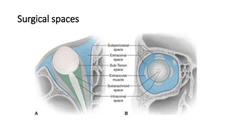
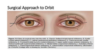
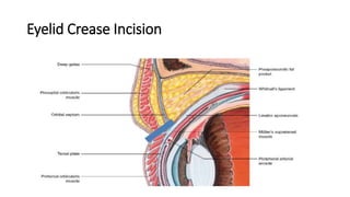
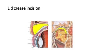
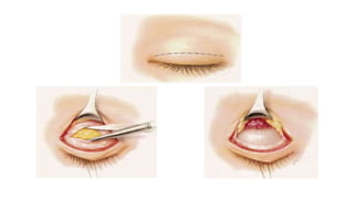
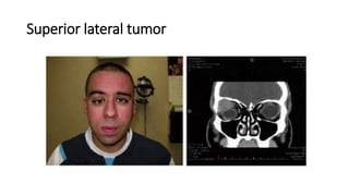
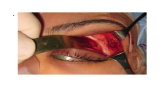
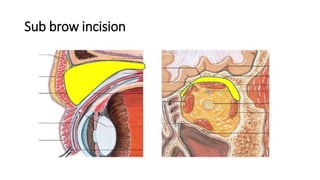
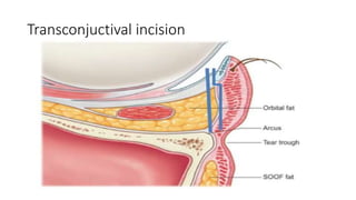
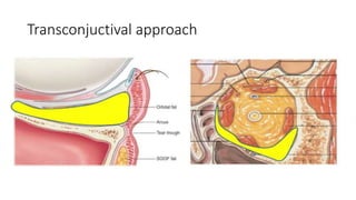
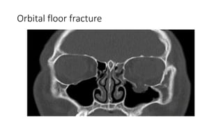
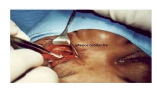
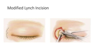
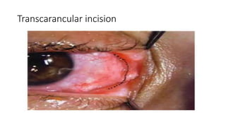
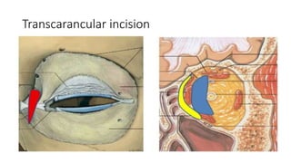
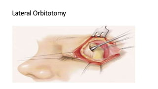
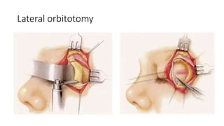
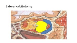
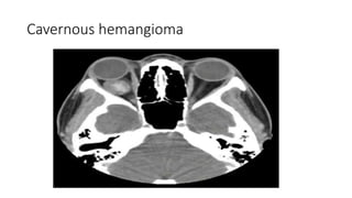
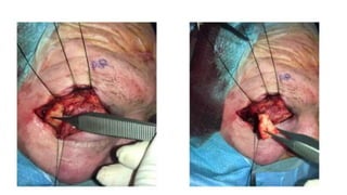
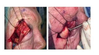
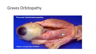
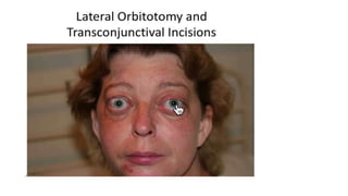
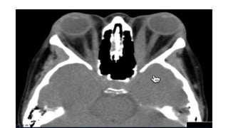
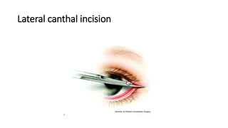
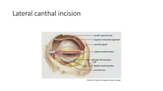
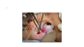
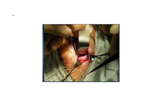
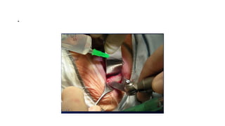
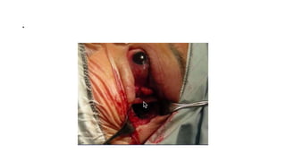
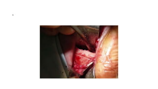
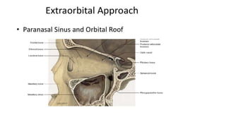
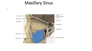
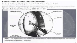
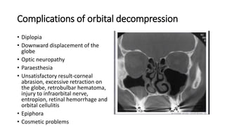
This document summarizes a presentation on orbital surgery. It discusses various surgical approaches to the orbit including lid crease incisions, lateral orbitotomies, and endoscopic decompression. It also covers orbital decompression techniques like superior, medial, inferior and lateral decompression to treat conditions like Graves' orbitopathy. Potential complications of orbital surgery are discussed such as diplopia, optic neuropathy, and hypoesthesia, as well as techniques to avoid complications by careful patient evaluation, approach selection, exposure and hemostasis.
We can ultimately aim to obtain “optical biopsies” of cornea with the introduction of high resolution AS-OCT .
AS-OCT helps assess tissue anatomy and evaluate differences in cellular morphology and patterns to distinguish between divergent anterior segment conditions.
We can ultimately aim to obtain “optical biopsies” of cornea with the introduction of high resolution AS-OCT .
AS-OCT helps assess tissue anatomy and evaluate differences in cellular morphology and patterns to distinguish between divergent anterior segment conditions.
Recent advances in diagnosis of glaucoma includes all the newer trends in the fields of measuring increased IOP, anterior chamber angle and depth assessment and optic nerve head assessment including RNFL thickness.
Ophthalmology Lectures ; Anterior segment OCT has been used widely in diagnosis of corneal disease, & in assessment of anterior segment surgery & keratoplasty
Maxillectomy and craniofacial resection Mamoon Ameen
all maxillectomy types in detail and maxillofacial resection ,indications ,contraindications ,preoperative asssessment and detail techniques and rehabilitations
Surgical excision of congenital Dermoid cysts in the orbitiosrjce
IOSR Journal of Dental and Medical Sciences is one of the speciality Journal in Dental Science and Medical Science published by International Organization of Scientific Research (IOSR). The Journal publishes papers of the highest scientific merit and widest possible scope work in all areas related to medical and dental science. The Journal welcome review articles, leading medical and clinical research articles, technical notes, case reports and others.
Title: Sense of Smell
Presenter: Dr. Faiza, Assistant Professor of Physiology
Qualifications:
MBBS (Best Graduate, AIMC Lahore)
FCPS Physiology
ICMT, CHPE, DHPE (STMU)
MPH (GC University, Faisalabad)
MBA (Virtual University of Pakistan)
Learning Objectives:
Describe the primary categories of smells and the concept of odor blindness.
Explain the structure and location of the olfactory membrane and mucosa, including the types and roles of cells involved in olfaction.
Describe the pathway and mechanisms of olfactory signal transmission from the olfactory receptors to the brain.
Illustrate the biochemical cascade triggered by odorant binding to olfactory receptors, including the role of G-proteins and second messengers in generating an action potential.
Identify different types of olfactory disorders such as anosmia, hyposmia, hyperosmia, and dysosmia, including their potential causes.
Key Topics:
Olfactory Genes:
3% of the human genome accounts for olfactory genes.
400 genes for odorant receptors.
Olfactory Membrane:
Located in the superior part of the nasal cavity.
Medially: Folds downward along the superior septum.
Laterally: Folds over the superior turbinate and upper surface of the middle turbinate.
Total surface area: 5-10 square centimeters.
Olfactory Mucosa:
Olfactory Cells: Bipolar nerve cells derived from the CNS (100 million), with 4-25 olfactory cilia per cell.
Sustentacular Cells: Produce mucus and maintain ionic and molecular environment.
Basal Cells: Replace worn-out olfactory cells with an average lifespan of 1-2 months.
Bowman’s Gland: Secretes mucus.
Stimulation of Olfactory Cells:
Odorant dissolves in mucus and attaches to receptors on olfactory cilia.
Involves a cascade effect through G-proteins and second messengers, leading to depolarization and action potential generation in the olfactory nerve.
Quality of a Good Odorant:
Small (3-20 Carbon atoms), volatile, water-soluble, and lipid-soluble.
Facilitated by odorant-binding proteins in mucus.
Membrane Potential and Action Potential:
Resting membrane potential: -55mV.
Action potential frequency in the olfactory nerve increases with odorant strength.
Adaptation Towards the Sense of Smell:
Rapid adaptation within the first second, with further slow adaptation.
Psychological adaptation greater than receptor adaptation, involving feedback inhibition from the central nervous system.
Primary Sensations of Smell:
Camphoraceous, Musky, Floral, Pepperminty, Ethereal, Pungent, Putrid.
Odor Detection Threshold:
Examples: Hydrogen sulfide (0.0005 ppm), Methyl-mercaptan (0.002 ppm).
Some toxic substances are odorless at lethal concentrations.
Characteristics of Smell:
Odor blindness for single substances due to lack of appropriate receptor protein.
Behavioral and emotional influences of smell.
Transmission of Olfactory Signals:
From olfactory cells to glomeruli in the olfactory bulb, involving lateral inhibition.
Primitive, less old, and new olfactory systems with different path
Anti ulcer drugs and their Advance pharmacology ||
Anti-ulcer drugs are medications used to prevent and treat ulcers in the stomach and upper part of the small intestine (duodenal ulcers). These ulcers are often caused by an imbalance between stomach acid and the mucosal lining, which protects the stomach lining.
||Scope: Overview of various classes of anti-ulcer drugs, their mechanisms of action, indications, side effects, and clinical considerations.
ARTIFICIAL INTELLIGENCE IN HEALTHCARE.pdfAnujkumaranit
Artificial intelligence (AI) refers to the simulation of human intelligence processes by machines, especially computer systems. It encompasses tasks such as learning, reasoning, problem-solving, perception, and language understanding. AI technologies are revolutionizing various fields, from healthcare to finance, by enabling machines to perform tasks that typically require human intelligence.
Pulmonary Thromboembolism - etilogy, types, medical- Surgical and nursing man...VarunMahajani
Disruption of blood supply to lung alveoli due to blockage of one or more pulmonary blood vessels is called as Pulmonary thromboembolism. In this presentation we will discuss its causes, types and its management in depth.
TEST BANK for Operations Management, 14th Edition by William J. Stevenson, Ve...kevinkariuki227
TEST BANK for Operations Management, 14th Edition by William J. Stevenson, Verified Chapters 1 - 19, Complete Newest Version.pdf
micro teaching on communication m.sc nursing.pdfAnurag Sharma
Microteaching is a unique model of practice teaching. It is a viable instrument for the. desired change in the teaching behavior or the behavior potential which, in specified types of real. classroom situations, tends to facilitate the achievement of specified types of objectives.
The prostate is an exocrine gland of the male mammalian reproductive system
It is a walnut-sized gland that forms part of the male reproductive system and is located in front of the rectum and just below the urinary bladder
Function is to store and secrete a clear, slightly alkaline fluid that constitutes 10-30% of the volume of the seminal fluid that along with the spermatozoa, constitutes semen
A healthy human prostate measures (4cm-vertical, by 3cm-horizontal, 2cm ant-post ).
It surrounds the urethra just below the urinary bladder. It has anterior, median, posterior and two lateral lobes
It’s work is regulated by androgens which are responsible for male sex characteristics
Generalised disease of the prostate due to hormonal derangement which leads to non malignant enlargement of the gland (increase in the number of epithelial cells and stromal tissue)to cause compression of the urethra leading to symptoms (LUTS
MANAGEMENT OF ATRIOVENTRICULAR CONDUCTION BLOCK.pdfJim Jacob Roy
Cardiac conduction defects can occur due to various causes.
Atrioventricular conduction blocks ( AV blocks ) are classified into 3 types.
This document describes the acute management of AV block.
2. Outline of presentation
• Surgical Spaces
• Orbitotomy
• Orbital Decompression
• Postoperative Care for Orbital Surgery
• Special Surgical Techniques in the Orbit
• Complications of Orbital Surgery
80. Orbital decompression
Indications
• Compressive optic neuropathy
• Globe subluxation
• Uncontrolled elevation of intraocular pressure
• Disfiguring proptosis secondary to thyroid eye disease
• Cosmesis
• Sphenoid wing meningiomas
• Large myopic globes
81. Goal of orbital decompression
• To expand the bony orbit confines to
make room for the increased volume
of the orbital contents
• First described by Dollinger who
advocated fort removal of the lateral
orbital wall for decompression into
temporal fossa
• Naffziger reported removal of the
orbital roof with decompression into
the anterior cranial fossa via a
transcranial apprack.
• .
82. Preoperative assessment
• Physical examination confirms the
upper and lower eyelid retraction,
proptosis and other physical signs
of hyperthyroidism
• Complete ophthalmologic exam
• Complete head and neck exam
including thyroid status
• Ultrasound scan demonstrates
thickening of extraocular muscles
• CT scans of the orbit are essential,
should include PNS and rule out
any sinus disease.
• Thyroid profile
• .
84. Surgical techniques
• Superior orbital decompression
• Medial orbital decompression
• Inferior orbital decompression
• Lateral orbital decompression
• Endoscopic orbital
decompression
• Orbital fat removal
• .
85. Superior orbital decompression
• Unroofing the entire superior orbital wall by a craniotomy
• Exposes the orbit by a frontal craniotomy
• After the optic nerve has been identified, the bony roof of the orbit is
removed from anterior to the optic foramen to the anterosuperior
orbital rim
• Superior periosteum is then incised in an H-shaped fashion and the
orbital fat allowed to herniate into the cranial vualt
86. .
• Titanium mesh and pericranial
flap are used to close the defect
• This approach is used for only
very severe cases due to
associated morbidity
• Complications, including;
-Meningitis
-CSF leak
-Pulsatile proptosis
• .
87. Medial orbital decompression
• External ethmpoidectomy
incision or through a coronal
forehead approach
• Ethmoidectomy approach
displaces the medial canthal
tendon and elevates the lacrimal
sac out of its fossa
• Anterior and posterior ethmpid
artery are identified and clipped
• ,
88. .
• A complete ethmoidectomy is
performed removing all the
mucosa bearing septa
• Posterior ethmoid cells are
removed back to the posterior
ethmoid plate
• Medial orbital periosteum is
incised longitudinally
• .
89. Inferior orbital decompression
• Done through subciliary,
transconjuctival or Caldwell-Luc
incision or combined approach
• A skin-muscle flap is elevated in the
lower eyelid and the orbital rim is
visualized
• The periosteum is incised and
elevated from the orbital floor for
approximately 4cm
• Caldwell-Luc incision is made
sublabially and wide antrostomy is
formed
• .
90. course of the infraorbital nerve is visualized and the bone
medial and lateral to the nerve is removed
the remainder of the floor is remoived under direct
visualization, 3 cm anteroposterior range or bone removal is
safe, medially remioved to lacrimal fossa and laterally removed
to the zygoma
91. .
• Periorbita is incised longitudinally, number of incisions determined
intraoperatively, 4 to 6 usually adequate
• Fat herniates into the defects on either side of the nerve
• Middle meatal ostium enlarged to provide for ventilation and
drainage of the sinus
92. Lateral orbital decompression
• Coronal, direct rim incision or
extended lateral canthotomy
• Periosteum over the lateral
orbital rim is exposed and
incised widely
• It is elevated from the orbital
side of the infratemporal fossa
for approximately 3-3.5 cm
posteriorly
• .
93. ,
• Lateral orbital rim can be cut and mobilized leaving its attachment to
the periosteum to assist with closure
• Much of the lateral orbital wall can be removed (about 2.5-3.5cm)
• Periorbita is incised and fat teased out into newly created space
94. Endoscopic orbital decompression
• Medial and medioinferior floors of
the orbit can be removed through
a transnasal approach
• Uncinate process is taken down
and large antrostomy is created
opening superiorly to the level of
the orbital floor and inferior
turbinate
• Middle turbinate is resected
• .
95. .
• Ethmoidectomy is performed and the anterior and posterior ethmoid
arteries are identified
• Medial orbital wall is exposed from the fovea ethmoidalis to the
anterior face of the sphenoid sinus
• Trocar inserted through the canine fossa can allow visualization
through the puncture while working through the nose
96. .
• Infraorbital nerve is identified and
mucosa elevated from the roof of the
maxillary sinus
Lamina papyracea is fractured and
removed to the level of the ethmoid
arteries
Bone removal is carried superiorly
within 2 mm of the fovea ethmoidalis,
posteriorly to the face of the sphenoid,
and laterally to the nerve
A buttress of bone is preserved
anteriorly at the junction of the
inferiorly and medial orbital walls to
avoid excessive inferior displacement of
the globe
• .
97. .
• Orbital periosteum is incised superiorly in a posterior to anterior
direction with a sickle knife taking care to avoid excessive penetration
with the knife
• Orbital fat protrudes into the ethmoid cavity
• Silastic splint is placed to avoid postoperative adhesions and packing
is not used
• Endoscopic approach allows a mean reduction of proptosis of 3mm.
99. Orbital fat decompression
• Orbital fat decompression or removal can be used either in isolation, or in
conjunction with other bony decompression procedures
• First described by Trokel in 1993
• This procedure involves opening the periosteum using an extended
transconjuctival approach
100. .
• Under direct visualization, using the bipolar cautery dissolve the fat
between and around the extraocular muscles
• 2-3mm decrease in proptosis can be expected
• A significant reduction in diplopia and an improvement in visual
acuity
101. Complications of orbital decompression
• Diplopia
• Downward displacement of the
globe
• Optic neuropathy
• Paraesthesia
• Unsatisfactory result-corneal
abrasion, excessive retraction on
the globe, retrobulbar hematoma,
injury to infraorbital nerve,
entropion, retinal hemorrhage and
orbital cellulitis
• Epiphora
• Cosmetic problems
• .
102. Post operative care for orbital surgery
• Head elevation
• Iced compresses on the eyelids
• Systemic steroids: Tabs Prednisolone 20mg bd for 5 days
• Optional placement of a drain (removed in 24-48 hours)
• VA checked in the first 12 hours after surgery
• Systemic antibiotics: I.V Cef 1g od for 3 days, then Tabs Cefixime
400mg od for 5 days
103. Special surgical techniques in the orbit
• FNAB used for lymphoid lesions, secondary tumors invading the orbit
from the sinuses, suspected metastatic tumors, and blind eyes with
optic nerve tumors.
• Limitation of FNAB: not very effective for obtaining tissue from
fibrous lesions because of difficulty in successfully aspirating cells
• Procedure: using a 4cm 22- or 23-gauge needle attached to a syringe
in a pistol-grip syringe holder. Can be guided by US or CT. specimen
studied by cytologist
104. .
• Frontal craniotomy or frontotemporal-orbitozygomatic approach. For
meningiomas, fibrous dysplasia, hemangioma, hemangiopericytomas,
schwannomas, gliomas
105. Complications of orbital surgery
How can the complications be avoided?
• Complete preoperative evaluation with orbital imaging when
indicated
• Choosing the appropriate surgical approach
• Obtaining adequate exposure
• Carefully manipulating the tissues
• Employing proper instrumentation and illumination
• Maintaining good hemostasis
• Using a team approach when appropriate
106. .
• Decreased or lost vision. Causes:
-Excessive traction on the globe
and optic nerve
-Contusion of the optic nerve
-Postoperative infection or
hemorrhage
• Hypoesthesia in the distribution
of the infraorbital nerve, in
orbital floor decompression
• Superior division of CN 3
susceptible to injury in tumor
resection in superior orbit
• Ciliary ganglion at risk in lateral
approaches to intraconal space
107. Other complications of orbital surgeries
• Ptosis
• Neuroparalyitc keratopathy
• Pupillary changes
• Vitreous hemorrhage
• Retinal detachment
• Hypoesthesia of forehead
• Keratitis sicca
• CSF leak
• Infection
108. References
• AAO Bk 7
• Scott-Brown’s Otorhinolaryngology, Head and Neck Surgery, Vol-2, 7th edition
• Cummings, Otorhinolaryngology, Head and Neck Surgery, Vol-2, 4th edition.
• Purgason PA, Hornblass A. Complications of surgery for orbital tumors. Ophthal
Plast Reconstr Surg. 1992;8(2):88-93
• Kacker A, Kazim M, Murphy M, Trokel S, Close LG. "Balanced" orbital
decompression for severe Graves’ orbitopathy: technique with treatment
algorithm. Otolaryngol Head Neck Surg. 2003;128(2):228-235.
• Perry JD, Kadakia A, Foster JA. Transcaruncular orbital decompression for
dysthyroid optic neuropathy. Ophthal Plast Reconstr Surg. 2003;19(5):353-358.
• White WA, White WL, Shapiro PE. Combined endoscopic medial and inferior
orbital decompression with transcutaneous lateral orbital decompression in
Graves' orbitopathy. Ophthalmology. 2003;110(9):1827-1832