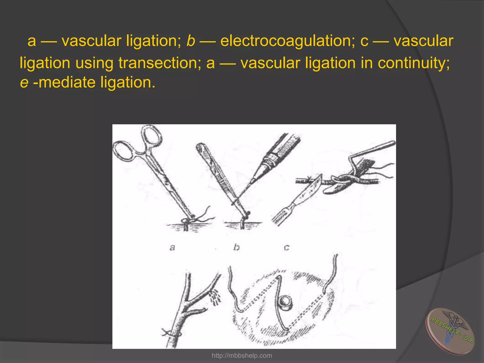
Bleeding
http://mbbshelp.com
Bleeding or hemorrhage , is the escape of
blood from the blood vessels as a result of
an injury or defect in the permeability of
the vascular walls.
http://mbbshelp.com
Types of bleeding:
 A) Haemorrhage per rexin (as a result of
mechanical damage to the vessel )
 B) Haemorrhage per diabrosin (results
from erosion of blood vessels)
 С) Haemorrhage per diapedesin (due to
a defect in the permeability of the vascular
walls)
http://mbbshelp.com
Bleeding can be:
 1.Arterial
 2.Arterio-venous
 3.Venous
 4.Capillary
 5.Parenchymal bleeding
http://mbbshelp.com
 1. External bleeding (when blood oozes
outside the body)
 2.Internal bleeding (when blood
accumulates in an enclosed space of
the body or in the cavity of a hollow
organ).
http://mbbshelp.com
Traumatic bleeding can also
be:
 1.primary
 2.secondary
http://mbbshelp.com
Acute bleeding
http://mbbshelp.com
Clinical picture
 The signs include skin pallor, diaphoresis, facial
cyanosis, weak and fast pulse, tachypnoea (periodic
breathing, or Cheyne-Stokes phenomenon)
 The symptoms involve headache, dryness in the
mouth and thirst, nausea, blurred vision and
progressive malaise.
 In contrast, if the blood is being lost slowly, the
symptoms and signs may not reflect the amount of
the blood lost.
http://mbbshelp.com
The four degrees of blood loss are
identified:
 1.mild - a reduction in blood circulating
volume of 10—12%. or 500-700 ml of blood;
 2.moderate - a reduction in blood circulating
volume of 15-20%, or 1,000-1,400 ml of
blood;
 3.severe — a reduction in blood circulating
volume of 20-30%, or 1,500-2,000 ml of
blood:
 4.massive - a reduction in blood circulating
volume of more than 30%, or more than
2,000 ml of blood. 3
http://mbbshelp.com
Laboratory investigations:
 Checking for levels of:
 1. The red blood cells
 2. Haemoglobin and
 3. Haematocrit
http://mbbshelp.com
Special diagnostic methods:
 1. Diagnostic puncture ( thoracocentesis,
laparocentesis)
 2. X-ray
 3. ultrasound
 4 scanning
 5.computerised tomography
 6.Endoscopic methods ( gastroscopy,
rectoscopy, cystoscopy and arthroscopy,
laporoscopy)
http://mbbshelp.com
Treatment
 1.infusions of blood substitutes
 2.Blood transfusion should be considered
whenever haemoglobin and haematocrit
levels fall as low as 80 g/l and 30,
respectively.
 In severe acute bleeding blood transfusion
should be started by the fast flow method
through one, two or even three veins, while
slow infusion can be justifiable only after
the systolic blood pressure has at least
risen to as high as 80 mm Hg.
http://mbbshelp.com
 3.Acidosis is corrected by giving sodium
bicarbonate, trisamin and lactasol
 4.Oxygen therapy should also be
considered; especially effective is hyperbaric
oxygenation, which is used after bleeding
has stopped.
http://mbbshelp.com
External bleeding
The colour of the escaping blood depends on
the type of the vessel affected: it is bright red
in arterial bleeding and dark red in venous
haemorrhage. It is noteworthy that the lethal
bleeding within a few minutes after injury may
result not only from a damage to the aorta but
also from that to the femoral or axillary arteries
or even larger veins. Injury to the major
cervical or thoracic vessels can lead to a very
serious complication — air embolism.
http://mbbshelp.com
Internal bleeding :
 Acute anaemia (due to a ruptured ectopic
pregnancy or raptured spleen with
subcapsular haematoma)
 Blood escapes into a hollow organ (result of
bleeding from the Lung, trachea, pharynx,
oesophagus, stomach or duodenum)
 Bleeding into an entrapped body cavity (the
cranium, spinal canal, thoracic and abdominal
cavities, pericardium and synovial space)
tends to be the most complicated.
http://mbbshelp.com
HAEMOSTASIS
( or control of hemorrhage)
http://mbbshelp.com
Temporary methods of haemostasis:
 1.Aplication of a tourniquet
 2.Digital compression
 3.Flexion of the limb in a joint
 4 Wound package combined with
application of a pressure bandage.
 5. Using artery forceps or vessel clamps to
stop blooding vessels located deeply
inside.
 6Temporary bypass of vessels.
http://mbbshelp.com
Application of a tourniquet
. Application of the facial tourniquet.
http://mbbshelp.com
Application of the cervical
<military> tourniquet.
http://mbbshelp.com
Application of the limb tourniquet:
a — preparing the area for application: b —
the start of the procedure
http://mbbshelp.com
c — fixing the first round: d — the view
after application.
http://mbbshelp.com
Application of the tourniquet in a damage to the femoral <a) and
axillary (b) arteries.
http://mbbshelp.com
1. It is a must to write down the time when the
tourniquet has been applied.
2.Keeping a tourniquet for more than 2 hours on the
lower limb and for above I'/, hours on the upper
one can result in ischaemic necrosis.
3. If the patient's transportation takes more than 1,5
hours, the tourniquet should periodically (even 10-
15 minutes) be released until the reappearance of
the arterial blood flow. At that time using pressing
on the blooding vessel.
4 The removal and reapplication of the tourniquet
can be repeated (in winter time every 30 minutes,
in summer each 50-60 minutes).
http://mbbshelp.com
Topography of the arteries
which can be pressed
http://mbbshelp.com
Digital pressure on an artery to temporarily arrest the bleeding:: a —
the carotid artery; b — the sublingual artery; c — the temporal artery;
d — the subclavian artery; e — the humeral artery; f — the axillary
rtery; g - the femoral artery
http://mbbshelp.com
Temporary control of femoral (a), popliteal (b).
humeral and ulnar (c) arterial hemorrhage by
means of maximal bending.
http://mbbshelp.com
DEFINITIVE
HAEMOSTASiS
 The methods of definitive haemostasis
divide into the four groups:
 mechanical;
 physical:
 chemical and biological;
 combined.
http://mbbshelp.com
Mechanical methods.
 1.ligating the bleeding vessel inside the
wound or somewhere along it.
 2.Twisting of the bleeding vessel.
 3.Wound package.
 4.Vascular sutures
.
http://mbbshelp.com
a — vascular ligation; b — electrocoagulation; c — vascular
ligation using transection; a — vascular ligation in continuity;
e -mediate ligation.
http://mbbshelp.com
Vascular sutures: a — Interrupted
(Carrel-type); b — mattress; C-blanket;
d-continuous mattress; e-mechanical
A B C D
http://mbbshelp.com
Physical methods:
 1.Surgical diathermy
 2.Laser (focused beam of electronic
rays)
 3.Cryosurgery
http://mbbshelp.com
Chemical and biological
methods:
 1. resorptive
 2. topical action
http://mbbshelp.com
Haemostatics with resorptive action:
 1. direct blood transfusion, transfusion of
small amount of freshly frozen blood,
plasma, fibrinogen, prothrombin
complex .
 2. Inhibitors of fibrinolysis: contrycal,
aprotinin, aminocapronic acid.
 3. Analogue of vitamin K: vicasol
http://mbbshelp.com
Topical haemostatics:
 Fibrin sponge, biological antiseptic pack,
haemostatic and gelatin sponges,
thrombin.
http://mbbshelp.com
Thank you for your attention .
http://mbbshelp.com

Bleeding

  • 1.
  • 2.
    Bleeding or hemorrhage, is the escape of blood from the blood vessels as a result of an injury or defect in the permeability of the vascular walls. http://mbbshelp.com
  • 3.
    Types of bleeding: A) Haemorrhage per rexin (as a result of mechanical damage to the vessel )  B) Haemorrhage per diabrosin (results from erosion of blood vessels)  С) Haemorrhage per diapedesin (due to a defect in the permeability of the vascular walls) http://mbbshelp.com
  • 4.
    Bleeding can be: 1.Arterial  2.Arterio-venous  3.Venous  4.Capillary  5.Parenchymal bleeding http://mbbshelp.com
  • 5.
     1. Externalbleeding (when blood oozes outside the body)  2.Internal bleeding (when blood accumulates in an enclosed space of the body or in the cavity of a hollow organ). http://mbbshelp.com
  • 6.
    Traumatic bleeding canalso be:  1.primary  2.secondary http://mbbshelp.com
  • 7.
  • 8.
    Clinical picture  Thesigns include skin pallor, diaphoresis, facial cyanosis, weak and fast pulse, tachypnoea (periodic breathing, or Cheyne-Stokes phenomenon)  The symptoms involve headache, dryness in the mouth and thirst, nausea, blurred vision and progressive malaise.  In contrast, if the blood is being lost slowly, the symptoms and signs may not reflect the amount of the blood lost. http://mbbshelp.com
  • 9.
    The four degreesof blood loss are identified:  1.mild - a reduction in blood circulating volume of 10—12%. or 500-700 ml of blood;  2.moderate - a reduction in blood circulating volume of 15-20%, or 1,000-1,400 ml of blood;  3.severe — a reduction in blood circulating volume of 20-30%, or 1,500-2,000 ml of blood:  4.massive - a reduction in blood circulating volume of more than 30%, or more than 2,000 ml of blood. 3 http://mbbshelp.com
  • 10.
    Laboratory investigations:  Checkingfor levels of:  1. The red blood cells  2. Haemoglobin and  3. Haematocrit http://mbbshelp.com
  • 11.
    Special diagnostic methods: 1. Diagnostic puncture ( thoracocentesis, laparocentesis)  2. X-ray  3. ultrasound  4 scanning  5.computerised tomography  6.Endoscopic methods ( gastroscopy, rectoscopy, cystoscopy and arthroscopy, laporoscopy) http://mbbshelp.com
  • 12.
    Treatment  1.infusions ofblood substitutes  2.Blood transfusion should be considered whenever haemoglobin and haematocrit levels fall as low as 80 g/l and 30, respectively.  In severe acute bleeding blood transfusion should be started by the fast flow method through one, two or even three veins, while slow infusion can be justifiable only after the systolic blood pressure has at least risen to as high as 80 mm Hg. http://mbbshelp.com
  • 13.
     3.Acidosis iscorrected by giving sodium bicarbonate, trisamin and lactasol  4.Oxygen therapy should also be considered; especially effective is hyperbaric oxygenation, which is used after bleeding has stopped. http://mbbshelp.com
  • 14.
    External bleeding The colourof the escaping blood depends on the type of the vessel affected: it is bright red in arterial bleeding and dark red in venous haemorrhage. It is noteworthy that the lethal bleeding within a few minutes after injury may result not only from a damage to the aorta but also from that to the femoral or axillary arteries or even larger veins. Injury to the major cervical or thoracic vessels can lead to a very serious complication — air embolism. http://mbbshelp.com
  • 15.
    Internal bleeding : Acute anaemia (due to a ruptured ectopic pregnancy or raptured spleen with subcapsular haematoma)  Blood escapes into a hollow organ (result of bleeding from the Lung, trachea, pharynx, oesophagus, stomach or duodenum)  Bleeding into an entrapped body cavity (the cranium, spinal canal, thoracic and abdominal cavities, pericardium and synovial space) tends to be the most complicated. http://mbbshelp.com
  • 16.
    HAEMOSTASIS ( or controlof hemorrhage) http://mbbshelp.com
  • 17.
    Temporary methods ofhaemostasis:  1.Aplication of a tourniquet  2.Digital compression  3.Flexion of the limb in a joint  4 Wound package combined with application of a pressure bandage.  5. Using artery forceps or vessel clamps to stop blooding vessels located deeply inside.  6Temporary bypass of vessels. http://mbbshelp.com
  • 18.
    Application of atourniquet . Application of the facial tourniquet. http://mbbshelp.com
  • 19.
    Application of thecervical <military> tourniquet. http://mbbshelp.com
  • 20.
    Application of thelimb tourniquet: a — preparing the area for application: b — the start of the procedure http://mbbshelp.com
  • 21.
    c — fixingthe first round: d — the view after application. http://mbbshelp.com
  • 22.
    Application of thetourniquet in a damage to the femoral <a) and axillary (b) arteries. http://mbbshelp.com
  • 23.
    1. It isa must to write down the time when the tourniquet has been applied. 2.Keeping a tourniquet for more than 2 hours on the lower limb and for above I'/, hours on the upper one can result in ischaemic necrosis. 3. If the patient's transportation takes more than 1,5 hours, the tourniquet should periodically (even 10- 15 minutes) be released until the reappearance of the arterial blood flow. At that time using pressing on the blooding vessel. 4 The removal and reapplication of the tourniquet can be repeated (in winter time every 30 minutes, in summer each 50-60 minutes). http://mbbshelp.com
  • 24.
    Topography of thearteries which can be pressed http://mbbshelp.com
  • 25.
    Digital pressure onan artery to temporarily arrest the bleeding:: a — the carotid artery; b — the sublingual artery; c — the temporal artery; d — the subclavian artery; e — the humeral artery; f — the axillary rtery; g - the femoral artery http://mbbshelp.com
  • 26.
    Temporary control offemoral (a), popliteal (b). humeral and ulnar (c) arterial hemorrhage by means of maximal bending. http://mbbshelp.com
  • 27.
    DEFINITIVE HAEMOSTASiS  The methodsof definitive haemostasis divide into the four groups:  mechanical;  physical:  chemical and biological;  combined. http://mbbshelp.com
  • 28.
    Mechanical methods.  1.ligatingthe bleeding vessel inside the wound or somewhere along it.  2.Twisting of the bleeding vessel.  3.Wound package.  4.Vascular sutures . http://mbbshelp.com
  • 29.
    a — vascularligation; b — electrocoagulation; c — vascular ligation using transection; a — vascular ligation in continuity; e -mediate ligation. http://mbbshelp.com
  • 30.
    Vascular sutures: a— Interrupted (Carrel-type); b — mattress; C-blanket; d-continuous mattress; e-mechanical A B C D http://mbbshelp.com
  • 31.
    Physical methods:  1.Surgicaldiathermy  2.Laser (focused beam of electronic rays)  3.Cryosurgery http://mbbshelp.com
  • 32.
    Chemical and biological methods: 1. resorptive  2. topical action http://mbbshelp.com
  • 33.
    Haemostatics with resorptiveaction:  1. direct blood transfusion, transfusion of small amount of freshly frozen blood, plasma, fibrinogen, prothrombin complex .  2. Inhibitors of fibrinolysis: contrycal, aprotinin, aminocapronic acid.  3. Analogue of vitamin K: vicasol http://mbbshelp.com
  • 34.
    Topical haemostatics:  Fibrinsponge, biological antiseptic pack, haemostatic and gelatin sponges, thrombin. http://mbbshelp.com
  • 35.
    Thank you foryour attention . http://mbbshelp.com