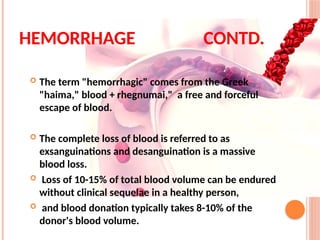
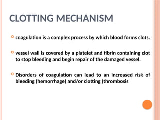
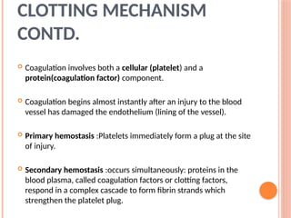
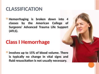
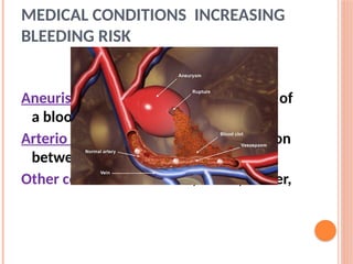
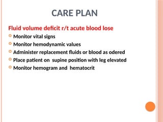
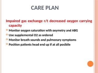
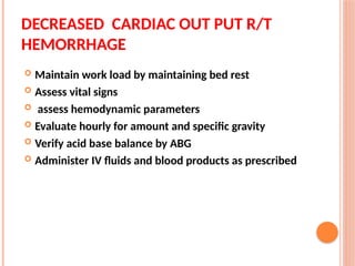
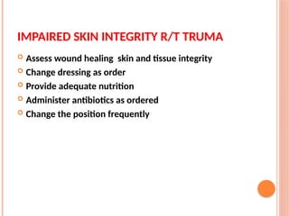
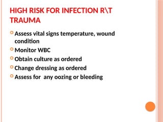
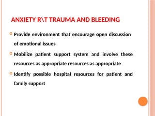
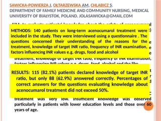
HEMORRHAGE
• Bleeding, technically known as hemorrhage. It is the
loss of blood from the circulatory system.
• Bleeding can occur internally, where blood leaks
from blood vessels inside the body or externally,
either through a natural opening such as the vagina,
mouth or anus, or through a break in the skin.
 The term "hemorrhagic" comes from the Greek
"haima," blood + rhegnumai," a free and forceful
escape of blood.
 The complete loss of blood is referred to as
exsanguinations and desanguination is a massive
blood loss.
 Loss of 10-15% of total blood volume can be endured
without clinical sequelae in a healthy person,
 and blood donation typically takes 8-10% of the
donor's blood volume.
HEMORRHAGE CONTD.
CLOTTING MECHANISM
 coagulation is a complex process by which blood forms clots.
 vessel wall is covered by a platelet and fibrin containing clot
to stop bleeding and begin repair of the damaged vessel.
 Disorders of coagulation can lead to an increased risk of
bleeding (hemorrhage) and/or clotting (thrombosis
 Coagulation involves both a cellular (platelet) and a
protein(coagulation factor) component.
 Coagulation begins almost instantly after an injury to the blood
vessel has damaged the endothelium (lining of the vessel).
 Primary hemostasis :Platelets immediately form a plug at the site
of injury.
 Secondary hemostasis :occurs simultaneously: proteins in the
blood plasma, called coagulation factors or clotting factors,
respond in a complex cascade to form fibrin strands which
strengthen the platelet plug.
CLOTTING MECHANISM
CONTD.
CLASSIFICATION
 Hemorrhaging is broken down into 4
classes by the American College of
Surgeons' Advanced Trauma Life Support
(ATLS).
Class I Hemorrhage
 involves up to 15% of blood volume. There
is typically no change in vital signs and
fluid resuscitation is not usually necessary.
Class II Hemorrhage
 involves 15-30% of total blood volume.
 A patient is often tachycardic (rapid heart beat) with a narrowing
of the difference between the systolic and diastolic blood
pressures.
 The body attempts to compensate with peripheral
vasoconstriction. Skin may start to look pale and be cool to the
touch.. Volume resuscitation with crystalloids (Saline solution or
Lactated Ringer solution) is all that is typically required. Blood
transfusion is not typically required.
Class III Hemorrhage
 Involves loss of 30-40% of circulating blood volume.
 The patient's blood pressure drops, the heart rate
increases, peripheral perfusion, such as capillary
refill worsens, and the mental status worsens.
 Fluid resuscitation with crystalloid and blood
transfusion are usually necessary.
Class IV Hemorrhage
 Involves loss of >40% of circulating blood
volume.
 The limit of the body's compensation is reached
 Aggressive resuscitation is required to prevent
death.
CAUSES
Traumatic
• Traumatic bleeding is caused by some type of injury. There are
different types of wound which may cause traumatic bleeding.
• Abrasion - Also called a graze, this is caused by transverse
action of a foreign object against the skin, and usually does
not penetrate below the epidermis
• Excoriation - In common with Abrasion, this is caused by
mechanical destruction of the skin, although it usually has an
underlying medical cause
• Hematoma - - Caused by damage to a blood vessel that in
turn causes blood to collect under the skin.
CAUSES
• Contusion - Also known as a bruise, this is a blunt
trauma damaging tissue under the surface of the skin
• Crushing Injuries - Caused by a great or extreme
amount of force applied over a period of time. The
extent of a crushing injury may not immediately
present itself.
• Ballistic Trauma - Caused by a projectile weapon, this
may include two external wounds (entry and exit)
and a contiguous wound between the two
CAUSES
• Laceration - Irregular wound caused by blunt
impact to soft tissue overlying hard tissue or
tearing such as in childbirth. In some instances,
this can also be used to describe an incision.
• Incision - A cut into a body tissue or organ, such as
by a scalpel, made during surgery.
• Puncture Wound - Caused by an object that
penetrated the skin and underlying layers, such as
a nail, needle or knife
MEDICAL CONDITIONS INCREASING
BLEEDING RISK
Anatomical deformities
Aneurism:localised blood filled dilation of
a blood vessel
Arterio venous malformation: connection
between arteries and veins
Other conditions:infection, ulcers, cancer,
CONDITION THAT CAN AFFECT NORMAL
HEMOSTASIS
• Thrombocytopenia: a decrease in number of
platelet.
• Thrombasthenia: A decreased function of platelets
• Hemophilia: x linked recessive disorder in factor VIII
is deficient
• Christmas disease: deficiency of factor IX
• Von Willebrand disease: deficiency of Von
Willebrand factor which help in platelet activation
DRUGS
NSAIDs: inhibit platelet activation
eg:aspirine
Warfarin: inhibit vitamin K which is
needed for the production of factors II,
VII, IX,and X
MANAGEMENT
Minor bleeding
• Minor bleeding is bleeding that falls under a Class I
hemorrhage and the bleeding is easily stopped with pressure.
• The largest danger in a minor wound is infection
• Bleeding can be stopped with direct pressure and elevation,
and the wound should be washed well with soap and water.
• A dressing, typically made of gauze, should be applied. or
iodine solutions (such as Betadine) can also be used
PRESSURE POINTS
 In situations where direct pressure and elevation are
either not possible or proving ineffective, the use of
pressure to constrict the major artery which feeds the
point of the bleed is advocated .
 This is usually performed at a place where a pulse can
be found.
 There are risks involved in performing pressure point
constriction, including necrosis of the area below the
constriction.
PRESSURE POINTS
 Application of pressure in carotid artery may cause
damage to brain
 Pressure on the carotid artery can also can cause vagal
tone induced bradycardia.
 Other dangers in use of a constricting method include
rhabdomyolysis, which is a build up of toxins below the
pressure point.
Tourniquet
 Another method of achieving constriction of the supplying
artery is via the use of a tourniquet- a tightly tied band which
goes around a limb to restrict blood flow.
 Their use in emergency medicine is more limited, and is
restricted in most countries.
 An emergency tourniquet should in any case never be
applied to the forearm or lower leg since the arteries in these
locations run between bones and can not be compressed
Tourniquet
• Improvised tourniquets, however, usually fail to achieve
force enough to compress the arteries of the limb and thus
do not only fail to stop arterial bleeding but actually increase
bleeding due to the impaired venous blood flow.
•
PRESSURE DRESSING
 A sterile dressing applied with pressure to a
hemorrhaging wound enhances clot formation,
 compresses open blood vessels, and protects
the injury from further invasion by infectious
organisms.
 Sometimes more harm is done to the patient by
secondary infection than the trauma itself
ANTIHEMORRHAGICS
 Vitamin K/Inj .Phytomenadione /Menadione
: 5-10 mg/day orally for deficiency
 Fibrinogen: 0.5g infused in acute fibrinogenic state
 Inj. Tranexa 1000mg IV stat fb 500mg IV / oral TDS
 Factor IX: It is a synthetic protein prescribed for
control and prevention of hemorrhagic episodes in
patients with hemophilia B.
 Avatrombopag tablets are prescribed for treating
abnormally low platelet counts or a condition of
thrombocytopenia in adult patients with severe liver
dysfunction who are planning to undergo a medical or
dental procedure.
ANTIHEMORRHAGICS
 Antifibrinolytics amino acids (Aminocaproic acid, Tranexamic
acid, Aminomethylbenzoic acid)
 Local hemostatics Absorbable gelatin sponge · Oxidized
cellulose · Tetragalacturonic acid hydroxymethylester ·
Adrenalone · Thrombin · Collagen · Calcium alginate ·
Epinephrine
 Other systemic hemostatics Etamsylate · Carbazochrome ·
Batroxobin · Romiplostim
ANTIHEMORRHAGICS
CARE PLAN
Fluid volume deficit r/t acute blood lose
 Monitor vital signs
 Monitor hemodynamic values
 Administer replacement fluids or blood as odered
 Place patient on supine position with leg elevated
 Monitor hemogram and hematocrit
Impaired gas exchange r/t decreased oxygen carrying
capacity
 Monitor oxygen saturation with oxymetry and ABG
 Use supplemental O2 as ordered
 Monitor breath sounds and pulmonary symptoms
 Position patients head end up if at all posibile
CARE PLAN
DECREASED CARDIAC OUT PUT R/T
HEMORRHAGE
 Maintain work load by maintaining bed rest
 Assess vital signs
 assess hemodynamic parameters
 Evaluate hourly for amount and specific gravity
 Verify acid base balance by ABG
 Administer IV fluids and blood products as prescribed
IMPAIRED SKIN INTEGRITY R/T TRUMA
 Assess wound healing skin and tissue integrity
 Change dressing as order
 Provide adequate nutrition
 Administer antibiotics as ordered
 Change the position frequently
HIGH RISK FOR INFECTION RT
TRAUMA
 Assess vital signs temperature, wound
condition
 Monitor WBC
 Obtain culture as ordered
 Change dressing as ordered
 Assess for any oozing or bleeding
ANXIETY RT TRAUMA AND BLEEDING
 Provide environment that encourage open discussion
of emotional issues
 Mobilize patient support system and involve these
resources as appropriate resources as appropriate
 Identify possible hospital resources for patient and
family support
SAWICKA-POWIERZA J, OŁTARZEWSKA AM, CHLABICZ S.
DEPARTMENT OF FAMILY MEDICINE AND COMMUNITY NURSING, MEDICAL
UNIVERSITY OF BIAŁYSTOK, POLAND. JOLASAWICKA@GMAIL.COM
AIM; to evaluate patients' knowledge about the safety of acenocumarol
treatment, the therapeutic range of International Normalized Ratio (INR)
and its interactions with other medications and food.
METHODS: 140 patients on long-term acenocumarol treatment were
included in the study. They were interviewed using a questionnaire . The
questions concerned their understanding of the reasons for the
treatment, knowledge of target INR ratio, frequency of INR examination,
factors influencing INR values e.g. drugs, food, alcohol and the like.
RESULTS: 115 (82.1%) patients declared knowledge of target INR ratio, but
only 88 (62.9%) answered correctly. Percentages of correct answers for
the questions evaluating knowledge about acenocumarol treatment did
not exceed 50%.
CONCLUSIONS: Level of patients' knowledge about the safety of OAC
treatment was very low. Insufficient knowledge was observed
particularly in patients with lower education levels and those over 60
years of age.
RESULTS: 115 (82.1%) patients declared knowledge of target INR
ratio, but only 88 (62.9%) answered correctly. Percentages of
correct answers for the questions evaluating knowledge about
acenocumarol treatment did not exceed 50%.
METHODS: 140 patients on long-term acenocumarol treatment were
included in the study. They were interviewed using a questionnaire . The
questions concerned their understanding of the reasons for the
treatment, knowledge of target INR ratio, frequency of INR examination,
factors influencing INR values e.g. drugs, food and alcohol
SUMMARY
 Definition
 Causes
 Types
 Management
REFERENCES :
 Smeltzer S.C ,Bare B.G Medical surgical nursing 10th
edition
 Tripati K.D Essentials of medical pharmacology 1st
edition
new1.pptx haemorrhage power point presentations

new1.pptx haemorrhage power point presentations

  • 2.
    HEMORRHAGE • Bleeding, technicallyknown as hemorrhage. It is the loss of blood from the circulatory system. • Bleeding can occur internally, where blood leaks from blood vessels inside the body or externally, either through a natural opening such as the vagina, mouth or anus, or through a break in the skin.
  • 3.
     The term"hemorrhagic" comes from the Greek "haima," blood + rhegnumai," a free and forceful escape of blood.  The complete loss of blood is referred to as exsanguinations and desanguination is a massive blood loss.  Loss of 10-15% of total blood volume can be endured without clinical sequelae in a healthy person,  and blood donation typically takes 8-10% of the donor's blood volume. HEMORRHAGE CONTD.
  • 4.
    CLOTTING MECHANISM  coagulationis a complex process by which blood forms clots.  vessel wall is covered by a platelet and fibrin containing clot to stop bleeding and begin repair of the damaged vessel.  Disorders of coagulation can lead to an increased risk of bleeding (hemorrhage) and/or clotting (thrombosis
  • 5.
     Coagulation involvesboth a cellular (platelet) and a protein(coagulation factor) component.  Coagulation begins almost instantly after an injury to the blood vessel has damaged the endothelium (lining of the vessel).  Primary hemostasis :Platelets immediately form a plug at the site of injury.  Secondary hemostasis :occurs simultaneously: proteins in the blood plasma, called coagulation factors or clotting factors, respond in a complex cascade to form fibrin strands which strengthen the platelet plug. CLOTTING MECHANISM CONTD.
  • 6.
    CLASSIFICATION  Hemorrhaging isbroken down into 4 classes by the American College of Surgeons' Advanced Trauma Life Support (ATLS). Class I Hemorrhage  involves up to 15% of blood volume. There is typically no change in vital signs and fluid resuscitation is not usually necessary.
  • 7.
    Class II Hemorrhage involves 15-30% of total blood volume.  A patient is often tachycardic (rapid heart beat) with a narrowing of the difference between the systolic and diastolic blood pressures.  The body attempts to compensate with peripheral vasoconstriction. Skin may start to look pale and be cool to the touch.. Volume resuscitation with crystalloids (Saline solution or Lactated Ringer solution) is all that is typically required. Blood transfusion is not typically required.
  • 8.
    Class III Hemorrhage Involves loss of 30-40% of circulating blood volume.  The patient's blood pressure drops, the heart rate increases, peripheral perfusion, such as capillary refill worsens, and the mental status worsens.  Fluid resuscitation with crystalloid and blood transfusion are usually necessary.
  • 9.
    Class IV Hemorrhage Involves loss of >40% of circulating blood volume.  The limit of the body's compensation is reached  Aggressive resuscitation is required to prevent death.
  • 10.
    CAUSES Traumatic • Traumatic bleedingis caused by some type of injury. There are different types of wound which may cause traumatic bleeding. • Abrasion - Also called a graze, this is caused by transverse action of a foreign object against the skin, and usually does not penetrate below the epidermis • Excoriation - In common with Abrasion, this is caused by mechanical destruction of the skin, although it usually has an underlying medical cause • Hematoma - - Caused by damage to a blood vessel that in turn causes blood to collect under the skin.
  • 12.
    CAUSES • Contusion -Also known as a bruise, this is a blunt trauma damaging tissue under the surface of the skin • Crushing Injuries - Caused by a great or extreme amount of force applied over a period of time. The extent of a crushing injury may not immediately present itself. • Ballistic Trauma - Caused by a projectile weapon, this may include two external wounds (entry and exit) and a contiguous wound between the two
  • 13.
    CAUSES • Laceration -Irregular wound caused by blunt impact to soft tissue overlying hard tissue or tearing such as in childbirth. In some instances, this can also be used to describe an incision. • Incision - A cut into a body tissue or organ, such as by a scalpel, made during surgery. • Puncture Wound - Caused by an object that penetrated the skin and underlying layers, such as a nail, needle or knife
  • 14.
    MEDICAL CONDITIONS INCREASING BLEEDINGRISK Anatomical deformities Aneurism:localised blood filled dilation of a blood vessel Arterio venous malformation: connection between arteries and veins Other conditions:infection, ulcers, cancer,
  • 15.
    CONDITION THAT CANAFFECT NORMAL HEMOSTASIS • Thrombocytopenia: a decrease in number of platelet. • Thrombasthenia: A decreased function of platelets • Hemophilia: x linked recessive disorder in factor VIII is deficient • Christmas disease: deficiency of factor IX • Von Willebrand disease: deficiency of Von Willebrand factor which help in platelet activation
  • 16.
    DRUGS NSAIDs: inhibit plateletactivation eg:aspirine Warfarin: inhibit vitamin K which is needed for the production of factors II, VII, IX,and X
  • 17.
    MANAGEMENT Minor bleeding • Minorbleeding is bleeding that falls under a Class I hemorrhage and the bleeding is easily stopped with pressure. • The largest danger in a minor wound is infection • Bleeding can be stopped with direct pressure and elevation, and the wound should be washed well with soap and water. • A dressing, typically made of gauze, should be applied. or iodine solutions (such as Betadine) can also be used
  • 18.
    PRESSURE POINTS  Insituations where direct pressure and elevation are either not possible or proving ineffective, the use of pressure to constrict the major artery which feeds the point of the bleed is advocated .  This is usually performed at a place where a pulse can be found.  There are risks involved in performing pressure point constriction, including necrosis of the area below the constriction.
  • 19.
    PRESSURE POINTS  Applicationof pressure in carotid artery may cause damage to brain  Pressure on the carotid artery can also can cause vagal tone induced bradycardia.  Other dangers in use of a constricting method include rhabdomyolysis, which is a build up of toxins below the pressure point.
  • 20.
    Tourniquet  Another methodof achieving constriction of the supplying artery is via the use of a tourniquet- a tightly tied band which goes around a limb to restrict blood flow.  Their use in emergency medicine is more limited, and is restricted in most countries.  An emergency tourniquet should in any case never be applied to the forearm or lower leg since the arteries in these locations run between bones and can not be compressed
  • 21.
    Tourniquet • Improvised tourniquets,however, usually fail to achieve force enough to compress the arteries of the limb and thus do not only fail to stop arterial bleeding but actually increase bleeding due to the impaired venous blood flow. •
  • 22.
    PRESSURE DRESSING  Asterile dressing applied with pressure to a hemorrhaging wound enhances clot formation,  compresses open blood vessels, and protects the injury from further invasion by infectious organisms.  Sometimes more harm is done to the patient by secondary infection than the trauma itself
  • 23.
    ANTIHEMORRHAGICS  Vitamin K/Inj.Phytomenadione /Menadione : 5-10 mg/day orally for deficiency  Fibrinogen: 0.5g infused in acute fibrinogenic state  Inj. Tranexa 1000mg IV stat fb 500mg IV / oral TDS  Factor IX: It is a synthetic protein prescribed for control and prevention of hemorrhagic episodes in patients with hemophilia B.
  • 24.
     Avatrombopag tabletsare prescribed for treating abnormally low platelet counts or a condition of thrombocytopenia in adult patients with severe liver dysfunction who are planning to undergo a medical or dental procedure. ANTIHEMORRHAGICS
  • 25.
     Antifibrinolytics aminoacids (Aminocaproic acid, Tranexamic acid, Aminomethylbenzoic acid)  Local hemostatics Absorbable gelatin sponge · Oxidized cellulose · Tetragalacturonic acid hydroxymethylester · Adrenalone · Thrombin · Collagen · Calcium alginate · Epinephrine  Other systemic hemostatics Etamsylate · Carbazochrome · Batroxobin · Romiplostim ANTIHEMORRHAGICS
  • 26.
    CARE PLAN Fluid volumedeficit r/t acute blood lose  Monitor vital signs  Monitor hemodynamic values  Administer replacement fluids or blood as odered  Place patient on supine position with leg elevated  Monitor hemogram and hematocrit
  • 27.
    Impaired gas exchanger/t decreased oxygen carrying capacity  Monitor oxygen saturation with oxymetry and ABG  Use supplemental O2 as ordered  Monitor breath sounds and pulmonary symptoms  Position patients head end up if at all posibile CARE PLAN
  • 28.
    DECREASED CARDIAC OUTPUT R/T HEMORRHAGE  Maintain work load by maintaining bed rest  Assess vital signs  assess hemodynamic parameters  Evaluate hourly for amount and specific gravity  Verify acid base balance by ABG  Administer IV fluids and blood products as prescribed
  • 29.
    IMPAIRED SKIN INTEGRITYR/T TRUMA  Assess wound healing skin and tissue integrity  Change dressing as order  Provide adequate nutrition  Administer antibiotics as ordered  Change the position frequently
  • 30.
    HIGH RISK FORINFECTION RT TRAUMA  Assess vital signs temperature, wound condition  Monitor WBC  Obtain culture as ordered  Change dressing as ordered  Assess for any oozing or bleeding
  • 31.
    ANXIETY RT TRAUMAAND BLEEDING  Provide environment that encourage open discussion of emotional issues  Mobilize patient support system and involve these resources as appropriate resources as appropriate  Identify possible hospital resources for patient and family support
  • 32.
    SAWICKA-POWIERZA J, OŁTARZEWSKAAM, CHLABICZ S. DEPARTMENT OF FAMILY MEDICINE AND COMMUNITY NURSING, MEDICAL UNIVERSITY OF BIAŁYSTOK, POLAND. JOLASAWICKA@GMAIL.COM AIM; to evaluate patients' knowledge about the safety of acenocumarol treatment, the therapeutic range of International Normalized Ratio (INR) and its interactions with other medications and food. METHODS: 140 patients on long-term acenocumarol treatment were included in the study. They were interviewed using a questionnaire . The questions concerned their understanding of the reasons for the treatment, knowledge of target INR ratio, frequency of INR examination, factors influencing INR values e.g. drugs, food, alcohol and the like. RESULTS: 115 (82.1%) patients declared knowledge of target INR ratio, but only 88 (62.9%) answered correctly. Percentages of correct answers for the questions evaluating knowledge about acenocumarol treatment did not exceed 50%. CONCLUSIONS: Level of patients' knowledge about the safety of OAC treatment was very low. Insufficient knowledge was observed particularly in patients with lower education levels and those over 60 years of age. RESULTS: 115 (82.1%) patients declared knowledge of target INR ratio, but only 88 (62.9%) answered correctly. Percentages of correct answers for the questions evaluating knowledge about acenocumarol treatment did not exceed 50%. METHODS: 140 patients on long-term acenocumarol treatment were included in the study. They were interviewed using a questionnaire . The questions concerned their understanding of the reasons for the treatment, knowledge of target INR ratio, frequency of INR examination, factors influencing INR values e.g. drugs, food and alcohol
  • 33.
  • 34.
    REFERENCES :  SmeltzerS.C ,Bare B.G Medical surgical nursing 10th edition  Tripati K.D Essentials of medical pharmacology 1st edition