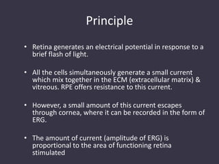
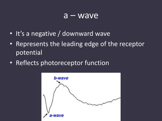
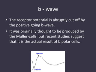
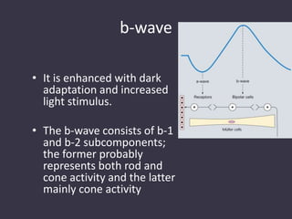
Electrophysiological Tests in
ophthalmology
Introduction
• These are used for objective assessment of
the retinal function & visual pathway.
• Diagnosis of various retinal disease can be
aided by their use
Why objective assessment of retinal
function is important?
1) It gives an idea about the site affected (within the eye
– macula/entire retina or visual pathway)
2) It also permits quantitative assessment of the degree
of malfunction (that can be followed up over time for
projecting long term prognosis)
3) For documentation purpose. The printout can be
handed over to the patient, so that they also can
appreciate the magnitude of their visual impairment.
Electrophysiological Tests
1) Electroretinography (ERG)
2) Electrooculography (EOG)
3) Visually Evoked Potential (VEP)
ELECTRORETINOGRAPHY (ERG)
Principle
• Retina generates an electrical potential in response to a
brief flash of light.
• All the cells simultaneously generate a small current
which mix together in the ECM (extracellular matrix) &
vitreous. RPE offers resistance to this current.
• However, a small amount of this current escapes
through cornea, where it can be recorded in the form of
ERG.
• The amount of current (amplitude of ERG) is
proportional to the area of functioning retina
stimulated
Different wave forms & their origin
• The clinical ERG consists of easily recognizable
waves that are generated by different retinal
elements & may be differently affected in
retinal diseases:
1) a – wave
2) b – wave
3) Oscillatory potentials
a – wave
• It’s a negative / downward wave
• Represents the leading edge of the receptor
potential
• Reflects photoreceptor function
b - wave
• The receptor potential is abruptly cut off by
the positive going b-wave.
• It was originally thought to be produced by
the Muller-cells, but recent studies suggest
that it is the actual result of bipolar cells.
b-wave
• It is enhanced with dark
adaptation and increased
light stimulus.
• The b-wave consists of b-1
and b-2 subcomponents;
the former probably
represents both rod and
cone activity and the latter
mainly cone activity
Oscillatory potentials
• These are produced by processes of IPL
(Amacrine cells) in response to bright flash of
light.
• These oscillatory potentials are actually
wavelets on the b-wave & are best seen with
stimuli that excite both the rods & the cones;
however they can also be detected in either
rod or cone isolated responses
Oscillatory potentials
• Frequency: 100 – 160 Hz
• Decreased in ischemic disorders like:
– Diabetic retinopathy
– CRVO
– Sickle cell retinopathy
Other waves
• Few authors also describe:
– c-wave:
• Present in scotopic ERG response
• Represents RPE hyperpolarisation
– d-wave:
• Present in scotopic ERG response
• Represents off response of the stimulus
Standard ERG
• The normal ERG consists of five recordings .
• The first three are elicited after 30 minutes of dark
adaptation (scotopic), and the last two after 10
minutes of adaptation to moderately bright diffuse
illumination (photopic).
• It may be difficult to dark adapt children for 30 minutes
and therefore dim light (mesopic) conditions can be
utilized to evoke predominantly rod-mediated
responses to low intensity white or blue light stimuli.
Standard ERG
• Scotopic ERG:
– Rod responses are elicited with a very dim flash of
white or blue light, resulting in a large b-wave and a
small or non-recordable a-wave
– Combined rod and cone responses are elicited with a
very bright white flash, resulting in a prominent a-wave
and b-wave
– Oscillatory potentials are elicited by using a bright
flash and changing the recording parameters. The
oscillatory wavelets occur on the ascending limb of the
b-wave and are generated by cells in the inner retina.
Standard ERG
• Photopic ERG:
– Cone responses are elicited with a single bright
flash, resulting in an a-wave and a b-wave with
small oscillations
– Cone flicker is used to isolate cones by using a
flickering light stimulus at a frequency of 30 Hz to
which rods cannot respond. It provides a measure
of the amplitude and implicit time of the cone b-
wave. Cone responses can be elicited in normal
eyes up to 50 Hz, after which point individual
responses are no longer recordable (‘critical flicker
fusion’).
Measurements
• Amplitude :
– a-wave: from baseline to trough of a wave
– b-wave: from trough of a-wave to peak of b-wave
• Latency: time taken from stimulus onset to
beginning of a response
• Implicit time: time from onset of stimulus to
peak of a response.
Rod Vs Cone
• Rods are 10 times more sensitive than cones
& are more sensitive to blue-green light
• Their sensitivity increases to 1 lac times after
dark adaptation
• Rods recover from light adaptation more
slowly than cones.
• Cones are sensitive to red light, to which rods
hardly respond
Rod Vs Cone
• These differences forms the basis of scotopic
& mesopic response, which are particularly
useful in distinguishing rod-cone from cone-
rod retinal degeneration
• At low intensity of stimulus, the scotopic ERG
is a long latency, slow rising monophasic
positive wave.
• With increase in intensity of stimulus, the
latency decreases & the amplitude of the
scotopic b-wave increases.
• With further increase in intensity, the b-wave
is preceded by a small negative a-wave.
• The cone threshold corresponds
approximately to the intensity required to
produce an a-wave, so an ERG without an a-
wave is purely due to rods, whereas an ERG
with both a & b-waves results from both rods
& cones in higher range of stimulus intensities
in dark adapted retina
• In light adapted retina, a pure photopic ERG is
produced, where the a-wave may become so
large that it may overwhelm the b-wave
Electrodes used for ERG
• Different types of ERG electrodes are
commercially available. The choice is based on
international standards or on practical
considerations such as size of the eye, recent
trauma, general cooperation, & presence or
absence of ocular infection
ERG Electrodes
• Burian-Allen Corneal
contact lens electrodes:
– Most commonly used
electrodes
– Placed over the cornea after
proper anaesthesia
– These lenses come in
various sizes
– Expensive but long lasting
ERG electrodes
• Jet disposable
electrodes:
– Disposable electrode for
one time use
– Mounted on a small hard
contact lens
– Monopolar electrode &
requires a skin electrode
as a reference (which is
kept over the forehead)
ERG Electrodes
• HK Loops:
– It’s a plastic coated wire electrode which is kept in
the lower lid (fornix)
– The wire is pliable & can be bent in any direction
& shape to fit the eye
– Therefore it can be used comfortably in
uncooperative patients esp. children & patients
with recent trauma
– It is an ideal electrode when corneal electrode can
not be used.
ERG Electrodes
• DTL Electrode:
– It is a fine silver nylon fiber electrode, with small
adhesive disc at each end
– The silver fiber rests in the palpebral sac & the
disc is attached to the canthi to secure the
electrode in place.
– Designed for single use, but is expensive
ERG Electrodes
• Gold-plated Mylar Electrodes:
– Gold foil single use electrodes
– Rests in the lower fornix
– Gold comes in direct contact with the skin
ERG Electrodes
• Skin Electrodes:
– These are silver chloride cup electrodes, which are
used for ground or reference & active electrodes.
– This is specially sensitive to facial movements, so
is of limited value in crying children
– Moisture below the electrode further reduces the
ERG signal
– Esp. used in trauma patients, where routine
placement in eyes is not possible.
Evaluation of ERG
• Rules to evaluate ERG:
– Focal affection of retina causes reduction in the
amplitude but little effect on its waveform if the
involved area is electrically silent e.g. Retinal
detachment
– Extensive inner retinal layer pathologies result in
loss of oscillatory potentials & b-wave with
relative preservation of the a-wave e.g. Diabetic
Retinopathy
Evaluation of ERG
– Disease that involve the photoreceptors results in
reduction of the ERG amplitude & slowing of a & b-
waves e.g Tapetoretinal degeneration &
Choroideremia
• Factors that affect ERG:
– Infants & elderly have a low amplitude ERG as
compared to adults
– High myopes & males have low amplitude ERG as
compared to the females of same age group
– Patients with oculocutaneous albinism have
supernormal ERG amplitudes
Recording Procedure
• Three electrodes:
– Active (Corneal)
– Reference (outer canthus/forehead/ within lid
speculum)
– Ground (Earlobe)
Patient preparation
• Pupils should be dilated
• 30 min dark adaptation - Scotopic Response
• 10 min light adaptation – Photopic Response
• No FFA before test, if done dark adaptation for
1 hour
Clinical Examples Affecting ERG
• Diabetic Retinopathy:
– Reduction of amplitude & delay of peak implicit
time relates to severity of DR
– Bresnick & co-workers found that the amplitude
of oscillatory potentials is a good predictor of the
progression of retinopathy from NPDR or early
PDR to severe PDR
Clinical Examples Affecting ERG
• POAG:
– POAG affects ganglion cells
– On pattern ERG, glaucoma patients show
reduction in ERG amplitude
Clinical Examples Affecting ERG
• Chronic Retinal Toxicity (Lead,
Chloroquine,any Drug):
– Results in reduction in amplitude & incresed
implicit time of the b-wave of ERG
Clinical Examples Affecting ERG
• Inflammatory condition:
– ERG findings parallel the ophthalmoscopic findings
– Focal inflammation (toxoplasmosis,
histoplasmosis) generally show normal ERG
– Diffuse, chronic inflammation shows reduction in
amplitudes & increase in the implicit time
Clinical Examples Affecting ERG
• Central Serous Chorioretinopathy:
– On focal foveal ERG, CSR results in reduction in a-
wave, b-wave & oscillatory potentials with
prolongation of implicit times of these responses
on ERG
Clinical Examples Affecting ERG
• Vitamin-A Deficiency:
– Reduced rod & cone responses with normal
implicit times, but rod ERG is affected before cone
ERG.
– With vitamin-A supplementation complete
recovery in the ERG findings occur
Indications & Clinical uses of ERG
• Determining presence or absence of retinal function
• Evaluating the progression of retinal degeneration
• Confirming diagnosis of a specific disease
• Gives idea about the prognosis of a disease
• Helpful particularly in cases of unexplained visual loss
• Evaluation of visual function in infants & children
(abnormal visual behavior, sensory type nystagmus,
excessive oculodigital stimulation)
Specialized Forms of ERG
• Bright Flash ERG:
– Assessment of retinal function in case of media
opacities or severe trauma
– The flash used is 100,000 times more brighter
than the flash used in standard flash ERG
– The goal is to produce a standard saturated
response when standard ERG results are severely
abnormal or are non-recordable.
Specialized Forms of ERG
• Successive responses are obtained with
flashes of increasing intensity, allowing time
for readaptation between flashes.
• A response that does not increase with a
tenfold increase in the intensity of stimulus is
considered as saturated response &
represents the maximum output of the
functional retina.
Specialized Forms of ERG
• VH / recent trauma can cause reversible
decrease of the response, but this reduction is
rarely to the point of extinction in non-
penetrated eyes.
• So a non-recordable bright flash ERG is an
ominous sign for visual prognosis
Specialized Forms of ERG
• Focal ERG:
– It evaluates small/focal areas of the retina
– It is useful in cases where there is focal involvement of the
retina (where full field ERG gives a normal response)
– A very small stimulus size (4 degrees) is used, which is
surrounded by an annulus (10 degree) of slightly larger size
to reduce the light scatter to other parts of retina.
– A computer average is used to record numerous microvolt
macular responses, the affect of eye movements &
blinking is nullified & the final results are then analysed.
Specialized Forms of ERG
– Useful in detecting cases of early maular /cone
diasease, even before fundoscopic changes are
evident.
– FERG can also differentiate between early macular
& optic nerve pathology.
Specialized Forms of ERG
• Multifocal ERG:
– For recording multifocal ERG (MFERG), the stimuli
consist of densely arranged black or white
hexagonal elements that are displayed over a CRT
monitor. The hexagonal elements change from
light to dark independently, which causes a
change in luminance & MFERGs can then be
recorded.
– Burian-Allen contact lens electrode are used for
recording this.
Specialized Forms of ERG
• The recorded MFERG appears as both
topographic map & an actual printout of small
ERGs from various portions of the retina.
• Both rod & cone MFERG can be recorded.
• MFERG findings correlate with visual field
defects, but small scotomas can go
undetected by this method
Specialized Forms of ERG
• Pattern ERG:
– Riggs et. al first described about recording the
electrical response by using the alternating
checkerboards or bar gratings of constant mean
luminance & termed it as PERG
– PERG mainly represents inner retinal activity (esp.
ganglion cell activity)
– For recording PERG, refractive errors should be
corrected. Patient is asked to watch a patterned
checkerboard, & approx 200-300 responses are
averaged. The machine automatically adjusts for the
errors caused by eye blinking & eye movements
Specialized Forms of ERG
• PERG is found to be abnormal/non recordable
in some maculopathies, with PERG amplitudes
correlating to the visual acuity in patients
• It is useful in testing patients with optic nerve
disease.
• It is abnormal /non recordable in optic
atrophy, glaucoma.
ELECTRO-OCULOGRAM (EOG)
Standing Potential
• Eye in some ways behaves like a “battery”, with
“standing potential”, resulting in cornea being positive
in relation to the retina.
• This standing potential is in the region of 6mV, & it
varies according to the adaptive state of the eye & is
also dependant upon the function of RPE
• Factors like retinal illumination with a steady light raises
the level of the standing potential
• Apart from light, chemicals like acetazolamide, &
alcohol can also induce changes in the standing
potential.
What EOG Measures ? ?
• EOG measures changes in the standing
potential to light and dark conditions & thus it
measures the photoreceptor-mediated RPE
function.
Principle
• The development of a normal light peak requires
normally functioning photoreceptors in contact
with a normally functioning RPE, leading to
progressive depolarization of the RPE basal
membrane by mechanisms not fully understood.
• Clinically, EOG measures the standing potential
indirectly with the patient making lateral eye
movements with skin electrodes placed nasal and
temporal to the eye, at the inner and outer
canthi.
Recording Technique
1) Pupillary dilatation
2) Electrodes
3) Ganzfeld dome
• Standard silver-silver chloride or gold surface
electrodes are placed at both inner and outer
canthi, taking care that all electrodes fall in the
same horizontal plane to minimise artefact
contamination from vertical eye movements
which can occur when the patient blinks
Recording Technique
• The electrodes can be connected so that an
eye movement to the right results in an
upwards deflection on the screen.
• The ground electrode is typically placed on the
forehead.
• The skin electrodes are applied using standard
techniques to ensure that impedance is =5KΩ.
Recording Technique
• Avoid too bright or dim illumination before EOG
recording; the patient should be in room light for
approximately 15 minutes before recording.
• Consistent ocular saccades are induced by
alternatively illuminating the fixation lights (LED)
which are 30 degrees apart within the Ganzfeld
dome, and asking the patient to follow the
alternating targets from right to left, and then
from left to right
Recording Technique
• The patient should be instructed to make sure that only
the eyes move; the head should remain still
throughout the procedure.
• Saccades are recorded for first 10-15 seconds of each
minute, allowing 45-50 seconds rest.
• After a baseline adaptation in the Ganzfeld the lights
are turned out, recordings take place for 20 minutes in
the dark phase and 15 minutes after the Ganzfeld
background is illuminated (the light phase).
Calculation
• The EOG is quantified by calculating the ratio
of the maximum amplitude in light (the light
peak) to the minimum amplitude in darkness
(the dark trough).
• It is usually expressed as a percentage, the
Arden index. A normal EOG light rise will be
>175% for most laboratories
Interpretation
• Only a normal functioning RPE will have a
normal EOG light rise.
• However, as the EOG light rise also depends
upon photoreceptor function, patients with
abnormal rod ERGs will usually have reduction
in the EOG light rise that parallels the extent
of rod photoreceptor disease.
Interpretation
• Any discrepancy, such that there is profound
EOG light rise reduction, but relatively good
rod function, then localises the dysfunction to
the RPE.
• The classic use of EOG is in the diagnosis of
Best’s macular dystrophy, where the EOG light
rise is virtually abolished in the presence of
normal ERGs.
Interpretation
• Even the asymptomatic genetic carriers of the
mutation (of Best’s disease) show EOG
abnormality.
• if Best’s disease is suspected in a young child who
is not capable of doing the EOG, testing of both
parents should be performed; as Best’s disease is
dominantly inherited, one of the parents must
carry the mutation if the child is affected and will
manifest the characteristic EOG abnormality.
VISUAL EVOKED POTENTIAL (VEP)
• The VEP is an evoked electrophysiological
potential that can be extracted, using signal
averaging, from the ongoing EEG
(electroencephalographic activity) activity
recorded at the scalp
• The VEP is assumed largely to arise in the
occipital cortices and allow assessment of the
functional integrity of the visual pathways.
• Although of high importance in the
assessment of the intracranial visual
pathways, it should always be remembered
that as a “downstream” response the VEP can
be affected by a lesion anywhere in the visual
path, including the eye, and that VEP
abnormalities in themselves are often non-
specific.

Electrophysiological tests in ophthalmology

  • 1.
  • 2.
    Introduction • These areused for objective assessment of the retinal function & visual pathway. • Diagnosis of various retinal disease can be aided by their use
  • 3.
    Why objective assessmentof retinal function is important? 1) It gives an idea about the site affected (within the eye – macula/entire retina or visual pathway) 2) It also permits quantitative assessment of the degree of malfunction (that can be followed up over time for projecting long term prognosis) 3) For documentation purpose. The printout can be handed over to the patient, so that they also can appreciate the magnitude of their visual impairment.
  • 4.
    Electrophysiological Tests 1) Electroretinography(ERG) 2) Electrooculography (EOG) 3) Visually Evoked Potential (VEP)
  • 5.
  • 6.
    Principle • Retina generatesan electrical potential in response to a brief flash of light. • All the cells simultaneously generate a small current which mix together in the ECM (extracellular matrix) & vitreous. RPE offers resistance to this current. • However, a small amount of this current escapes through cornea, where it can be recorded in the form of ERG. • The amount of current (amplitude of ERG) is proportional to the area of functioning retina stimulated
  • 7.
    Different wave forms& their origin • The clinical ERG consists of easily recognizable waves that are generated by different retinal elements & may be differently affected in retinal diseases: 1) a – wave 2) b – wave 3) Oscillatory potentials
  • 8.
    a – wave •It’s a negative / downward wave • Represents the leading edge of the receptor potential • Reflects photoreceptor function
  • 9.
    b - wave •The receptor potential is abruptly cut off by the positive going b-wave. • It was originally thought to be produced by the Muller-cells, but recent studies suggest that it is the actual result of bipolar cells.
  • 10.
    b-wave • It isenhanced with dark adaptation and increased light stimulus. • The b-wave consists of b-1 and b-2 subcomponents; the former probably represents both rod and cone activity and the latter mainly cone activity
  • 11.
    Oscillatory potentials • Theseare produced by processes of IPL (Amacrine cells) in response to bright flash of light. • These oscillatory potentials are actually wavelets on the b-wave & are best seen with stimuli that excite both the rods & the cones; however they can also be detected in either rod or cone isolated responses
  • 12.
    Oscillatory potentials • Frequency:100 – 160 Hz • Decreased in ischemic disorders like: – Diabetic retinopathy – CRVO – Sickle cell retinopathy
  • 13.
    Other waves • Fewauthors also describe: – c-wave: • Present in scotopic ERG response • Represents RPE hyperpolarisation – d-wave: • Present in scotopic ERG response • Represents off response of the stimulus
  • 14.
    Standard ERG • Thenormal ERG consists of five recordings . • The first three are elicited after 30 minutes of dark adaptation (scotopic), and the last two after 10 minutes of adaptation to moderately bright diffuse illumination (photopic). • It may be difficult to dark adapt children for 30 minutes and therefore dim light (mesopic) conditions can be utilized to evoke predominantly rod-mediated responses to low intensity white or blue light stimuli.
  • 15.
    Standard ERG • ScotopicERG: – Rod responses are elicited with a very dim flash of white or blue light, resulting in a large b-wave and a small or non-recordable a-wave – Combined rod and cone responses are elicited with a very bright white flash, resulting in a prominent a-wave and b-wave – Oscillatory potentials are elicited by using a bright flash and changing the recording parameters. The oscillatory wavelets occur on the ascending limb of the b-wave and are generated by cells in the inner retina.
  • 17.
    Standard ERG • PhotopicERG: – Cone responses are elicited with a single bright flash, resulting in an a-wave and a b-wave with small oscillations – Cone flicker is used to isolate cones by using a flickering light stimulus at a frequency of 30 Hz to which rods cannot respond. It provides a measure of the amplitude and implicit time of the cone b- wave. Cone responses can be elicited in normal eyes up to 50 Hz, after which point individual responses are no longer recordable (‘critical flicker fusion’).
  • 18.
    Measurements • Amplitude : –a-wave: from baseline to trough of a wave – b-wave: from trough of a-wave to peak of b-wave • Latency: time taken from stimulus onset to beginning of a response • Implicit time: time from onset of stimulus to peak of a response.
  • 20.
    Rod Vs Cone •Rods are 10 times more sensitive than cones & are more sensitive to blue-green light • Their sensitivity increases to 1 lac times after dark adaptation • Rods recover from light adaptation more slowly than cones. • Cones are sensitive to red light, to which rods hardly respond
  • 21.
    Rod Vs Cone •These differences forms the basis of scotopic & mesopic response, which are particularly useful in distinguishing rod-cone from cone- rod retinal degeneration
  • 22.
    • At lowintensity of stimulus, the scotopic ERG is a long latency, slow rising monophasic positive wave. • With increase in intensity of stimulus, the latency decreases & the amplitude of the scotopic b-wave increases. • With further increase in intensity, the b-wave is preceded by a small negative a-wave.
  • 23.
    • The conethreshold corresponds approximately to the intensity required to produce an a-wave, so an ERG without an a- wave is purely due to rods, whereas an ERG with both a & b-waves results from both rods & cones in higher range of stimulus intensities in dark adapted retina
  • 24.
    • In lightadapted retina, a pure photopic ERG is produced, where the a-wave may become so large that it may overwhelm the b-wave
  • 25.
    Electrodes used forERG • Different types of ERG electrodes are commercially available. The choice is based on international standards or on practical considerations such as size of the eye, recent trauma, general cooperation, & presence or absence of ocular infection
  • 26.
    ERG Electrodes • Burian-AllenCorneal contact lens electrodes: – Most commonly used electrodes – Placed over the cornea after proper anaesthesia – These lenses come in various sizes – Expensive but long lasting
  • 27.
    ERG electrodes • Jetdisposable electrodes: – Disposable electrode for one time use – Mounted on a small hard contact lens – Monopolar electrode & requires a skin electrode as a reference (which is kept over the forehead)
  • 28.
    ERG Electrodes • HKLoops: – It’s a plastic coated wire electrode which is kept in the lower lid (fornix) – The wire is pliable & can be bent in any direction & shape to fit the eye – Therefore it can be used comfortably in uncooperative patients esp. children & patients with recent trauma – It is an ideal electrode when corneal electrode can not be used.
  • 29.
    ERG Electrodes • DTLElectrode: – It is a fine silver nylon fiber electrode, with small adhesive disc at each end – The silver fiber rests in the palpebral sac & the disc is attached to the canthi to secure the electrode in place. – Designed for single use, but is expensive
  • 30.
    ERG Electrodes • Gold-platedMylar Electrodes: – Gold foil single use electrodes – Rests in the lower fornix – Gold comes in direct contact with the skin
  • 31.
    ERG Electrodes • SkinElectrodes: – These are silver chloride cup electrodes, which are used for ground or reference & active electrodes. – This is specially sensitive to facial movements, so is of limited value in crying children – Moisture below the electrode further reduces the ERG signal – Esp. used in trauma patients, where routine placement in eyes is not possible.
  • 32.
    Evaluation of ERG •Rules to evaluate ERG: – Focal affection of retina causes reduction in the amplitude but little effect on its waveform if the involved area is electrically silent e.g. Retinal detachment – Extensive inner retinal layer pathologies result in loss of oscillatory potentials & b-wave with relative preservation of the a-wave e.g. Diabetic Retinopathy
  • 33.
    Evaluation of ERG –Disease that involve the photoreceptors results in reduction of the ERG amplitude & slowing of a & b- waves e.g Tapetoretinal degeneration & Choroideremia • Factors that affect ERG: – Infants & elderly have a low amplitude ERG as compared to adults – High myopes & males have low amplitude ERG as compared to the females of same age group – Patients with oculocutaneous albinism have supernormal ERG amplitudes
  • 34.
    Recording Procedure • Threeelectrodes: – Active (Corneal) – Reference (outer canthus/forehead/ within lid speculum) – Ground (Earlobe)
  • 36.
    Patient preparation • Pupilsshould be dilated • 30 min dark adaptation - Scotopic Response • 10 min light adaptation – Photopic Response • No FFA before test, if done dark adaptation for 1 hour
  • 37.
    Clinical Examples AffectingERG • Diabetic Retinopathy: – Reduction of amplitude & delay of peak implicit time relates to severity of DR – Bresnick & co-workers found that the amplitude of oscillatory potentials is a good predictor of the progression of retinopathy from NPDR or early PDR to severe PDR
  • 39.
    Clinical Examples AffectingERG • POAG: – POAG affects ganglion cells – On pattern ERG, glaucoma patients show reduction in ERG amplitude
  • 40.
    Clinical Examples AffectingERG • Chronic Retinal Toxicity (Lead, Chloroquine,any Drug): – Results in reduction in amplitude & incresed implicit time of the b-wave of ERG
  • 41.
    Clinical Examples AffectingERG • Inflammatory condition: – ERG findings parallel the ophthalmoscopic findings – Focal inflammation (toxoplasmosis, histoplasmosis) generally show normal ERG – Diffuse, chronic inflammation shows reduction in amplitudes & increase in the implicit time
  • 42.
    Clinical Examples AffectingERG • Central Serous Chorioretinopathy: – On focal foveal ERG, CSR results in reduction in a- wave, b-wave & oscillatory potentials with prolongation of implicit times of these responses on ERG
  • 43.
    Clinical Examples AffectingERG • Vitamin-A Deficiency: – Reduced rod & cone responses with normal implicit times, but rod ERG is affected before cone ERG. – With vitamin-A supplementation complete recovery in the ERG findings occur
  • 45.
    Indications & Clinicaluses of ERG • Determining presence or absence of retinal function • Evaluating the progression of retinal degeneration • Confirming diagnosis of a specific disease • Gives idea about the prognosis of a disease • Helpful particularly in cases of unexplained visual loss • Evaluation of visual function in infants & children (abnormal visual behavior, sensory type nystagmus, excessive oculodigital stimulation)
  • 46.
    Specialized Forms ofERG • Bright Flash ERG: – Assessment of retinal function in case of media opacities or severe trauma – The flash used is 100,000 times more brighter than the flash used in standard flash ERG – The goal is to produce a standard saturated response when standard ERG results are severely abnormal or are non-recordable.
  • 47.
    Specialized Forms ofERG • Successive responses are obtained with flashes of increasing intensity, allowing time for readaptation between flashes. • A response that does not increase with a tenfold increase in the intensity of stimulus is considered as saturated response & represents the maximum output of the functional retina.
  • 48.
    Specialized Forms ofERG • VH / recent trauma can cause reversible decrease of the response, but this reduction is rarely to the point of extinction in non- penetrated eyes. • So a non-recordable bright flash ERG is an ominous sign for visual prognosis
  • 49.
    Specialized Forms ofERG • Focal ERG: – It evaluates small/focal areas of the retina – It is useful in cases where there is focal involvement of the retina (where full field ERG gives a normal response) – A very small stimulus size (4 degrees) is used, which is surrounded by an annulus (10 degree) of slightly larger size to reduce the light scatter to other parts of retina. – A computer average is used to record numerous microvolt macular responses, the affect of eye movements & blinking is nullified & the final results are then analysed.
  • 50.
    Specialized Forms ofERG – Useful in detecting cases of early maular /cone diasease, even before fundoscopic changes are evident. – FERG can also differentiate between early macular & optic nerve pathology.
  • 51.
    Specialized Forms ofERG • Multifocal ERG: – For recording multifocal ERG (MFERG), the stimuli consist of densely arranged black or white hexagonal elements that are displayed over a CRT monitor. The hexagonal elements change from light to dark independently, which causes a change in luminance & MFERGs can then be recorded. – Burian-Allen contact lens electrode are used for recording this.
  • 52.
    Specialized Forms ofERG • The recorded MFERG appears as both topographic map & an actual printout of small ERGs from various portions of the retina. • Both rod & cone MFERG can be recorded. • MFERG findings correlate with visual field defects, but small scotomas can go undetected by this method
  • 53.
    Specialized Forms ofERG • Pattern ERG: – Riggs et. al first described about recording the electrical response by using the alternating checkerboards or bar gratings of constant mean luminance & termed it as PERG – PERG mainly represents inner retinal activity (esp. ganglion cell activity) – For recording PERG, refractive errors should be corrected. Patient is asked to watch a patterned checkerboard, & approx 200-300 responses are averaged. The machine automatically adjusts for the errors caused by eye blinking & eye movements
  • 54.
    Specialized Forms ofERG • PERG is found to be abnormal/non recordable in some maculopathies, with PERG amplitudes correlating to the visual acuity in patients • It is useful in testing patients with optic nerve disease. • It is abnormal /non recordable in optic atrophy, glaucoma.
  • 55.
  • 56.
    Standing Potential • Eyein some ways behaves like a “battery”, with “standing potential”, resulting in cornea being positive in relation to the retina. • This standing potential is in the region of 6mV, & it varies according to the adaptive state of the eye & is also dependant upon the function of RPE • Factors like retinal illumination with a steady light raises the level of the standing potential • Apart from light, chemicals like acetazolamide, & alcohol can also induce changes in the standing potential.
  • 57.
    What EOG Measures? ? • EOG measures changes in the standing potential to light and dark conditions & thus it measures the photoreceptor-mediated RPE function.
  • 58.
    Principle • The developmentof a normal light peak requires normally functioning photoreceptors in contact with a normally functioning RPE, leading to progressive depolarization of the RPE basal membrane by mechanisms not fully understood. • Clinically, EOG measures the standing potential indirectly with the patient making lateral eye movements with skin electrodes placed nasal and temporal to the eye, at the inner and outer canthi.
  • 59.
    Recording Technique 1) Pupillarydilatation 2) Electrodes 3) Ganzfeld dome • Standard silver-silver chloride or gold surface electrodes are placed at both inner and outer canthi, taking care that all electrodes fall in the same horizontal plane to minimise artefact contamination from vertical eye movements which can occur when the patient blinks
  • 60.
    Recording Technique • Theelectrodes can be connected so that an eye movement to the right results in an upwards deflection on the screen. • The ground electrode is typically placed on the forehead. • The skin electrodes are applied using standard techniques to ensure that impedance is =5KΩ.
  • 61.
    Recording Technique • Avoidtoo bright or dim illumination before EOG recording; the patient should be in room light for approximately 15 minutes before recording. • Consistent ocular saccades are induced by alternatively illuminating the fixation lights (LED) which are 30 degrees apart within the Ganzfeld dome, and asking the patient to follow the alternating targets from right to left, and then from left to right
  • 62.
    Recording Technique • Thepatient should be instructed to make sure that only the eyes move; the head should remain still throughout the procedure. • Saccades are recorded for first 10-15 seconds of each minute, allowing 45-50 seconds rest. • After a baseline adaptation in the Ganzfeld the lights are turned out, recordings take place for 20 minutes in the dark phase and 15 minutes after the Ganzfeld background is illuminated (the light phase).
  • 63.
    Calculation • The EOGis quantified by calculating the ratio of the maximum amplitude in light (the light peak) to the minimum amplitude in darkness (the dark trough). • It is usually expressed as a percentage, the Arden index. A normal EOG light rise will be >175% for most laboratories
  • 64.
    Interpretation • Only anormal functioning RPE will have a normal EOG light rise. • However, as the EOG light rise also depends upon photoreceptor function, patients with abnormal rod ERGs will usually have reduction in the EOG light rise that parallels the extent of rod photoreceptor disease.
  • 65.
    Interpretation • Any discrepancy,such that there is profound EOG light rise reduction, but relatively good rod function, then localises the dysfunction to the RPE. • The classic use of EOG is in the diagnosis of Best’s macular dystrophy, where the EOG light rise is virtually abolished in the presence of normal ERGs.
  • 66.
    Interpretation • Even theasymptomatic genetic carriers of the mutation (of Best’s disease) show EOG abnormality. • if Best’s disease is suspected in a young child who is not capable of doing the EOG, testing of both parents should be performed; as Best’s disease is dominantly inherited, one of the parents must carry the mutation if the child is affected and will manifest the characteristic EOG abnormality.
  • 67.
  • 68.
    • The VEPis an evoked electrophysiological potential that can be extracted, using signal averaging, from the ongoing EEG (electroencephalographic activity) activity recorded at the scalp • The VEP is assumed largely to arise in the occipital cortices and allow assessment of the functional integrity of the visual pathways.
  • 69.
    • Although ofhigh importance in the assessment of the intracranial visual pathways, it should always be remembered that as a “downstream” response the VEP can be affected by a lesion anywhere in the visual path, including the eye, and that VEP abnormalities in themselves are often non- specific.