Download to read offline


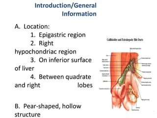
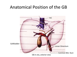
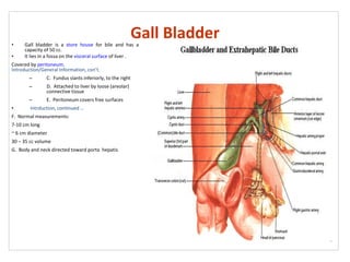


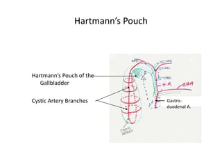
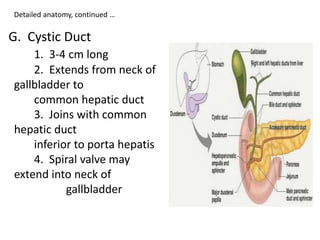


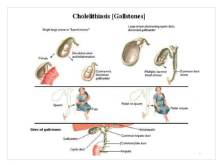


The biliary apparatus consists of the gallbladder, cystic duct, and hepatic ducts. The gallbladder stores and concentrates bile produced by the liver. It is located in the right hypochondriac region on the inferior surface of the liver between the quadrate and right lobes. The cystic duct joins the common hepatic duct to form the common bile duct which carries bile to the duodenum. The gallbladder receives its blood supply from the cystic artery and drains into the portal vein. It is innervated by both the parasympathetic and sympathetic nervous systems. Diseases of the gallbladder include cholelithiasis, in which stones form within it, and cholecystitis,