RADIOLOGY/Imaging of The Small Intestine
•Download as PPTX, PDF•
0 likes•2 views

RADIOLOGY(Imaging of The Small Intestine).pptx HUMAN ANATOMY RADIOLOGY(Imaging of The Small Intestine).pptx HUMAN ANATOMY RADIOLOGY(Imaging of The Small Intestine).pptx HUMAN ANATOMY RADIOLOGY(Imaging of The Small Intestine).pptx HUMAN ANATOMY

Recommended
More Related Content
Similar to RADIOLOGY/Imaging of The Small Intestine
Similar to RADIOLOGY/Imaging of The Small Intestine (20)
More from nidhi sharma
More from nidhi sharma (20)
Recently uploaded
Recently uploaded (20)
RADIOLOGY/Imaging of The Small Intestine
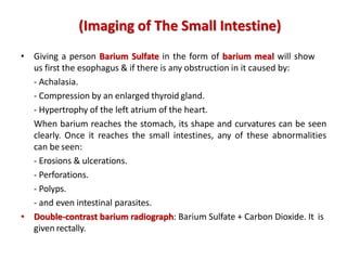
- 1. • Giving a person Barium Sulfate in the form of barium meal will show us first the esophagus & if there is any obstruction in it caused by: - Achalasia. - Compression by an enlarged thyroid gland. - Hypertrophy of the left atrium of the heart. When barium reaches the stomach, its shape and curvatures can be seen clearly. Once it reaches the small intestines, any of these abnormalities can be seen: - Erosions & ulcerations. - Perforations. - Polyps. - and even intestinal parasites. • Double-contrast barium radiograph: Barium Sulfate + Carbon Dioxide. It is given rectally. (Imaging of The Small Intestine)
- 2. Barium showing the stomach and the small intestine Barium showing the stomach and the duodenum Bird-beak seen in achalasia (Imaging of The Small Intestine)
- 3. Double contrast barium of the small intestine Double bubble sign seen in case of duodenal atresia caused by annular pancreas (Imaging of The Small Intestine)
- 4. • Esophagojejunostomy: no pancreatic enzymes will be released in the duodenum because it has been removed. Therefore, these enzymes must be gibe in synthetic form. • Overgrowth of the head of the pancreas might lead to compression of inferior vena cava which is located posterior to it. Therefore, resulting in lower limbs edema. • In acute pancreatitis, the pain will be referred to the back (dorsolumbar region). • Carcinoma in the head of the pancreas will lead to the obstruction of the bile duct resulting in jaundice. • Overgrowth or carcinoma in the neck of the pancreas will lead to the compression of the portal vein at the level of L1 vertebra. (Imaging of The Small Intestine)
- 13. GOOD LUCK!