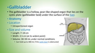
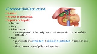
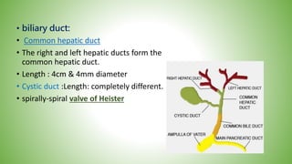
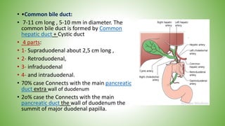
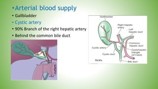
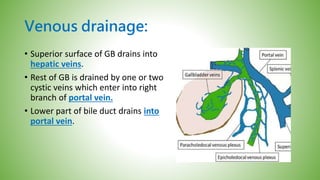
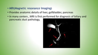
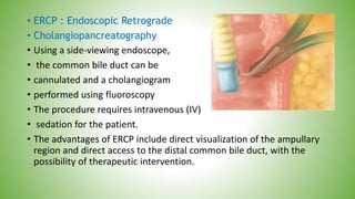
Gallbladders & bile duct:
•Gallbladder
• The gallbladder is a hollow, pear-like-shaped organ that lies on the
cystic plate (gallbladder bed) under the surface of the liver.
• Anatomy
• Location
• Intraperitoneal organ
• Size and volume
• Length: 7–10 cm
• Width: 2.5 cm (at its widest point)
• Volume: 30–50 mL under normal conditions
• Can hold up to 300 mL if the cystic duct is obstructed
•Composition/structure
• Surface:
• Inferior or peritoneal.
• Superior or hepatic
• Fundus
• Body
• Infundibulum
• Narrow portion of the body that is continuous with the neck of the
gallbladder
• Neck
• Connects to the cystic duct → common hepatic duct → common bile
duct.
• Most common site of gallstone impaction
• biliary duct:
• Common hepatic duct
• The right and left hepatic ducts form the
common hepatic duct.
• Length : 4cm & 4mm diameter
• Cystic duct :Length: completely different.
• spirally-spiral valve of Heister
• •Common bile duct:
• 7-11 cm long , 5-10 mm in diameter. The
common bile duct is formed by Common
hepatic duct + Cystic duct
• 4 parts:
• 1- Supraduodenal about 2,5 cm long ,
• 2- Retroduodenal,
• 3- infraduodenal
• 4- and intraduodenal.
• 70% case Connects with the main pancreatic
duct extra wall of duodenum
• 2o% case the Connects with the main
pancreatic duct the wall of duodenum the
summit of major duodenal papilla.
•Arterial blood supply
• Gallbladder
• Cystic artery
• 90% Branch of the right hepatic artery
• Behind the common bile duct
Venous drainage:
• Superior surface of GB drains into
hepatic veins.
• Rest of GB is drained by one or two
cystic veins which enter into right
branch of portal vein.
• Lower part of bile duct drains into
portal vein.
Lymphatic drainage:
• Lymphatic drainage:
• Lymph of sub serosa &sub mucosal
• drained to Cystic lymph node
• locate in the neck of GB.
• Sub serosa vessel of GB Connected
• with sub capsular lymph channel of the liver.
• Innervation
• innervation
• Sympathetic: via the celiac plexus
• Parasympathetic: vagus nerve
• Sensory: fibers from Rt phrenic nerve
Gallbladder surgical physiology:
• Bile is a secretion produced by the liver and stored in the gallbladder
that aids in digestion, neutralization of gastric acid, fat absorption,
and excretion of bilirubin and cholesterol.
• Bile composition:
• Bile is mainly composed of
• water 97%,
• bile salts 1-2%,
• bile pigments ,cholesterol & fatty acid 1%.
• Secretion of bile by liver 40ml/hr.
Continue:
• The effect of cholecystokinin on the
gallbladder and the sphincter of Oddi.
• 1-Storage:
• A. During fasting, with the sphincter of
Oddi contracted and the gallbladder
filling.
• B. In response to a meal, the sphincter
of Oddi relaxed and the gallbladder
emptying.
Continue:
• 2- concentration of bile:
• Bile is secreted in liver, store and concentrate into the gallbladder,
• Na , Cl , Hco3 absorbed from bile.
• Bile is concentrated ( 5- 10)
• Bile salt , bile pigment , cholesterol and calcium.
• 3- secretion of mucus ( 20 ml in 24 hour )
• Congenital anomalies
• Developmentally, a diverticulum grows out from the ventral wall of
the foregut (primitive duodenum), which differentiates into the
hepatic ducts and the liver.
• A lateral bud from this diverticulum becomes the gallbladder and
cystic duct.
• Anomalies are found in 10% of subjects and these are of importance
to the surgeon during cholecystectomy.
1.the gallbladder anomalies:
1. Congenital absence (agenesis) of the gallbladder:
occur 0,03%.
that important when pathologic process effected one are both GB
2. Duplication of the gallbladder: one in 4000.
A- Each GB connect to bile separately
B- shared
3. Left sided gallbladder :
drained to Lt hepatic duct OR CBD.
4. Posterior gallbladder
5.Phrygian Cap
• The Phrygian cap is present in 2–6
per cent of cholecystograms and
may be mistaken for a pathological
• deformity of the organ.
6. Floating gall bladder
• The gall bladder may hang on a
mesentery, which makes it
• liable to undergo torsion
Bile duct anomalies:
1.Small ducts may drain directly from the liver into the body of the
gallbladder.
If present, but not recognized at the time of a cholecystectomy, a
bile leak with the accumulation of bile (biloma) may occur in the
abdomen.
2. Absence of the cystic duct:
the gallbladder opening directly into the side of the common
bile duct.
3. choledochal cyst:
Cystic dilatation of the main bile ducts.
4. Congenital biliary atresia :
biliary atresia, one of the causes of neonatal jaundice.
5.A long cystic duct:
travelling alongside the
common hepatic duct to open near the
duodenal orice.
This occurs in 10% of cases.
• DIAGNOSTIC STUDIES
• Blood Tests
• When patients with suspected diseases of the gallbladder or the
• extrahepatic biliary tree are evaluated, a (CBC), LFT are routinely
• requested.
1. An elevated white blood cell (WBC) count may indicate or raise
suspicion of cholecystitis.
2. If associated with an elevation of
 bilirubin,
 alkaline phosphatase,
 and aminotransferase, cholangitis should be suspected.
• Blood Tests
3. Cholestasis, an obstruction to bile flow, is characterized by an
elevation of bilirubin (i.e., the conjugated form) and a rise in
alkaline phosphatase.
Serum aminotransferases may be normal or mildly elevated.
4. In patients with biliary colic or chronic cholecystitis, blood tests
will typically be normal.
u/s
• Initial investigation of choice in suspected in gallstones disease.
• Non invasive & • Painless
• Accurate identification of gall stones
• Not limited by jaundice or pregnancy
• Allows of GB size ,
• wall thickness ,
• inflammatory changes around GB
• Detects very small stones
• Accurate identification of dilated common bile ducts.
Endoscopic ultrasound:
• Endoscope with ultrasound transducer at tip
• Visualizes biliary tree from within stomach & duodenum
• Accurate to detect stones in CBD.
Plain radiography:
• X- RAY
1. 10-15 % of GB stones are radio opaque
2. Calcification (porcelain GB)
3. Bile lime
4. Air in bile duct.
IV & Oral cholecystography:
• IV Oral cholecystography
• Once considered the diagnostic procedure of choice for gallstones
• replaced by ultrasonography
Radioisotope scan:
• noninvasive evaluation of the liver, gallbladder, bile ducts, and
duodenum
• 99mTechnetium-labeled derivatives of
• iminodiacetic acid (HIDA) are injected IV.
• cleared by the Kupffer cells in the liver,
• and excreted in the bile.
• Uptake by the liver is detected within 10 minutes,
• the gallbladder, the bile ducts, and the duodenum are
• visualized within 60 minutes in fasting subjects
Ct scan:
• Computerized tomography:
• test of choice in suspected
• malignancy of the gallbladder,
• the extra hepatic biliary system,
• the tumor staging ,
• extent ,
• metastasis
• & lymph node
Percutaneous Transhepatic Cholangiography(PTC)
Intrahepatic bile ducts are accessed percutaneously with a
small needle under fluoroscopic guidance.
a guide wire is passed, and subsequently, a catheter is passed over the
wire .
Through the catheter, a cholangiogram
can be performed and therapeutic
interventions done,
such as biliary drain insertions
and stent placements.
Also tumors & stenosis
• MRI(Magnetic resonance Imaging)
• Provides anatomic details of liver, gallbladder, pancreas
• In many centers , MRI is first performed for diagnosis of biliary and
pancreatic duct pathology,
• ERCP : Endoscopic Retrograde
• Cholangiopancreatography
• Using a side-viewing endoscope,
• the common bile duct can be
• cannulated and a cholangiogram
• performed using fluoroscopy
• The procedure requires intravenous (IV)
• sedation for the patient.
• The advantages of ERCP include direct visualization of the ampullary
region and direct access to the distal common bile duct, with the
possibility of therapeutic intervention.
• CONTINUE…
• The test is rarely needed for uncomplicated gallstone
• disease, but for stones in the common bile duct, in particular,
• when associated with
• obstructive jaundice,
• cholangitis,
• or gallstone pancreatitis,
• ERC is the diagnostic and often therapeutic procedure of choice.
• Once the endoscopic cholangiogram has shown ductal stones,
Sphincterotomy and stone extraction can be performed,
• ERCP:
• Complications include:
• Pancreatitis
• Biliary tracts perforation
• Minor bleeding
• & cholangitis
Continue:

222222.pptx

  • 1.
  • 2.
    •Gallbladder • The gallbladderis a hollow, pear-like-shaped organ that lies on the cystic plate (gallbladder bed) under the surface of the liver. • Anatomy • Location • Intraperitoneal organ • Size and volume • Length: 7–10 cm • Width: 2.5 cm (at its widest point) • Volume: 30–50 mL under normal conditions • Can hold up to 300 mL if the cystic duct is obstructed
  • 3.
    •Composition/structure • Surface: • Inferioror peritoneal. • Superior or hepatic • Fundus • Body • Infundibulum • Narrow portion of the body that is continuous with the neck of the gallbladder • Neck • Connects to the cystic duct → common hepatic duct → common bile duct. • Most common site of gallstone impaction
  • 4.
    • biliary duct: •Common hepatic duct • The right and left hepatic ducts form the common hepatic duct. • Length : 4cm & 4mm diameter • Cystic duct :Length: completely different. • spirally-spiral valve of Heister
  • 5.
    • •Common bileduct: • 7-11 cm long , 5-10 mm in diameter. The common bile duct is formed by Common hepatic duct + Cystic duct • 4 parts: • 1- Supraduodenal about 2,5 cm long , • 2- Retroduodenal, • 3- infraduodenal • 4- and intraduodenal. • 70% case Connects with the main pancreatic duct extra wall of duodenum • 2o% case the Connects with the main pancreatic duct the wall of duodenum the summit of major duodenal papilla.
  • 6.
    •Arterial blood supply •Gallbladder • Cystic artery • 90% Branch of the right hepatic artery • Behind the common bile duct
  • 7.
    Venous drainage: • Superiorsurface of GB drains into hepatic veins. • Rest of GB is drained by one or two cystic veins which enter into right branch of portal vein. • Lower part of bile duct drains into portal vein.
  • 8.
    Lymphatic drainage: • Lymphaticdrainage: • Lymph of sub serosa &sub mucosal • drained to Cystic lymph node • locate in the neck of GB. • Sub serosa vessel of GB Connected • with sub capsular lymph channel of the liver.
  • 9.
    • Innervation • innervation •Sympathetic: via the celiac plexus • Parasympathetic: vagus nerve • Sensory: fibers from Rt phrenic nerve
  • 10.
    Gallbladder surgical physiology: •Bile is a secretion produced by the liver and stored in the gallbladder that aids in digestion, neutralization of gastric acid, fat absorption, and excretion of bilirubin and cholesterol. • Bile composition: • Bile is mainly composed of • water 97%, • bile salts 1-2%, • bile pigments ,cholesterol & fatty acid 1%. • Secretion of bile by liver 40ml/hr.
  • 11.
    Continue: • The effectof cholecystokinin on the gallbladder and the sphincter of Oddi. • 1-Storage: • A. During fasting, with the sphincter of Oddi contracted and the gallbladder filling. • B. In response to a meal, the sphincter of Oddi relaxed and the gallbladder emptying.
  • 12.
    Continue: • 2- concentrationof bile: • Bile is secreted in liver, store and concentrate into the gallbladder, • Na , Cl , Hco3 absorbed from bile. • Bile is concentrated ( 5- 10) • Bile salt , bile pigment , cholesterol and calcium. • 3- secretion of mucus ( 20 ml in 24 hour )
  • 13.
    • Congenital anomalies •Developmentally, a diverticulum grows out from the ventral wall of the foregut (primitive duodenum), which differentiates into the hepatic ducts and the liver. • A lateral bud from this diverticulum becomes the gallbladder and cystic duct. • Anomalies are found in 10% of subjects and these are of importance to the surgeon during cholecystectomy.
  • 14.
    1.the gallbladder anomalies: 1.Congenital absence (agenesis) of the gallbladder: occur 0,03%. that important when pathologic process effected one are both GB 2. Duplication of the gallbladder: one in 4000. A- Each GB connect to bile separately B- shared 3. Left sided gallbladder : drained to Lt hepatic duct OR CBD. 4. Posterior gallbladder
  • 15.
    5.Phrygian Cap • ThePhrygian cap is present in 2–6 per cent of cholecystograms and may be mistaken for a pathological • deformity of the organ. 6. Floating gall bladder • The gall bladder may hang on a mesentery, which makes it • liable to undergo torsion
  • 16.
    Bile duct anomalies: 1.Smallducts may drain directly from the liver into the body of the gallbladder. If present, but not recognized at the time of a cholecystectomy, a bile leak with the accumulation of bile (biloma) may occur in the abdomen. 2. Absence of the cystic duct: the gallbladder opening directly into the side of the common bile duct.
  • 17.
    3. choledochal cyst: Cysticdilatation of the main bile ducts.
  • 18.
    4. Congenital biliaryatresia : biliary atresia, one of the causes of neonatal jaundice. 5.A long cystic duct: travelling alongside the common hepatic duct to open near the duodenal orice. This occurs in 10% of cases.
  • 19.
    • DIAGNOSTIC STUDIES •Blood Tests • When patients with suspected diseases of the gallbladder or the • extrahepatic biliary tree are evaluated, a (CBC), LFT are routinely • requested. 1. An elevated white blood cell (WBC) count may indicate or raise suspicion of cholecystitis. 2. If associated with an elevation of  bilirubin,  alkaline phosphatase,  and aminotransferase, cholangitis should be suspected.
  • 20.
    • Blood Tests 3.Cholestasis, an obstruction to bile flow, is characterized by an elevation of bilirubin (i.e., the conjugated form) and a rise in alkaline phosphatase. Serum aminotransferases may be normal or mildly elevated. 4. In patients with biliary colic or chronic cholecystitis, blood tests will typically be normal.
  • 21.
    u/s • Initial investigationof choice in suspected in gallstones disease. • Non invasive & • Painless • Accurate identification of gall stones • Not limited by jaundice or pregnancy • Allows of GB size , • wall thickness , • inflammatory changes around GB • Detects very small stones • Accurate identification of dilated common bile ducts.
  • 22.
    Endoscopic ultrasound: • Endoscopewith ultrasound transducer at tip • Visualizes biliary tree from within stomach & duodenum • Accurate to detect stones in CBD.
  • 23.
    Plain radiography: • X-RAY 1. 10-15 % of GB stones are radio opaque 2. Calcification (porcelain GB) 3. Bile lime 4. Air in bile duct.
  • 24.
    IV & Oralcholecystography: • IV Oral cholecystography • Once considered the diagnostic procedure of choice for gallstones • replaced by ultrasonography
  • 25.
    Radioisotope scan: • noninvasiveevaluation of the liver, gallbladder, bile ducts, and duodenum • 99mTechnetium-labeled derivatives of • iminodiacetic acid (HIDA) are injected IV. • cleared by the Kupffer cells in the liver, • and excreted in the bile. • Uptake by the liver is detected within 10 minutes, • the gallbladder, the bile ducts, and the duodenum are • visualized within 60 minutes in fasting subjects
  • 26.
    Ct scan: • Computerizedtomography: • test of choice in suspected • malignancy of the gallbladder, • the extra hepatic biliary system, • the tumor staging , • extent , • metastasis • & lymph node
  • 27.
    Percutaneous Transhepatic Cholangiography(PTC) Intrahepaticbile ducts are accessed percutaneously with a small needle under fluoroscopic guidance. a guide wire is passed, and subsequently, a catheter is passed over the wire . Through the catheter, a cholangiogram can be performed and therapeutic interventions done, such as biliary drain insertions and stent placements. Also tumors & stenosis
  • 28.
    • MRI(Magnetic resonanceImaging) • Provides anatomic details of liver, gallbladder, pancreas • In many centers , MRI is first performed for diagnosis of biliary and pancreatic duct pathology,
  • 29.
    • ERCP :Endoscopic Retrograde • Cholangiopancreatography • Using a side-viewing endoscope, • the common bile duct can be • cannulated and a cholangiogram • performed using fluoroscopy • The procedure requires intravenous (IV) • sedation for the patient. • The advantages of ERCP include direct visualization of the ampullary region and direct access to the distal common bile duct, with the possibility of therapeutic intervention.
  • 30.
    • CONTINUE… • Thetest is rarely needed for uncomplicated gallstone • disease, but for stones in the common bile duct, in particular, • when associated with • obstructive jaundice, • cholangitis, • or gallstone pancreatitis, • ERC is the diagnostic and often therapeutic procedure of choice. • Once the endoscopic cholangiogram has shown ductal stones, Sphincterotomy and stone extraction can be performed,
  • 31.
    • ERCP: • Complicationsinclude: • Pancreatitis • Biliary tracts perforation • Minor bleeding • & cholangitis
  • 32.