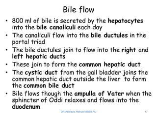
1
DR.Abdiaziz Hakiye MBBS AU
Gallbladder and Biliary tract
• The biliary ducts convey bile from the liver to the
duodenum
• Bile is produced continuously by the liver and
stored and concentrated in the gallbladder, which
releases it intermittently when fat enters the
duodenum
• Bile emulsifies the fat, so that it can be absorbed
in the distal intestine
• Bile canaliculi drain into small interlobular biliary
ducts and then into large collecting bile ducts of
the intrahepatic portal triad, which merges to
form the right and left hepatic ducts
• The right and left hepatic ducts drain the right
and left (parts of the) liver, respectively
• Shortly after leaving the porta hepatis, the right
and left hepatic ducts unite to form common
hepatic duct, which is joined on the right side by
the cystic duct to form the bile duct (part of the
extrahepatic portal triad of the lesser omentum),
which conveys the bile to the duodenum
2
DR.Abdiaziz Hakiye MBBS AU
Bile Duct
• The bile duct (common bile duct) forms in the
free edge of the lesser omentum by the union
of the cystic duct and the common hepatic
duct
• The length of the bile duct varies from 5 to 15
cm, depending on where the cystic duct joins
the common hepatic duct
• The bile duct descends posterior to the
superior part of the duodenum and lies in a
groove on the posterior surface of the head of
the pancreas
• On the left side of the descending part of the
duodenum, the bile duct comes into contact
with the main pancreatic duct
3
DR.Abdiaziz Hakiye MBBS AU
• These ducts run obliquely through the wall of
this part of the duodenum, where they unite
to form the hepatopancreatic ampulla, the
dilation within the major duodenal papilla
• The distal end of the ampulla opens into the
duodenum through the major duodenal papilla
• The circular muscle around the distal end of
the bile duct is thickened to form the
sphincter of the bile duct
• When this sphincter contracts, bile cannot
enter the ampulla and the duodenum; hence
bile backs up and passes along the cystic duct
to the gallbladder for concentration and
storage
4
DR.Abdiaziz Hakiye MBBS AU
5
DR.Abdiaziz Hakiye MBBS AU
• Arterial supply
– Cystic artery: supplying the proximal part of the duct
– Right hepatic artery: supplying the middle part of the
duct
– Posterior superior pancreaticoduodenal artery and
gastroduodenal artery: supplying the retroduodenal
part of the duct
• Venous drainage
– The veins from the proximal part of the bile duct and
the hepatic ducts usually enter the liver directly
– The posterior superior pancreaticoduodenal vein
drains the distal part of the bile duct and empties
into the portal vein or one of its tributaries
• Lymphatic vessels
– From the bile duct pass to the cystic lymph nodes
near the neck of the gallbladder, the node of the
omental foramen, and the hepatic lymph nodes
– Efferent lymphatic vessels from these nodes pass to
the celiac lymph nodes
6
DR.Abdiaziz Hakiye MBBS AU
7
DR.Abdiaziz Hakiye MBBS AU
Gallbladder
• Hollow, pear-shaped organ attached to the
lower surface of the liver
• Capable of storing 30–50 mL of bile
• 7 - 10 cm long
• Lies in the fossa for the gallbladder on the
visceral surface of the liver
• This shallow fossa lies at the junction of the
right and left (parts of the) liver
• The relationship of the gallbladder to the
duodenum is so intimate that the superior part
of the duodenum is usually stained with bile in
the cadaver
8
DR.Abdiaziz Hakiye MBBS AU
• The body of the gallbladder lies anterior to
the duodenum, and its neck and the cystic
duct are immediately superior to the
duodenum
• Peritoneum completely surrounds the fundus
of the gallbladder and binds its body and neck
to the liver
• The hepatic surface of the gallbladder
attaches to the liver by connective tissue of
the fibrous capsule of the liver
9
DR.Abdiaziz Hakiye MBBS AU
• Parts of gallbladder
– Fundus
• the wide end of the organ
• projects from the inferior border of the liver
• usually located at the tip of the right 9th costal
cartilage in the MCL
– Body
• contacts the visceral surface of the liver, the
transverse colon, and the superior part of the
duodenum
– Neck
• narrow and tapered
• directed toward the porta hepatis
• makes an S-shaped bend and joins the cystic duct
10
DR.Abdiaziz Hakiye MBBS AU
11
DR.Abdiaziz Hakiye MBBS AU
• Cystic duct
– 3 - 4 cm long
– Connects the neck of the gallbladder to the common
hepatic duct
– The mucosa of the neck spirals into the spiral fold
(spiral valve)
• helps keep the cystic duct open; thus bile can easily
be diverted into the gallbladder when the distal
end of the bile duct is closed by the sphincter of
the bile duct and/or hepatopancreatic sphincter, or
bile can pass to the duodenum as the gallbladder
contracts
• it also offers additional resistance to sudden
dumping of bile when the sphincters are closed, and
intra-abdominal pressure is suddenly increased, as
during a sneeze or cough
– The cystic duct passes between the layers of the
lesser omentum, usually parallel to the common
hepatic duct, which it joins to form the bile duct
12
DR.Abdiaziz Hakiye MBBS AU
Neurovasculature
• Cystic artery
– supply the gallbladder and cystic duct
– commonly arises from the right hepatic artery in the
angle between the common hepatic duct and the cystic
duct (cystohepatic triangle (of Calot))
– variations occur in the origin and course of the cystic
artery
• Cystic veins
– drain the neck of the gallbladder and cystic duct
– enter the liver directly or drain through the portal
vein to the liver, after joining the veins draining the
hepatic ducts and upper bile duct
• The veins from the fundus and body of the
gallbladder pass directly into the visceral
surface of the liver and drain into the
hepatic sinusoids 13
DR.Abdiaziz Hakiye MBBS AU
14
DR.Abdiaziz Hakiye MBBS AU
• Lymphatic drainage
– to the hepatic lymph nodes, often through cystic
lymph nodes located near the neck of the gallbladder
– efferent lymphatic vessels from these nodes pass to
the celiac lymph nodes
• Innervation
– The nerves to the gallbladder and cystic duct pass
along the cystic artery from the celiac nerve plexus
(sympathetic and visceral afferent [pain] fibers), the
vagus nerve (parasympathetic), and the right phrenic
nerve (somatic afferent fibers)
– Parasympathetic stimulation causes contractions of
the gallbladder and relaxation of the sphincters at
the hepatopancreatic ampulla
– However, these responses are generally stimulated
by the hormone cholecystokinin (CCK), produced by
the duodenal walls (in response to the arrival of a
fatty meal) and circulated through the bloodstream
15
DR.Abdiaziz Hakiye MBBS AU
16
DR.Abdiaziz Hakiye MBBS AU
Bile flow
• 800 ml of bile is secreted by the hepatocytes
into the bile canaliculi each day
• The canaliculi flow into the bile ductules in the
portal triad
• The bile ductules join to flow into the right and
left hepatic ducts
• These join to form the common hepatic duct
• The cystic duct from the gall bladder joins the
common hepatic duct outside the liver to form
the common bile duct
• Bile flows though the ampulla of Vater when the
sphincter of Oddi relaxes and flows into the
duodenum
17
DR.Abdiaziz Hakiye MBBS AU
18
DR.Abdiaziz Hakiye MBBS AU

A.CAVITY 5.pptx

  • 1.
  • 2.
    Gallbladder and Biliarytract • The biliary ducts convey bile from the liver to the duodenum • Bile is produced continuously by the liver and stored and concentrated in the gallbladder, which releases it intermittently when fat enters the duodenum • Bile emulsifies the fat, so that it can be absorbed in the distal intestine • Bile canaliculi drain into small interlobular biliary ducts and then into large collecting bile ducts of the intrahepatic portal triad, which merges to form the right and left hepatic ducts • The right and left hepatic ducts drain the right and left (parts of the) liver, respectively • Shortly after leaving the porta hepatis, the right and left hepatic ducts unite to form common hepatic duct, which is joined on the right side by the cystic duct to form the bile duct (part of the extrahepatic portal triad of the lesser omentum), which conveys the bile to the duodenum 2 DR.Abdiaziz Hakiye MBBS AU
  • 3.
    Bile Duct • Thebile duct (common bile duct) forms in the free edge of the lesser omentum by the union of the cystic duct and the common hepatic duct • The length of the bile duct varies from 5 to 15 cm, depending on where the cystic duct joins the common hepatic duct • The bile duct descends posterior to the superior part of the duodenum and lies in a groove on the posterior surface of the head of the pancreas • On the left side of the descending part of the duodenum, the bile duct comes into contact with the main pancreatic duct 3 DR.Abdiaziz Hakiye MBBS AU
  • 4.
    • These ductsrun obliquely through the wall of this part of the duodenum, where they unite to form the hepatopancreatic ampulla, the dilation within the major duodenal papilla • The distal end of the ampulla opens into the duodenum through the major duodenal papilla • The circular muscle around the distal end of the bile duct is thickened to form the sphincter of the bile duct • When this sphincter contracts, bile cannot enter the ampulla and the duodenum; hence bile backs up and passes along the cystic duct to the gallbladder for concentration and storage 4 DR.Abdiaziz Hakiye MBBS AU
  • 5.
  • 6.
    • Arterial supply –Cystic artery: supplying the proximal part of the duct – Right hepatic artery: supplying the middle part of the duct – Posterior superior pancreaticoduodenal artery and gastroduodenal artery: supplying the retroduodenal part of the duct • Venous drainage – The veins from the proximal part of the bile duct and the hepatic ducts usually enter the liver directly – The posterior superior pancreaticoduodenal vein drains the distal part of the bile duct and empties into the portal vein or one of its tributaries • Lymphatic vessels – From the bile duct pass to the cystic lymph nodes near the neck of the gallbladder, the node of the omental foramen, and the hepatic lymph nodes – Efferent lymphatic vessels from these nodes pass to the celiac lymph nodes 6 DR.Abdiaziz Hakiye MBBS AU
  • 7.
  • 8.
    Gallbladder • Hollow, pear-shapedorgan attached to the lower surface of the liver • Capable of storing 30–50 mL of bile • 7 - 10 cm long • Lies in the fossa for the gallbladder on the visceral surface of the liver • This shallow fossa lies at the junction of the right and left (parts of the) liver • The relationship of the gallbladder to the duodenum is so intimate that the superior part of the duodenum is usually stained with bile in the cadaver 8 DR.Abdiaziz Hakiye MBBS AU
  • 9.
    • The bodyof the gallbladder lies anterior to the duodenum, and its neck and the cystic duct are immediately superior to the duodenum • Peritoneum completely surrounds the fundus of the gallbladder and binds its body and neck to the liver • The hepatic surface of the gallbladder attaches to the liver by connective tissue of the fibrous capsule of the liver 9 DR.Abdiaziz Hakiye MBBS AU
  • 10.
    • Parts ofgallbladder – Fundus • the wide end of the organ • projects from the inferior border of the liver • usually located at the tip of the right 9th costal cartilage in the MCL – Body • contacts the visceral surface of the liver, the transverse colon, and the superior part of the duodenum – Neck • narrow and tapered • directed toward the porta hepatis • makes an S-shaped bend and joins the cystic duct 10 DR.Abdiaziz Hakiye MBBS AU
  • 11.
  • 12.
    • Cystic duct –3 - 4 cm long – Connects the neck of the gallbladder to the common hepatic duct – The mucosa of the neck spirals into the spiral fold (spiral valve) • helps keep the cystic duct open; thus bile can easily be diverted into the gallbladder when the distal end of the bile duct is closed by the sphincter of the bile duct and/or hepatopancreatic sphincter, or bile can pass to the duodenum as the gallbladder contracts • it also offers additional resistance to sudden dumping of bile when the sphincters are closed, and intra-abdominal pressure is suddenly increased, as during a sneeze or cough – The cystic duct passes between the layers of the lesser omentum, usually parallel to the common hepatic duct, which it joins to form the bile duct 12 DR.Abdiaziz Hakiye MBBS AU
  • 13.
    Neurovasculature • Cystic artery –supply the gallbladder and cystic duct – commonly arises from the right hepatic artery in the angle between the common hepatic duct and the cystic duct (cystohepatic triangle (of Calot)) – variations occur in the origin and course of the cystic artery • Cystic veins – drain the neck of the gallbladder and cystic duct – enter the liver directly or drain through the portal vein to the liver, after joining the veins draining the hepatic ducts and upper bile duct • The veins from the fundus and body of the gallbladder pass directly into the visceral surface of the liver and drain into the hepatic sinusoids 13 DR.Abdiaziz Hakiye MBBS AU
  • 14.
  • 15.
    • Lymphatic drainage –to the hepatic lymph nodes, often through cystic lymph nodes located near the neck of the gallbladder – efferent lymphatic vessels from these nodes pass to the celiac lymph nodes • Innervation – The nerves to the gallbladder and cystic duct pass along the cystic artery from the celiac nerve plexus (sympathetic and visceral afferent [pain] fibers), the vagus nerve (parasympathetic), and the right phrenic nerve (somatic afferent fibers) – Parasympathetic stimulation causes contractions of the gallbladder and relaxation of the sphincters at the hepatopancreatic ampulla – However, these responses are generally stimulated by the hormone cholecystokinin (CCK), produced by the duodenal walls (in response to the arrival of a fatty meal) and circulated through the bloodstream 15 DR.Abdiaziz Hakiye MBBS AU
  • 16.
  • 17.
    Bile flow • 800ml of bile is secreted by the hepatocytes into the bile canaliculi each day • The canaliculi flow into the bile ductules in the portal triad • The bile ductules join to flow into the right and left hepatic ducts • These join to form the common hepatic duct • The cystic duct from the gall bladder joins the common hepatic duct outside the liver to form the common bile duct • Bile flows though the ampulla of Vater when the sphincter of Oddi relaxes and flows into the duodenum 17 DR.Abdiaziz Hakiye MBBS AU
  • 18.