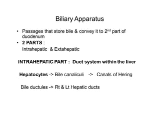
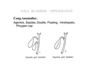
Biliary Apparatus
• Passages that store bile & convey it to 2nd part of
duodenum
• 2 PARTS :
Intrahepatic & Extahepatic
INTRAHEPATIC PART : Duct system within the liver
Hepatocytes -> Bile canaliculi -> Canals of Hering
Bile ductules -> Rt & Lt Hepatic ducts
Extrahepatic
• Rt and left hepatic ducts
• Common hepatic duct
• Cystic duct
• Bile duct
• Gall bladder
Rt and left hepatic ducts
– Emerge through Porta hepatis
– Relations : at Porta hepatis
Ducts - Arteries - Veins (Ant to post)
- Unite & form Common hepatic
duct
Anterior
Porta hepatis
Common Hepatic duct
PV
• Loc : In Rt free margin of Lsr Omentum
– In front of Portal vn & to the Rt of Hepatic A
• Length : 4cm, Diameter : 4mm
• Joins cystic duct 1cm above 1st part of
duodenum (variable)
Posterior
CYSTIC DUCT :
• Connects Neck of GB with CH Duct
• Joins CH Duct on the Rt, at an acute angle
• Length : 2 - 3cm, Diameter : 2-3mm
• Relations :
Cystic Artery
CHD
Rt HD Lt HD
B D
CYSTIC DUCT : Cont’d
• 5 -12 crescentic valves in lumen :
Spiral valves of Heister
• Hartmann’s pouch :
CHD
Spiral valves
CALOT’S TRIANGLE
BOUNDARIES :
– Inf surface of liver (Above & laterally)
– Cystic duct (Below & Lat)
– Common Hepatic Duct (Medially)
– Contents : Cystic artery with its variable dispositions
Cystic Lymph Node
APPLIED : Identification of Cystic artery - a prerequisite
in cholecyctectomy.
BILE DUCT :
Formation : CHD + Cystic duct
– Length : 8cm, Diameter : 8mm
Course :
– Passes down, back & to the Rt
– Joins Pancreatic duct-
– Forms Ampulla of Vater &
opens in Major duodenal papilla
4 Parts :
– Supra, Retro, Infra (Para) &
Intra duodenal parts :
S
R
I
I
Blood supply
• From Brs of Cystic, Hepatic and GD arteries
- Ligation of cystic duct - away from Bile duct
BILE DUCT : APPLIEDANATOMY
– Supra duodenal part : Most accessible part for surgery
Eg : Choledochotomy, Insertion of drainage tube
– Infra (Para) duodenal :
Obstructed in Carcinoma Hd of pancreas (Jaundice)
– Intra duodenal part :
Ampullary carcinoma (Recurrent – obstructive -
jaundice)
GALL BLADDER
• Pear shaped fibro-muscular hollow viscus
• Slate blue in colour
• Size : 7-10 x 3 cm, 30-50 ml
• Location :
Gall bladder fossa on visceral surf of Rt lobe of liver,
near quadrate lobe
G B
Quadrate lobe
GALL BLADDER
Parts : Fundus, Body & Neck
• Fundus - In lower bdr of liver
Gall bladder
SURFACE MARKING - Fundus of GB
• Transpyloric plane crosses costal margin at lat bdr of Rt
Rectus sheath
• At tip of Rt 9th costal cartilage
• Area of tenderness in GB disease
GALL BLADDER- cont’d
Functions :
•Concentration & storage of bile
•Reduces alkalinity
•Regulates ductal pressure
GALL BLADDER cont’d
• Arterial supply : Cystic A (Rt br of Hepatic A)
•Occassionally from Lt Br of HA/ HA / GDA
•Small vessels from hepatic bed of GB
• Venous : Cystic vn (multiple)
•Do not accompany Cystic Artery
•Drain into Intra hepatic part of portal vein
• Lymphatics : Hepatic nodes
GALL BLADDER- cont’d
• Nerve supply :
•Parasymp : Hepatic Br of Ant Vagal trunk
–Contraction of GB, Relaxation of Sphincters
•Hormonal : CCK
•Symp : C Ganglion- From T 7-9
•Refd pain : C3- 4 (Phrenic) / T 7-9
–Pain at tip of shoulder,
–Rt Hypochondrium, Epigastrium, Infrascapular
region
GALL BLADDER : APPLIEDANAT
CongAnomalies :
Agenisis, Septate, Double, Floating, Intrahepatic,
Phrygian cap
GALL BLADDER : APPLIEDANAT
• Cholecystitis,
• Cholangitis
• Cholelithiasis,
• White bile
• Biliary colic
• Cholecystectomy
• Cholidocotomy : incision of bile duct (upper part) for
removal of Gall stones
Oral/
Intravenous
Percutaneous Transhepatic cholangiography (PTC)
• Detect obstructions
• A thin needle is inserted into liver
to deliver dye to the bile ducts
• Movement of dye tracked using a fluoroscope
(specialized X-ray machine)
?

Biliary apparatus

  • 1.
    Biliary Apparatus • Passagesthat store bile & convey it to 2nd part of duodenum • 2 PARTS : Intrahepatic & Extahepatic INTRAHEPATIC PART : Duct system within the liver Hepatocytes -> Bile canaliculi -> Canals of Hering Bile ductules -> Rt & Lt Hepatic ducts
  • 2.
    Extrahepatic • Rt andleft hepatic ducts • Common hepatic duct • Cystic duct • Bile duct • Gall bladder
  • 3.
    Rt and lefthepatic ducts – Emerge through Porta hepatis – Relations : at Porta hepatis Ducts - Arteries - Veins (Ant to post) - Unite & form Common hepatic duct Anterior Porta hepatis
  • 4.
    Common Hepatic duct PV •Loc : In Rt free margin of Lsr Omentum – In front of Portal vn & to the Rt of Hepatic A • Length : 4cm, Diameter : 4mm • Joins cystic duct 1cm above 1st part of duodenum (variable) Posterior
  • 5.
    CYSTIC DUCT : •Connects Neck of GB with CH Duct • Joins CH Duct on the Rt, at an acute angle • Length : 2 - 3cm, Diameter : 2-3mm • Relations : Cystic Artery CHD Rt HD Lt HD B D
  • 6.
    CYSTIC DUCT :Cont’d • 5 -12 crescentic valves in lumen : Spiral valves of Heister • Hartmann’s pouch : CHD Spiral valves
  • 7.
    CALOT’S TRIANGLE BOUNDARIES : –Inf surface of liver (Above & laterally) – Cystic duct (Below & Lat) – Common Hepatic Duct (Medially) – Contents : Cystic artery with its variable dispositions Cystic Lymph Node APPLIED : Identification of Cystic artery - a prerequisite in cholecyctectomy.
  • 8.
    BILE DUCT : Formation: CHD + Cystic duct – Length : 8cm, Diameter : 8mm Course : – Passes down, back & to the Rt – Joins Pancreatic duct- – Forms Ampulla of Vater & opens in Major duodenal papilla 4 Parts : – Supra, Retro, Infra (Para) & Intra duodenal parts : S R I I
  • 9.
    Blood supply • FromBrs of Cystic, Hepatic and GD arteries - Ligation of cystic duct - away from Bile duct
  • 10.
    BILE DUCT :APPLIEDANATOMY – Supra duodenal part : Most accessible part for surgery Eg : Choledochotomy, Insertion of drainage tube – Infra (Para) duodenal : Obstructed in Carcinoma Hd of pancreas (Jaundice) – Intra duodenal part : Ampullary carcinoma (Recurrent – obstructive - jaundice)
  • 11.
    GALL BLADDER • Pearshaped fibro-muscular hollow viscus • Slate blue in colour • Size : 7-10 x 3 cm, 30-50 ml • Location : Gall bladder fossa on visceral surf of Rt lobe of liver, near quadrate lobe G B Quadrate lobe
  • 12.
    GALL BLADDER Parts :Fundus, Body & Neck • Fundus - In lower bdr of liver Gall bladder
  • 13.
    SURFACE MARKING -Fundus of GB • Transpyloric plane crosses costal margin at lat bdr of Rt Rectus sheath • At tip of Rt 9th costal cartilage • Area of tenderness in GB disease
  • 14.
    GALL BLADDER- cont’d Functions: •Concentration & storage of bile •Reduces alkalinity •Regulates ductal pressure
  • 15.
    GALL BLADDER cont’d •Arterial supply : Cystic A (Rt br of Hepatic A) •Occassionally from Lt Br of HA/ HA / GDA •Small vessels from hepatic bed of GB • Venous : Cystic vn (multiple) •Do not accompany Cystic Artery •Drain into Intra hepatic part of portal vein • Lymphatics : Hepatic nodes
  • 16.
    GALL BLADDER- cont’d •Nerve supply : •Parasymp : Hepatic Br of Ant Vagal trunk –Contraction of GB, Relaxation of Sphincters •Hormonal : CCK •Symp : C Ganglion- From T 7-9 •Refd pain : C3- 4 (Phrenic) / T 7-9 –Pain at tip of shoulder, –Rt Hypochondrium, Epigastrium, Infrascapular region
  • 17.
    GALL BLADDER :APPLIEDANAT CongAnomalies : Agenisis, Septate, Double, Floating, Intrahepatic, Phrygian cap
  • 18.
    GALL BLADDER :APPLIEDANAT • Cholecystitis, • Cholangitis • Cholelithiasis, • White bile • Biliary colic • Cholecystectomy • Cholidocotomy : incision of bile duct (upper part) for removal of Gall stones
  • 20.
  • 21.
    Percutaneous Transhepatic cholangiography(PTC) • Detect obstructions • A thin needle is inserted into liver to deliver dye to the bile ducts • Movement of dye tracked using a fluoroscope (specialized X-ray machine)
  • 22.