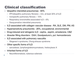
Interstitial Lung Disease
호흡기알레르기내과
실습 2조 강천지, 구윤수
Definition
 Heterogenous group of lower respiratory tract disorders
 Many potential causes
 Common features
 Exertional dyspnea, restrictive pattern on PFT, airflow
obstruction, decreased DLCO, increased alveolar-arterial
oxygen difference
 absence of pulmonary infection or neoplasm
 ILD = misnomer
 In pathology, Not restricted to the interstitium
 Gas exchange unit + beyond gas exchange unit are
involved
Clinical classification
 Idiopathic interstitial pneumonias : 40%
 IPF(idiopathic pulmonary fibrosis) – m/c, at least 30% of ILD
 nonspecific pulmonary fibrosis – 10%
 Respiratory bronchiolitis-associated ILD – 6%
 Desquamative interstitial pneumonia
 ILD associated with collagen vascular disease : RA, SLE, DM, PM, AS
 Hypersensitivity pneumonitis : 26%, occupational, environmental
 Drug-induced and iatrogenic ILD : cepha., aspirin, amodarone, CBZ
 Alveolar filling disorders : DAH, Goodpasture’s, pul. hemosiderosis
 ILD associated with pulmonary vasculitis
 Wegener’s syndrome, Churg-strauss syndrome
 Other specific forms of ILD
 sarcoidosis, lymphangioleiomyomatosis, histiocytosis X
 Inherited forms of ILD
 Neurofibromatosis, tuberous sclerosis
Epidemiology
 Prevalence in US
 Man : 81/100,000
 Woman : 61/100,000
 Overall incidence in US : man > woman
 Man : 31.5/100,000
 Woman : 26.1/100,000
Pathobiology
 Unknown tissue injury + attempted repair
 Fibroproliferattion ⇒ honeycombing
⇒ pulmonary vascular resistance↑ + 2nd pulmonary HTN
 Highly heterogeneous findings
 Normal, inflammation, fibrosis, granuloma, vasculitis,
secondary vascular change
 Gene : hTERT, hTR(telomerase), MUC5B promotor
Clinical manifestations
 Progressive dyspnea : typically Sx.
 Nonproductive cough, fatigue : common Sx.
 Pleuritic chest pain : collagen vascular, drug-induced
 Pneumothorax : LAM, neurofibromatosis, Histiocytosis X
 Hemoptysis : diffuse alveolar hemorrhagic synd., SLE,
LAM, Wegener, Goodpasture
 Coarse rale, crackle in ILD / wheezing is not common
 Cyanosis, clubbing
Diagnosis : History
 Age, sex, smoking
 IPF : >50 yrs, 75% smoking
 Sarcoidosis : young, middle-aged adult
 Langerhans cell histiocytosis : smoking, young man
 LAM : women of childbearing age
 Respiratory bronchiolitis associated ILD : smoking, all ages
 Environmental, occupational factor
 Framing, avian antigen, mold, mining, grinding, welding
 Medication, drug
 Immunosuppression, HIV, transplant
 Onset, duration, progression
 Extra-pul. Sx.
 dysphagia, arthritis, m. weakness, rec. sinusitis, hematuria
Diagnosis : Physical examination
 Rarely helpful on respiratory exam
 Rhonchi, rale, digital clubbing = non-specific
 Findings On cardiac exam, in Pts with advanced lung
disease
 P2, tricuspid insufficiency, pulmonary hypertension, cor
pulmonale
 Extra-thoracic findings
 Sarcoidosis : skin abNL, peripheral lymphadenopathy,
hepatosplenomegaly, arthritis
 Polymyositis : m. tenderness, proximal m. weakness
 Collagen vascular disease : arthritis
Diagnosis : Laboratory testing
 Not specific : CBC, chemistry panel, U/A
 Hypoxemia in moderate to severe ILD
 SLE : ANA
 RA : RF, anti-citrullinated peptide
 Scleroderma : Scl 70
 DM, PM : anti-Jo-1, aldolase, creatine kinase
 Wegener’s granulomatosis : ANCA
 Goodpasture’s syndrome: anti-BM
Diagnosis : Chest radiograph
 Normal in 10% pts of ILD
 Most ILDs
 Infiltration in the lower lung zones
 No hilar / mediastinal adenopathy
 Decreased lung volume
 Diffuse GGO -> reticulonodular infiltration
ill-defined nodules w/ air bronchograms
 Infiltration : recurrent, migratory
 Pleural thickening, pleural effusion, pneumothorax
Diagnosis : HRCT
 Essential in both the diagnosis and staging of ILD
 Earlier diagnosis than CXR
 Help narrow differential diagnosis
 Aid selecting the site for BAL, lung biopsy
 Assist in choosing among therapeutic options
 Assist in estimating the response to treatment
Diagnosis : Pulmonary function test
 Monitoring the progression, prognosis
 Restrictive lung defect
 Decreased : DLCO, FVC, FEV1, total lung capacity,
lung volume
 DDx
 Obstructive-restrictive : CSS, allergic bronchopulmonary
aspergillosis, endobronchial sarcoidosis, hypersensitivity
pneumonitis
 Respiratory m. weakness : PM, systemic sclerosis, SLE
Diagnosis : Exercise testing
 Increased PAO2-PaO2 ∝ severity of disease, degree
of fibrosis
 Decreased work rate, maximal oxygen consumption
 Abnormally high minute ventilation
 Decreased peak minute ventilation
 Failure of tidal volumes to increase
 6-min walk test : quantitative date on exercise
capacity and oxygen desaturation with exercise
Diagnosis : Invasive evaluation
 Bronchoalveolar lavage
 Lymphocyte, eosinophil, asbestos body, staining
surfactants
 Transbronchial lung biopsy
 Non-caseating granuloma, giant cell, smooth m.
proliferation
 Video-assisted thoracoscopic biopsy
 Open lung biopsy
 Most IPF do not need to have biopsy to confirm the
diagnosis
Treatment
 Avoidance of the inciting agent
 Systemic corticosteroid
 Unclear dosage and duration
 Supportive oxygen supplementation
 Treatments for pulmonary hypertension
 Lung transplantation
 End-stage ILD : significant pul. Fibrosis, pul. Hypertension
2014-07-1116
Idiopathic Interstitial Pneumonias
Idiopathic pulmonary fibrosis
Nonspecific interstitial pneumonia
Respiratory bronchiolitis-associated ILD
Desquamative interstitial pneumonia
Acute interstitial pneumonia
Cryptogenic organizing pneumonia
Lymphoid and lymphocytic interstitial pneumonia
Idiopathic Interstitial Pneumonias
 Unknown etiology
 Dyspnea, nonproductive cough, chest pain, wt. loss,
fatigue, crackle, clubbing
 HRCT, better than CXR
Idiopathic Pulmonary Fibrosis
2014-07-1118
 Epidemiology
 50~60% of IIP
 Mean age at onset : 62 yrs(older than 50 yrs)
 Clinical manifestation
 Cigarette smoking, coexisting emphysema
 Gradual dyspnea, restrictive pattern on PFT
 HRCT : bilateral pulmonary fibrosis
 Median survival : 3~5 yrs after diagnosis
 Acute exacerbation : 5~10%, admission & ICU
Diagnosis
2014-07-1119
 CXR : basal, reticular pattern, decreased lung volume
 HRCT
 basilar patchy intra-lobular reticulation
 Subpleural honeycomb cysts
 Traction bronchiectasis : advanced disease
 PFT : restrictive pattern
 Mild disease : normal lung volume, small decreased DLCO,
normal PFT
 BAL : nonspecific / neutrophil, eosinophil
 Histopathology : honeycombing, fibrosis, inflammatory
cell, collagen deposition, normal lung
Diagnosis
2014-07-1120
Treatment
2014-07-1121
 No treatment to improve survival rate
 All treatments are experimental
 Oral prednisone + azathioprine + N-acetylcysteine
 Better preservation of PFT
 Pirfenidone + anti-fibrotic agent
 Loss of lung function↓, progression-free survival↑
 Empirical IV corticosteroid
 Not beneficial
 INF-ɣ1b , cyclophosphamide, colchicine, D-penicillamine, oral
corticosteroid + immunosuppressive agent
 Supplemental O2, Tx of infection, PE, pul. HTN, GERD
 Immunization for influenza, herpes zoster, pneumococcus
 Lung transplantation : 2/3 of IPF is a contraindication
Prognosis
2014-07-1122
 Progressive impairment of lung function, gas exchange
 Median survival : 3~5 yrs
 Longer survival
 Less fibrosis, less functional impairment, no pul. HTN,
 no significant oxygen desaturation on the 6min walk test
 Shorter survival
 Emphysema, pul. HTN, acute exacerbation
Nonspecific Interstitial Pneumonia
2014-07-1123
 Two subgroup : cellular, fibrotic
 Fibrotic NIP is similar to early IPF
 Onset age : 50 yrs
 Diagnosis
 CXR : bilateral patchy infiltration in LLF
 HRCT
 Cellular : bilateral GGO, consolidation, subpleural reticulation, loss
of lower lobe volume
 Fibrotic : bilateral architectural derangement in lower lobe
 Bx.
 Cellular : chronic lymphoplasmacytic infiltration
 Fibrotic : dense interstitial fibrosis
 Treatment : corticosteroid
 Prognosis : better than IPF
RESPIRATORY BRONCHIOLITIS–
ASSOCIATED ILD
2014-07-1124
 Epidemiology : Smoking, 40th or 50th yrs
 Diagnosis
 PFTs
 airway obstruction, mildly decreased or preserved TLC, Dlco ↓
 CXR
 bronchial wall thickening, GGO
 BAL
 brown-pigmented alveolar macrophages w/ neutrophils
 HRCT
 Biopsy
Diagnosis
2014-07-1125
Treatment and Prognosis
2014-07-1126
 cessation of smoking
 low-dose corticosteroids
 (e.g., prednisone, 10 to 20 mg/day) for a few months
DESQUAMATIVE INTERSTITIAL
PNEUMONIA
2014-07-1127
 more extensive form of RB-ILD
 More affected in smoker
 Diagnosis
 PFTs
 restrictive pattern, Dlco ↓ w/ or w/o obstruction
 CXR
 patchy basal consolidation w/ a lower lobe & periphery
 BAL
 pigmented alveolar Mø, frequently w/ neutrophils ↑
 HRCT
 Biopsy
Diagnosis
2014-07-1128
Treatment and Prognosis
2014-07-1129
 cessation of smoking
 oral corticosteroid therapy
 lung transplantation for selected patients
 Good therapeutic outcome, survival rate : 70% at 10 yrs
Treatment and Prognosis
2014-07-1130
Treatment and Prognosis
2014-07-1131
ACUTE INTERSTITIAL PNEUMONIA
2014-07-1132
 Healthy person
 after viral URI
 mimics the ARDS
 Dx
 CT scan
CRYPTOGENIC ORGANIZING
PNEUMONIA (idiopathic BOOP)
2014-07-1133
 flulike illness w/ nonproductive cough, followed by
exertional dyspnea
 DX
 PFTs : restrictive defect
 CXR : peripheral or recurrent migratory alveolar opacities
 BAL : nonspecific
 HRCT
 LLF consolidation(subpleural or peribronchial),
 small nodules along bronchovascular bundles & GGO
 Biopsy : granulation tissue ↑↑
within the small airways & alveolar ducts
Treatment and Prognosis
2014-07-1134
 oral corticosteroids
 adjunct immunosuppressive agents (azathioprine)
 Spontaneous remissions can occur
LYMPHOID AND LYMPHOCYTIC
INTERSTITIAL PNEUMONIA
2014-07-1135
 Epidemiology
 women > men, especially in the 50th
 Clinical manifestation
 Concurrent collagen vascular dz. or an autoimmune dis (esp.
Sjogren’s synd.)
 gradual onset of cough & exertional dyspnea
 Dx
 CXR : Lower lung field reticular or reticulonodular pattern
 BAL : lymphocytes ↑
 HRCT : bilateral GGO, small or large nodules & scattered cysts
perivascular honeycombing & reticular abnormalities
 Biopsy : dense interstitial lymphocytic infiltrate
Treatment and Prognosis
2014-07-1136
 oral corticosteroids
 more than 1/3 of pts progressing to diffuse pul. fibrosis
ILD Associated with
Collagen Vascular Disease
2014-07-1137
Progressive systemic sclerosis
Rheumatoid arthritis
Systemic lupus erythematosus
Dermatomyositis and polymyositis
Sjogren’s syndrome
Mixed connective tissue disease
Ankylosing spondylitis
Progressive systemic sclerosis
2014-07-1138
 most frequently associated with ILD
 nonspecific interstitial pneumonia
 Clinical manifestation
 Pulmonary Sx → cutaneous or digital Sx
 chronic pulmonary fibrosis : bronchogenic ca. ↑
 Tx
 cyclophosphamide for 1 year
Rheumatoid arthritis
2014-07-1139
 more common in men (3:1 ratio) at 50th to 60th
 Clinical manifestation
 bronchiectasis, bronchiolitis,
 idiopathic interstitial pneumonias
 pleural effusions or pleural thickening
 lymphocytic infiltrate ↑ -> fibrous tissue, honeycomb
 Tx
 underlying RA
Systemic lupus erythematosus
2014-07-1140
 Pleural disease (m/c) or pleural effusions
 widespread GGO w/ consolidation, or DAH
 Shrinking lung
 Result of diaphragmatic weakness→Progressive lung
restriction →resistant to Tx
 Tx
 underlying SLE
Dermatomyositis and Polymyositis
2014-07-1141
 anti-Jo-1 antibody
 Clinical manifestation
 acute interstitial pneumonia–like syndrome
 ILD → muscular manifestations
 The severity of the muscular dis. does not corr. with ILD
 Tx
 underlying DM PM
Sjogren’s syndrome
2014-07-1142
 ILD is primary form
 Lymphocytic interstitial pneumonia
 Respiratory infections & bronchiectasis
 common in advanced stages
 Tx response is usually good
Mixed connective tissue disease
2014-07-1143
 overlap syndrome
 Pulmonary disease is common
 but most often subclinical & identified only radiographically
 Tx
 underlying disease
Ankylosing spondylitis
2014-07-1144
 upper lobe, bilateral reticulonodular infiltrates
w/ cyst formation
 Tx : no known effective therapy
for apical fibrobullous disease
Hypersensitivity Pneumonitis
2014-07-1145
Hypersensitivity Pneumonitis
2014-07-1146
 extrinsic allergic alveolitis
 Pathogenesis
 repeated inhalation of specific antigens
 inflammatory cell infiltration : bronchioles, alveoli & interstitium
 noncaseating, epithelioid cell granulomas
 A history of exposure is essential to Diagnosis & Tx
2014-07-1147
 Diagnosis
 CXR : focal patchy consolidation or diffuse GGO
→ micronodular & reticular shadowing
→ upper lung zone reticulation with honeycombing
 BAL : lymphocytes & plasma cells ↑↑
 HRCT : small centrilobular ill-defined nodules of GGO
 Biopsy : granulation ↑↑ (small airways & alveolar ducts)
& chronic inflammation in the surrounding alveoli
 Tx & Px
 avoidance of exposure to the antigen
 Corticosteroids
 Continued exposure : chronic HP & irreversible fibrosis
Occupational ILDs
2014-07-1148
silicosis inhalation of silica in crystalline form
or silicon dioxide,
sandblasting & working w/ granite
coal workers’
pneumoconiosis
inhalation of coal dust
asbestosis deposition of fibers
during mining, milling
welding & working in a shipyard
berylliosis seen in aerospace workers
& in electronic industries
Drug-Induced ILD
2014-07-1149
alveolar and interstitial abnormalities
granulomatous pneumonitis
chronic nitrofurantoin-induced ILD
Drug-Induced ILD
2014-07-1150
Nonspecific bilateral
alveolar & interstitial
inflammatory
& fibrotic abnormalities
sarcoid-like
granulomatous ILD
Alveolar Filling Disorders
2014-07-1151
IDIOPATHIC PULMONARY HEMOSIDEROSIS
CHRONIC EOSINOPHILIC PNEUMONIA
IDIOPATHIC PULMONARY
HEMOSIDEROSIS
2014-07-1152
 Epidemiology
 children and young adults : rare
 DAH w/o vasculitis, inflammation, granulomas, necrosis
 Dx
 BAL : Hemosiderin-laden macrophages
 CXR
 diffuse, bilateral alveolar infiltrates
 hilar & mediastinal adenopathy
 Tx
 Systemic corticosteroids
CHRONIC EOSINOPHILIC PNEUMONIA
2014-07-1153
 20th ~ 40th women, Peripheral blood eosinophilia : common, usually 10 to 40%
 Sx
 fevers, sweats, weight loss, fatigue, dyspnea, cough
 Cardinal feature
 CXR & HRCT : peripheral multifocal consolidation
predominantly in the upper and mid lung zones
 BAL : eosinophils > 40% during exacerbations
 Dx & Tx: corticosteroids
“photographic negative of pulmonary edema,”
 Px: Relapse rate is high
ILD associated
with Pulmonary Vasculitides
2014-07-1154
WEGENER’S GRANULOMATOSIS
CHURG-STRAUSS SYNDROME
IDIOPATHIC PULMONARY CAPILLARITIS
ILD associated
with Pulmonary Vasculitides
2014-07-1155
WEGENER’S
GRANULOMATOSIS
CHURG-STRAUSS
SYNDROME
IDIOPATHIC
PULMONARY
CAPILLARITIS
m/c form of vasculitis
Destruction of the
nasal cartilage
→ septal perforation
→ cavitating nodules
Vasculitis of both
respiratory tracts
allergic disorders,
eosinophilia, IgE ↑
bronchospasm
pulmonary vasculature
within the alveolar walls
Manifestation like ILD
CXR multiple nodular or
cavitating infiltrates
bilateral patchy,
diffuse nodular
infiltrates, diffuse
reticulonodular dis.
Dx: ACNA Histopathologic
examination
subclinical alveolar
hemorrhage associated
w/ p- ACNA
Tx cyclophosphamide
→ Rituximab
corticosteroid unclear
Corticosteroids
cyclophosphamide
or rituximab
Other Forms of ILD
2014-07-1156
SARCOIDOSIS
PULMONARY LANGERHANS CELL HISTIOCYTOSIS
LYMPHANGIOLEIOMYOMATOSIS
SARCOIDOSIS
2014-07-1157
 Idiopathic chronic multi- system granulomatous dis.
 Epidemiology : 20~ 40th , women
 Pathogenesis
 T helper 1 cell ↑↑ → non caseating granuloma
 Clinincal manifestation : Lung, Liver, skin, eye
 Lung : ILD, BHL
PULMONARY LANGERHANS CELL
HISTIOCYTOSIS
2014-07-1158
 pulmonary histiocytosis X or eosinophilic granuloma of the lung,
 idiopathic, granulomatous ILD 20 ~ 30th, male, smoke
 DIAGNOSIS
 CXR : diffuse symmetrical reticulonodular opacities + multiple small cysts (upper &
mid lung)
 HRCT : subpleural nodules, scattered GGO, irregular cysts in both lungs, with
spare lung bases
 PFTs : a mixed restrictive & obstructive pattern
 BAL : Langerhans cells (atypical histiocytes)
 TBLB. Open lung bx
 interstitial and peribronchiolar (histiocytes, eosinophils, and lymphocytes)
 peribronchiolar nodules + cysts + central stellate fibrosis.
 immunostaining : CD1, S-100
 Tx & Px
 prognosis is favorable
 discontinue smoking (75% of patients improving or stabilizing)
 corticosteroids
 w/ or w/o vincristine, cyclosporine, cyclophosphamide, azathioprine
LYMPHANGIOLEIOMYOMATOSIS
2014-07-1159
 Women>men, childbearing age.
 Sx
 Hemoptysis, pneumothorax(rupture of subpleural cysts) chylothorax (lymphatic ob.)
 CXR: Coarse reticulonodular infiltrates, often w/ bilat’ cysts or bullae, lung vol. ↑
 HRCT : diffuse thin-walled cysts <2 cm
 BAL : occult alveolar hemorrhage
 Lung bx : abNL sm. cells in airways, lymphatics, bv,
w/ concurrent airflow obstruction & replace parenchyma with cysts
 Tx & Px
 Sirolimus(RCT) progesterone or Tamoxifen(non-RCT)
 10 yrs survival after the onset of symptoms
Inherited Disorders
2014-07-1160
AD tuberous sclerosis indistinguishable from LAM
both radiographically & histopathologically
neurofibromatosis bilateral lower lobe fibrosis
& bullae or cystic changes
AR Gaucher’s disease interstitial infiltration
w/ fibrosis, alveolar consolidation,
& filling of alveolar spaces
Niemann-Pick disease infiltration of the characteristic "foam cell"
throughout the pulmonary lymphatics,
the pulmonary arteries,
& the pulmonary alveoli
Hermansky-Pudlak
syndrome
Pulmonary fibrosis;
onset in the 30th~ 40th
slowly progressive
경청해주셔서 감사합니다.

Cecil Chaper 92. ILD(interstitial lung disease)

  • 1.
  • 2.
    Definition  Heterogenous groupof lower respiratory tract disorders  Many potential causes  Common features  Exertional dyspnea, restrictive pattern on PFT, airflow obstruction, decreased DLCO, increased alveolar-arterial oxygen difference  absence of pulmonary infection or neoplasm  ILD = misnomer  In pathology, Not restricted to the interstitium  Gas exchange unit + beyond gas exchange unit are involved
  • 3.
    Clinical classification  Idiopathicinterstitial pneumonias : 40%  IPF(idiopathic pulmonary fibrosis) – m/c, at least 30% of ILD  nonspecific pulmonary fibrosis – 10%  Respiratory bronchiolitis-associated ILD – 6%  Desquamative interstitial pneumonia  ILD associated with collagen vascular disease : RA, SLE, DM, PM, AS  Hypersensitivity pneumonitis : 26%, occupational, environmental  Drug-induced and iatrogenic ILD : cepha., aspirin, amodarone, CBZ  Alveolar filling disorders : DAH, Goodpasture’s, pul. hemosiderosis  ILD associated with pulmonary vasculitis  Wegener’s syndrome, Churg-strauss syndrome  Other specific forms of ILD  sarcoidosis, lymphangioleiomyomatosis, histiocytosis X  Inherited forms of ILD  Neurofibromatosis, tuberous sclerosis
  • 4.
    Epidemiology  Prevalence inUS  Man : 81/100,000  Woman : 61/100,000  Overall incidence in US : man > woman  Man : 31.5/100,000  Woman : 26.1/100,000
  • 5.
    Pathobiology  Unknown tissueinjury + attempted repair  Fibroproliferattion ⇒ honeycombing ⇒ pulmonary vascular resistance↑ + 2nd pulmonary HTN  Highly heterogeneous findings  Normal, inflammation, fibrosis, granuloma, vasculitis, secondary vascular change  Gene : hTERT, hTR(telomerase), MUC5B promotor
  • 6.
    Clinical manifestations  Progressivedyspnea : typically Sx.  Nonproductive cough, fatigue : common Sx.  Pleuritic chest pain : collagen vascular, drug-induced  Pneumothorax : LAM, neurofibromatosis, Histiocytosis X  Hemoptysis : diffuse alveolar hemorrhagic synd., SLE, LAM, Wegener, Goodpasture  Coarse rale, crackle in ILD / wheezing is not common  Cyanosis, clubbing
  • 7.
    Diagnosis : History Age, sex, smoking  IPF : >50 yrs, 75% smoking  Sarcoidosis : young, middle-aged adult  Langerhans cell histiocytosis : smoking, young man  LAM : women of childbearing age  Respiratory bronchiolitis associated ILD : smoking, all ages  Environmental, occupational factor  Framing, avian antigen, mold, mining, grinding, welding  Medication, drug  Immunosuppression, HIV, transplant  Onset, duration, progression  Extra-pul. Sx.  dysphagia, arthritis, m. weakness, rec. sinusitis, hematuria
  • 8.
    Diagnosis : Physicalexamination  Rarely helpful on respiratory exam  Rhonchi, rale, digital clubbing = non-specific  Findings On cardiac exam, in Pts with advanced lung disease  P2, tricuspid insufficiency, pulmonary hypertension, cor pulmonale  Extra-thoracic findings  Sarcoidosis : skin abNL, peripheral lymphadenopathy, hepatosplenomegaly, arthritis  Polymyositis : m. tenderness, proximal m. weakness  Collagen vascular disease : arthritis
  • 9.
    Diagnosis : Laboratorytesting  Not specific : CBC, chemistry panel, U/A  Hypoxemia in moderate to severe ILD  SLE : ANA  RA : RF, anti-citrullinated peptide  Scleroderma : Scl 70  DM, PM : anti-Jo-1, aldolase, creatine kinase  Wegener’s granulomatosis : ANCA  Goodpasture’s syndrome: anti-BM
  • 10.
    Diagnosis : Chestradiograph  Normal in 10% pts of ILD  Most ILDs  Infiltration in the lower lung zones  No hilar / mediastinal adenopathy  Decreased lung volume  Diffuse GGO -> reticulonodular infiltration ill-defined nodules w/ air bronchograms  Infiltration : recurrent, migratory  Pleural thickening, pleural effusion, pneumothorax
  • 11.
    Diagnosis : HRCT Essential in both the diagnosis and staging of ILD  Earlier diagnosis than CXR  Help narrow differential diagnosis  Aid selecting the site for BAL, lung biopsy  Assist in choosing among therapeutic options  Assist in estimating the response to treatment
  • 12.
    Diagnosis : Pulmonaryfunction test  Monitoring the progression, prognosis  Restrictive lung defect  Decreased : DLCO, FVC, FEV1, total lung capacity, lung volume  DDx  Obstructive-restrictive : CSS, allergic bronchopulmonary aspergillosis, endobronchial sarcoidosis, hypersensitivity pneumonitis  Respiratory m. weakness : PM, systemic sclerosis, SLE
  • 13.
    Diagnosis : Exercisetesting  Increased PAO2-PaO2 ∝ severity of disease, degree of fibrosis  Decreased work rate, maximal oxygen consumption  Abnormally high minute ventilation  Decreased peak minute ventilation  Failure of tidal volumes to increase  6-min walk test : quantitative date on exercise capacity and oxygen desaturation with exercise
  • 14.
    Diagnosis : Invasiveevaluation  Bronchoalveolar lavage  Lymphocyte, eosinophil, asbestos body, staining surfactants  Transbronchial lung biopsy  Non-caseating granuloma, giant cell, smooth m. proliferation  Video-assisted thoracoscopic biopsy  Open lung biopsy  Most IPF do not need to have biopsy to confirm the diagnosis
  • 15.
    Treatment  Avoidance ofthe inciting agent  Systemic corticosteroid  Unclear dosage and duration  Supportive oxygen supplementation  Treatments for pulmonary hypertension  Lung transplantation  End-stage ILD : significant pul. Fibrosis, pul. Hypertension
  • 16.
    2014-07-1116 Idiopathic Interstitial Pneumonias Idiopathicpulmonary fibrosis Nonspecific interstitial pneumonia Respiratory bronchiolitis-associated ILD Desquamative interstitial pneumonia Acute interstitial pneumonia Cryptogenic organizing pneumonia Lymphoid and lymphocytic interstitial pneumonia
  • 17.
    Idiopathic Interstitial Pneumonias Unknown etiology  Dyspnea, nonproductive cough, chest pain, wt. loss, fatigue, crackle, clubbing  HRCT, better than CXR
  • 18.
    Idiopathic Pulmonary Fibrosis 2014-07-1118 Epidemiology  50~60% of IIP  Mean age at onset : 62 yrs(older than 50 yrs)  Clinical manifestation  Cigarette smoking, coexisting emphysema  Gradual dyspnea, restrictive pattern on PFT  HRCT : bilateral pulmonary fibrosis  Median survival : 3~5 yrs after diagnosis  Acute exacerbation : 5~10%, admission & ICU
  • 19.
    Diagnosis 2014-07-1119  CXR :basal, reticular pattern, decreased lung volume  HRCT  basilar patchy intra-lobular reticulation  Subpleural honeycomb cysts  Traction bronchiectasis : advanced disease  PFT : restrictive pattern  Mild disease : normal lung volume, small decreased DLCO, normal PFT  BAL : nonspecific / neutrophil, eosinophil  Histopathology : honeycombing, fibrosis, inflammatory cell, collagen deposition, normal lung
  • 20.
  • 21.
    Treatment 2014-07-1121  No treatmentto improve survival rate  All treatments are experimental  Oral prednisone + azathioprine + N-acetylcysteine  Better preservation of PFT  Pirfenidone + anti-fibrotic agent  Loss of lung function↓, progression-free survival↑  Empirical IV corticosteroid  Not beneficial  INF-ɣ1b , cyclophosphamide, colchicine, D-penicillamine, oral corticosteroid + immunosuppressive agent  Supplemental O2, Tx of infection, PE, pul. HTN, GERD  Immunization for influenza, herpes zoster, pneumococcus  Lung transplantation : 2/3 of IPF is a contraindication
  • 22.
    Prognosis 2014-07-1122  Progressive impairmentof lung function, gas exchange  Median survival : 3~5 yrs  Longer survival  Less fibrosis, less functional impairment, no pul. HTN,  no significant oxygen desaturation on the 6min walk test  Shorter survival  Emphysema, pul. HTN, acute exacerbation
  • 23.
    Nonspecific Interstitial Pneumonia 2014-07-1123 Two subgroup : cellular, fibrotic  Fibrotic NIP is similar to early IPF  Onset age : 50 yrs  Diagnosis  CXR : bilateral patchy infiltration in LLF  HRCT  Cellular : bilateral GGO, consolidation, subpleural reticulation, loss of lower lobe volume  Fibrotic : bilateral architectural derangement in lower lobe  Bx.  Cellular : chronic lymphoplasmacytic infiltration  Fibrotic : dense interstitial fibrosis  Treatment : corticosteroid  Prognosis : better than IPF
  • 24.
    RESPIRATORY BRONCHIOLITIS– ASSOCIATED ILD 2014-07-1124 Epidemiology : Smoking, 40th or 50th yrs  Diagnosis  PFTs  airway obstruction, mildly decreased or preserved TLC, Dlco ↓  CXR  bronchial wall thickening, GGO  BAL  brown-pigmented alveolar macrophages w/ neutrophils  HRCT  Biopsy
  • 25.
  • 26.
    Treatment and Prognosis 2014-07-1126 cessation of smoking  low-dose corticosteroids  (e.g., prednisone, 10 to 20 mg/day) for a few months
  • 27.
    DESQUAMATIVE INTERSTITIAL PNEUMONIA 2014-07-1127  moreextensive form of RB-ILD  More affected in smoker  Diagnosis  PFTs  restrictive pattern, Dlco ↓ w/ or w/o obstruction  CXR  patchy basal consolidation w/ a lower lobe & periphery  BAL  pigmented alveolar Mø, frequently w/ neutrophils ↑  HRCT  Biopsy
  • 28.
  • 29.
    Treatment and Prognosis 2014-07-1129 cessation of smoking  oral corticosteroid therapy  lung transplantation for selected patients  Good therapeutic outcome, survival rate : 70% at 10 yrs
  • 30.
  • 31.
  • 32.
    ACUTE INTERSTITIAL PNEUMONIA 2014-07-1132 Healthy person  after viral URI  mimics the ARDS  Dx  CT scan
  • 33.
    CRYPTOGENIC ORGANIZING PNEUMONIA (idiopathicBOOP) 2014-07-1133  flulike illness w/ nonproductive cough, followed by exertional dyspnea  DX  PFTs : restrictive defect  CXR : peripheral or recurrent migratory alveolar opacities  BAL : nonspecific  HRCT  LLF consolidation(subpleural or peribronchial),  small nodules along bronchovascular bundles & GGO  Biopsy : granulation tissue ↑↑ within the small airways & alveolar ducts
  • 34.
    Treatment and Prognosis 2014-07-1134 oral corticosteroids  adjunct immunosuppressive agents (azathioprine)  Spontaneous remissions can occur
  • 35.
    LYMPHOID AND LYMPHOCYTIC INTERSTITIALPNEUMONIA 2014-07-1135  Epidemiology  women > men, especially in the 50th  Clinical manifestation  Concurrent collagen vascular dz. or an autoimmune dis (esp. Sjogren’s synd.)  gradual onset of cough & exertional dyspnea  Dx  CXR : Lower lung field reticular or reticulonodular pattern  BAL : lymphocytes ↑  HRCT : bilateral GGO, small or large nodules & scattered cysts perivascular honeycombing & reticular abnormalities  Biopsy : dense interstitial lymphocytic infiltrate
  • 36.
    Treatment and Prognosis 2014-07-1136 oral corticosteroids  more than 1/3 of pts progressing to diffuse pul. fibrosis
  • 37.
    ILD Associated with CollagenVascular Disease 2014-07-1137 Progressive systemic sclerosis Rheumatoid arthritis Systemic lupus erythematosus Dermatomyositis and polymyositis Sjogren’s syndrome Mixed connective tissue disease Ankylosing spondylitis
  • 38.
    Progressive systemic sclerosis 2014-07-1138 most frequently associated with ILD  nonspecific interstitial pneumonia  Clinical manifestation  Pulmonary Sx → cutaneous or digital Sx  chronic pulmonary fibrosis : bronchogenic ca. ↑  Tx  cyclophosphamide for 1 year
  • 39.
    Rheumatoid arthritis 2014-07-1139  morecommon in men (3:1 ratio) at 50th to 60th  Clinical manifestation  bronchiectasis, bronchiolitis,  idiopathic interstitial pneumonias  pleural effusions or pleural thickening  lymphocytic infiltrate ↑ -> fibrous tissue, honeycomb  Tx  underlying RA
  • 40.
    Systemic lupus erythematosus 2014-07-1140 Pleural disease (m/c) or pleural effusions  widespread GGO w/ consolidation, or DAH  Shrinking lung  Result of diaphragmatic weakness→Progressive lung restriction →resistant to Tx  Tx  underlying SLE
  • 41.
    Dermatomyositis and Polymyositis 2014-07-1141 anti-Jo-1 antibody  Clinical manifestation  acute interstitial pneumonia–like syndrome  ILD → muscular manifestations  The severity of the muscular dis. does not corr. with ILD  Tx  underlying DM PM
  • 42.
    Sjogren’s syndrome 2014-07-1142  ILDis primary form  Lymphocytic interstitial pneumonia  Respiratory infections & bronchiectasis  common in advanced stages  Tx response is usually good
  • 43.
    Mixed connective tissuedisease 2014-07-1143  overlap syndrome  Pulmonary disease is common  but most often subclinical & identified only radiographically  Tx  underlying disease
  • 44.
    Ankylosing spondylitis 2014-07-1144  upperlobe, bilateral reticulonodular infiltrates w/ cyst formation  Tx : no known effective therapy for apical fibrobullous disease
  • 45.
  • 46.
    Hypersensitivity Pneumonitis 2014-07-1146  extrinsicallergic alveolitis  Pathogenesis  repeated inhalation of specific antigens  inflammatory cell infiltration : bronchioles, alveoli & interstitium  noncaseating, epithelioid cell granulomas  A history of exposure is essential to Diagnosis & Tx
  • 47.
    2014-07-1147  Diagnosis  CXR: focal patchy consolidation or diffuse GGO → micronodular & reticular shadowing → upper lung zone reticulation with honeycombing  BAL : lymphocytes & plasma cells ↑↑  HRCT : small centrilobular ill-defined nodules of GGO  Biopsy : granulation ↑↑ (small airways & alveolar ducts) & chronic inflammation in the surrounding alveoli  Tx & Px  avoidance of exposure to the antigen  Corticosteroids  Continued exposure : chronic HP & irreversible fibrosis
  • 48.
    Occupational ILDs 2014-07-1148 silicosis inhalationof silica in crystalline form or silicon dioxide, sandblasting & working w/ granite coal workers’ pneumoconiosis inhalation of coal dust asbestosis deposition of fibers during mining, milling welding & working in a shipyard berylliosis seen in aerospace workers & in electronic industries
  • 49.
    Drug-Induced ILD 2014-07-1149 alveolar andinterstitial abnormalities granulomatous pneumonitis chronic nitrofurantoin-induced ILD
  • 50.
    Drug-Induced ILD 2014-07-1150 Nonspecific bilateral alveolar& interstitial inflammatory & fibrotic abnormalities sarcoid-like granulomatous ILD
  • 51.
    Alveolar Filling Disorders 2014-07-1151 IDIOPATHICPULMONARY HEMOSIDEROSIS CHRONIC EOSINOPHILIC PNEUMONIA
  • 52.
    IDIOPATHIC PULMONARY HEMOSIDEROSIS 2014-07-1152  Epidemiology children and young adults : rare  DAH w/o vasculitis, inflammation, granulomas, necrosis  Dx  BAL : Hemosiderin-laden macrophages  CXR  diffuse, bilateral alveolar infiltrates  hilar & mediastinal adenopathy  Tx  Systemic corticosteroids
  • 53.
    CHRONIC EOSINOPHILIC PNEUMONIA 2014-07-1153 20th ~ 40th women, Peripheral blood eosinophilia : common, usually 10 to 40%  Sx  fevers, sweats, weight loss, fatigue, dyspnea, cough  Cardinal feature  CXR & HRCT : peripheral multifocal consolidation predominantly in the upper and mid lung zones  BAL : eosinophils > 40% during exacerbations  Dx & Tx: corticosteroids “photographic negative of pulmonary edema,”  Px: Relapse rate is high
  • 54.
    ILD associated with PulmonaryVasculitides 2014-07-1154 WEGENER’S GRANULOMATOSIS CHURG-STRAUSS SYNDROME IDIOPATHIC PULMONARY CAPILLARITIS
  • 55.
    ILD associated with PulmonaryVasculitides 2014-07-1155 WEGENER’S GRANULOMATOSIS CHURG-STRAUSS SYNDROME IDIOPATHIC PULMONARY CAPILLARITIS m/c form of vasculitis Destruction of the nasal cartilage → septal perforation → cavitating nodules Vasculitis of both respiratory tracts allergic disorders, eosinophilia, IgE ↑ bronchospasm pulmonary vasculature within the alveolar walls Manifestation like ILD CXR multiple nodular or cavitating infiltrates bilateral patchy, diffuse nodular infiltrates, diffuse reticulonodular dis. Dx: ACNA Histopathologic examination subclinical alveolar hemorrhage associated w/ p- ACNA Tx cyclophosphamide → Rituximab corticosteroid unclear Corticosteroids cyclophosphamide or rituximab
  • 56.
    Other Forms ofILD 2014-07-1156 SARCOIDOSIS PULMONARY LANGERHANS CELL HISTIOCYTOSIS LYMPHANGIOLEIOMYOMATOSIS
  • 57.
    SARCOIDOSIS 2014-07-1157  Idiopathic chronicmulti- system granulomatous dis.  Epidemiology : 20~ 40th , women  Pathogenesis  T helper 1 cell ↑↑ → non caseating granuloma  Clinincal manifestation : Lung, Liver, skin, eye  Lung : ILD, BHL
  • 58.
    PULMONARY LANGERHANS CELL HISTIOCYTOSIS 2014-07-1158 pulmonary histiocytosis X or eosinophilic granuloma of the lung,  idiopathic, granulomatous ILD 20 ~ 30th, male, smoke  DIAGNOSIS  CXR : diffuse symmetrical reticulonodular opacities + multiple small cysts (upper & mid lung)  HRCT : subpleural nodules, scattered GGO, irregular cysts in both lungs, with spare lung bases  PFTs : a mixed restrictive & obstructive pattern  BAL : Langerhans cells (atypical histiocytes)  TBLB. Open lung bx  interstitial and peribronchiolar (histiocytes, eosinophils, and lymphocytes)  peribronchiolar nodules + cysts + central stellate fibrosis.  immunostaining : CD1, S-100  Tx & Px  prognosis is favorable  discontinue smoking (75% of patients improving or stabilizing)  corticosteroids  w/ or w/o vincristine, cyclosporine, cyclophosphamide, azathioprine
  • 59.
    LYMPHANGIOLEIOMYOMATOSIS 2014-07-1159  Women>men, childbearingage.  Sx  Hemoptysis, pneumothorax(rupture of subpleural cysts) chylothorax (lymphatic ob.)  CXR: Coarse reticulonodular infiltrates, often w/ bilat’ cysts or bullae, lung vol. ↑  HRCT : diffuse thin-walled cysts <2 cm  BAL : occult alveolar hemorrhage  Lung bx : abNL sm. cells in airways, lymphatics, bv, w/ concurrent airflow obstruction & replace parenchyma with cysts  Tx & Px  Sirolimus(RCT) progesterone or Tamoxifen(non-RCT)  10 yrs survival after the onset of symptoms
  • 60.
    Inherited Disorders 2014-07-1160 AD tuberoussclerosis indistinguishable from LAM both radiographically & histopathologically neurofibromatosis bilateral lower lobe fibrosis & bullae or cystic changes AR Gaucher’s disease interstitial infiltration w/ fibrosis, alveolar consolidation, & filling of alveolar spaces Niemann-Pick disease infiltration of the characteristic "foam cell" throughout the pulmonary lymphatics, the pulmonary arteries, & the pulmonary alveoli Hermansky-Pudlak syndrome Pulmonary fibrosis; onset in the 30th~ 40th slowly progressive
  • 61.