This document provides an overview of cells and tissues. It begins by outlining the cell theory, which states that all living things are made of cells, cells are the basic units of structure and function, and cells come from preexisting cells. It then describes the basic components of cells, including the nucleus that controls cell activities, the plasma membrane that encloses the cell, and the cytoplasm containing organelles like mitochondria, ribosomes, and the endoplasmic reticulum. The document explains the structures and functions of these key cellular components.
Overview of the presentation focusing on cells and tissues.
Cell theory origins including ideas from Robert Hooke, Robert Brown, and the combined modern Cell Theory.
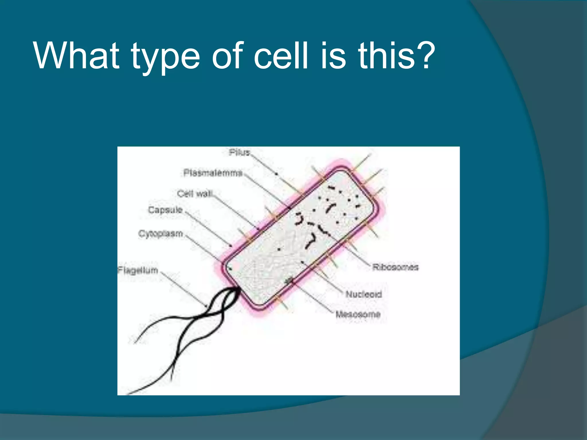
Cells consist of carbon, oxygen, nitrogen, hydrogen, with body cells in interstitial fluid. Types: Prokaryotic and Eukaryotic.
Discussion on generalized cell structures which share common features despite diversity.
Nucleus controls cell activities and contains DNA. Discusses nuclear envelope, nuclear pores, and chromatin.
Details about the plasma membrane, its semi-permeability, structure, and specialized membrane junctions.
Cytoplasm's role and major organelles like mitochondria and endoplasmic reticulum and their functions.
Additional organelles like Golgi apparatus, lysosomes, and peroxisomes, focusing on their specific roles.
Diversity of cells including fibroblasts, erythrocytes, neurons, and their specific roles in the body.
Membrane transport including passive and active transport, and vesicular transport methods.
Cell life cycle phases, mitosis stages, and cytokinesis processes with details on each phase.
Introduction to protein synthesis, involving DNA, mRNA, tRNA, and the transcription and translation processes.
Types of body tissues: Epithelial, connective, muscular, and nervous tissues and their functions.
Functions of epithelial tissues, types of epithelial cells categorized by layers and shapes.
Overview of connective tissues, their characteristics, types, and extracellular matrix.
Specific structures of bone and cartilage, their functions and types within connective tissues.
Muscle tissues types: Skeletal, cardiac, and smooth muscle with their roles and control mechanisms.Characteristics of nervous tissue, emphasizing neurons and their function in signal transmission.
Tissue repair processes: regeneration and fibrosis, detailing the steps involved in healing.
Key terms: Neoplasm, hyperplasia, atrophy, explaining their significance in tissue context.
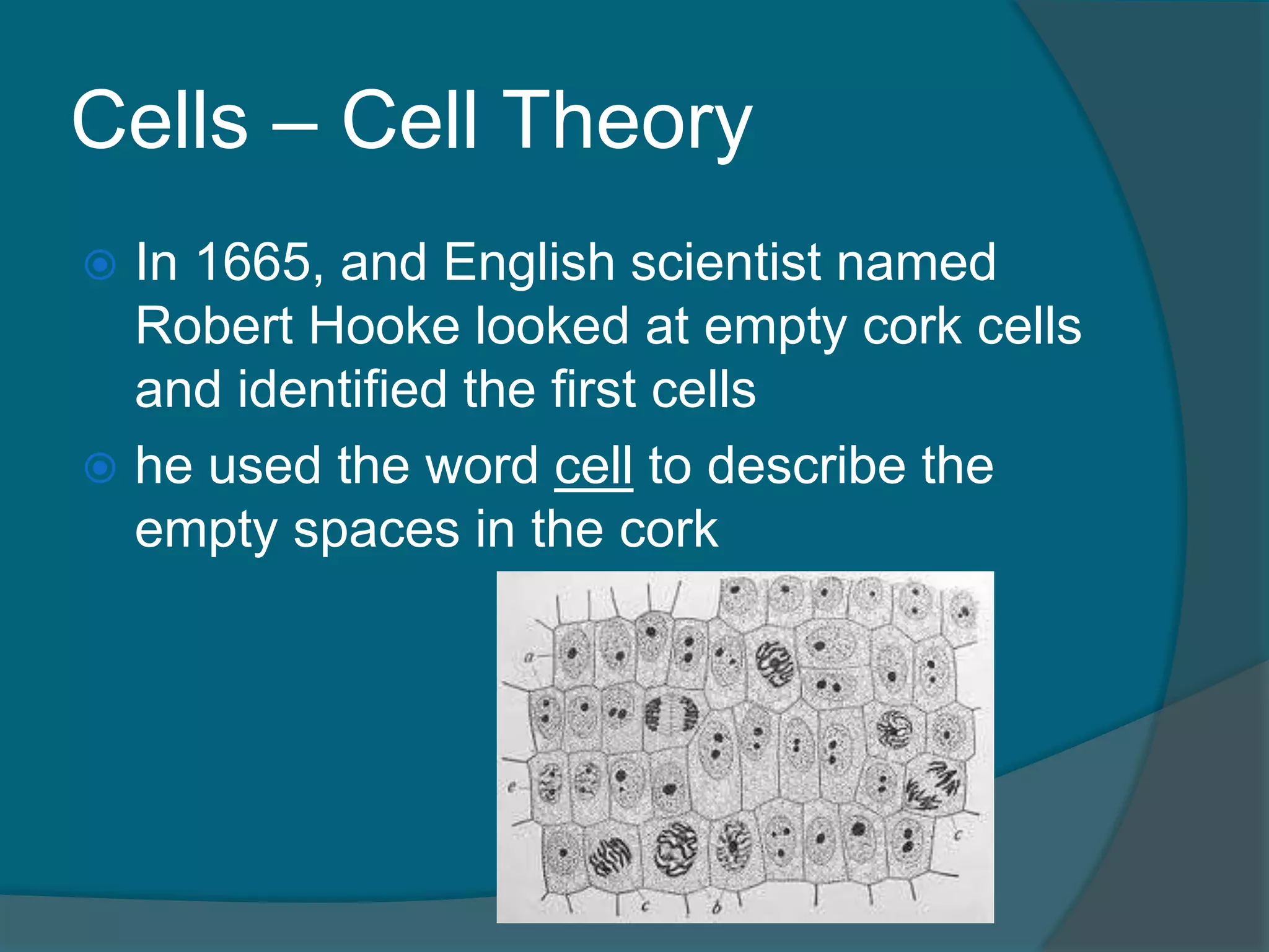
Cells – CellTheory
In 1665, and English scientist named
Robert Hooke looked at empty cork cells
and identified the first cells
he used the word cell to describe the
empty spaces in the cork
3.
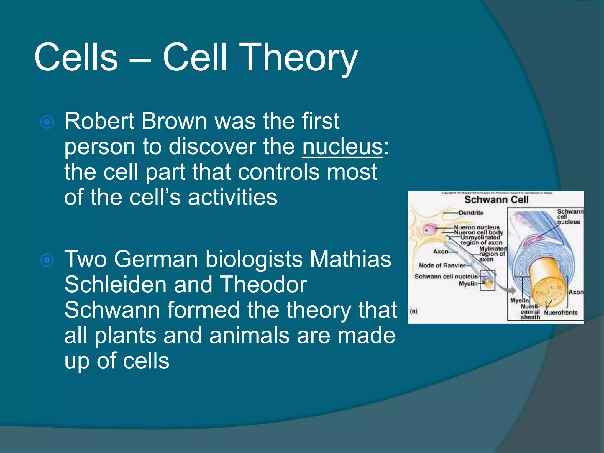
Cells – CellTheory
Robert Brown was the first
person to discover the nucleus:
the cell part that controls most
of the cell’s activities
Two German biologists Mathias
Schleiden and Theodor
Schwann formed the theory that
all plants and animals are made
up of cells
4.
Cells – CellTheory
All these ideas combined into the
modern Cell Theory:
1. All living things are made of one or
more cells
2. Cells are the basic units of structure
and function
3. All cells come from existing cells
5.
Cells – Thebasics
All cells are primarily made of four
elements: Carbon, Oxygen, Hydrogen,
Nitrogen
Living cells are about 60% water
6.
Cells – InterstitialFluid
In addition to large amounts of water,
the body cells are constantly covered in
a dilute saltwater solution called
interstitial fluid
This fluid is derived from blood
7.
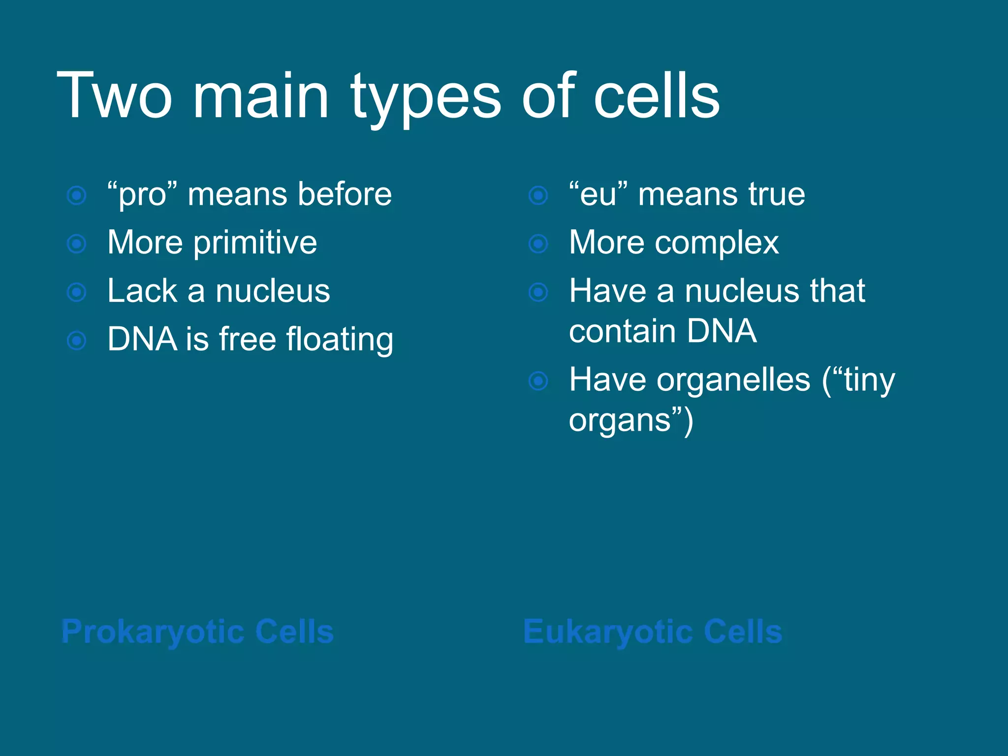
Two main typesof cells
Prokaryotic Cells Eukaryotic Cells
“pro” means before
More primitive
Lack a nucleus
DNA is free floating
“eu” means true
More complex
Have a nucleus that
contain DNA
Have organelles (“tiny
organs”)
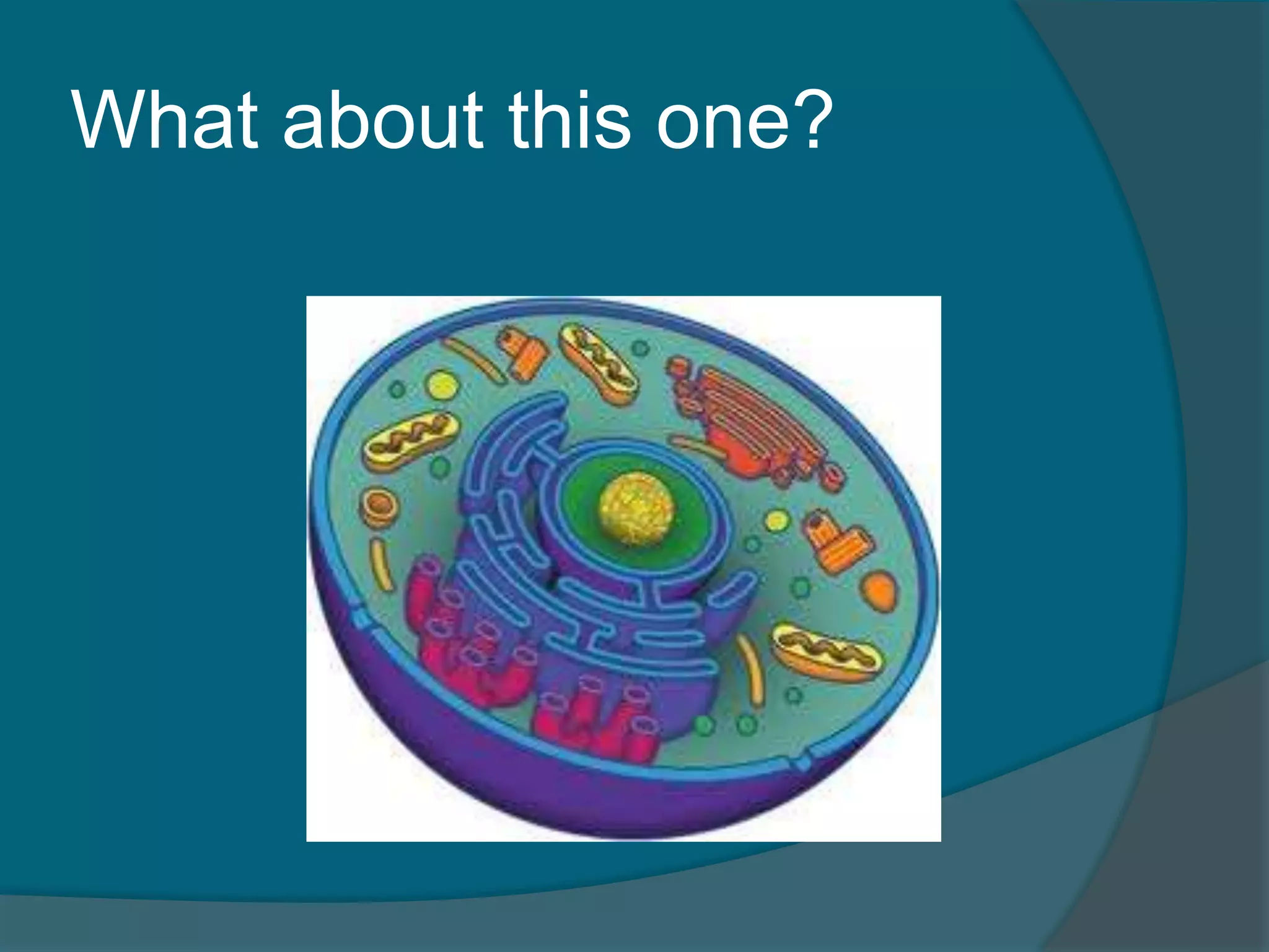
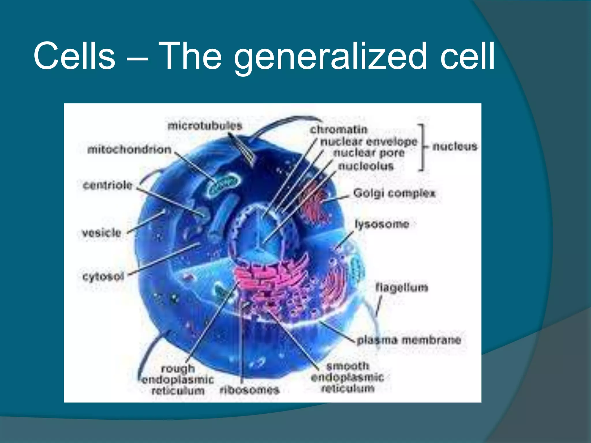
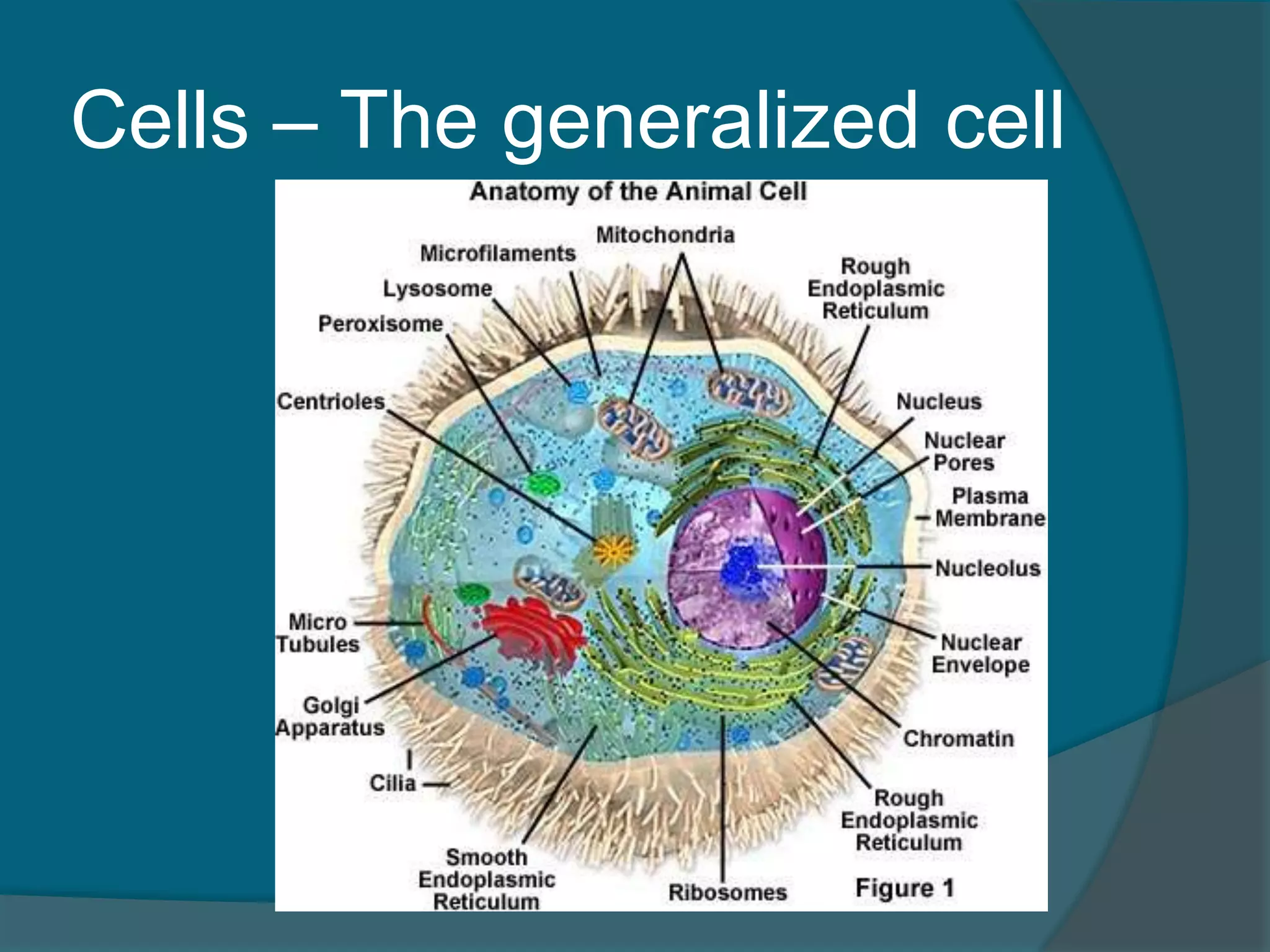
Cells – Thegeneralized cell
No one cell type is exactly like another
Most do have the same parts
Let’s talk about a generalized cell: a
basic cell used to demonstrate most cell
features
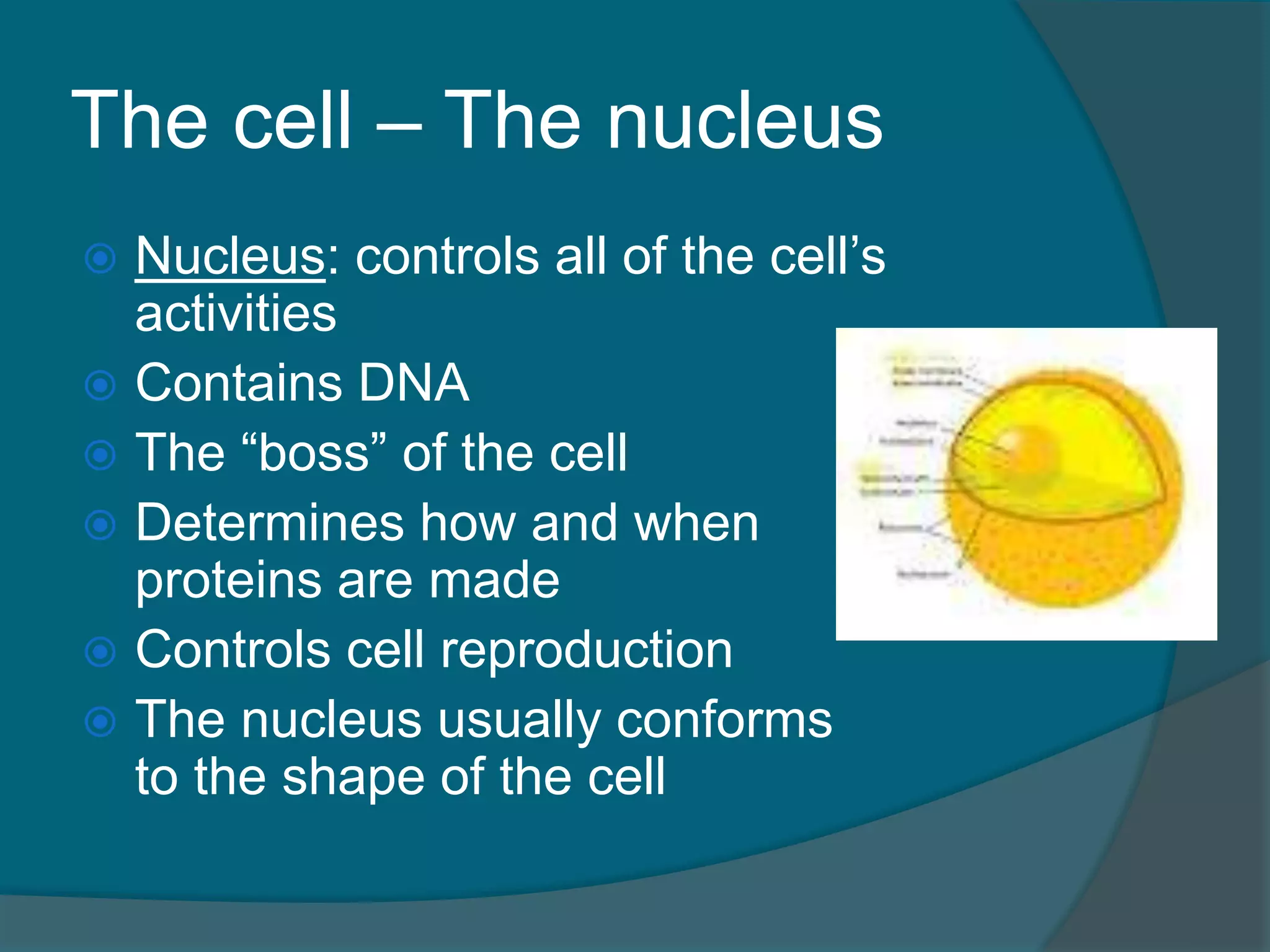
The cell –The nucleus
Nucleus: controls all of the cell’s
activities
Contains DNA
The “boss” of the cell
Determines how and when
proteins are made
Controls cell reproduction
The nucleus usually conforms
to the shape of the cell
14.
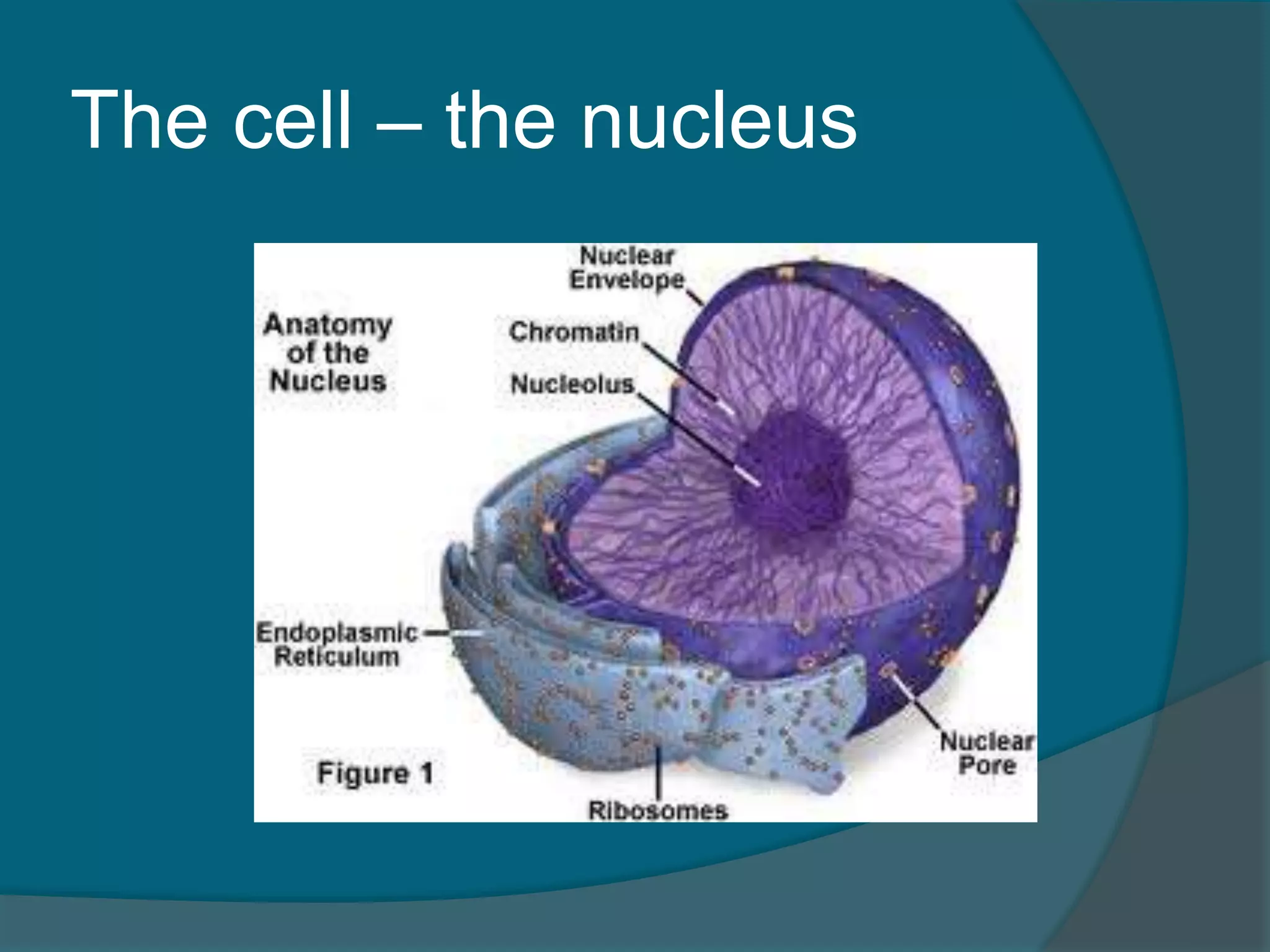
The cell –the nucleus
Is enclosed by a nuclear membrane (or
nuclear envelope)
Nuclear membrane: structure that
surrounds the nucleus and separates it
from the rest of the cell
Nuclear pores: openings in the nuclear
membrane that allows molecules to pass
Nucleoplasm: the jelly-like fluid between
the two layers of the nuclear membrane
15.
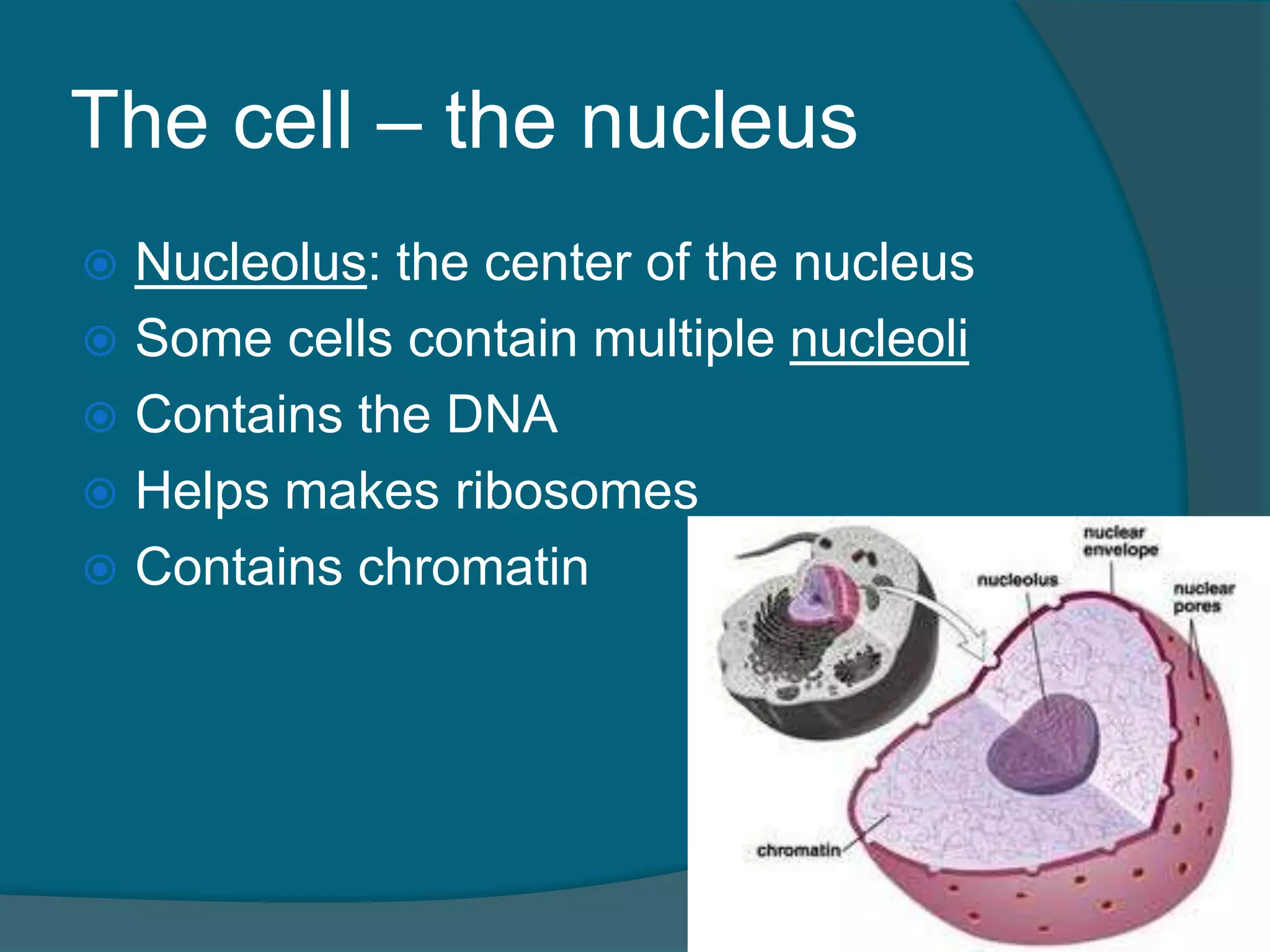
The cell –the nucleus
Nucleolus: the center of the nucleus
Some cells contain multiple nucleoli
Contains the DNA
Helps makes ribosomes
Contains chromatin
16.
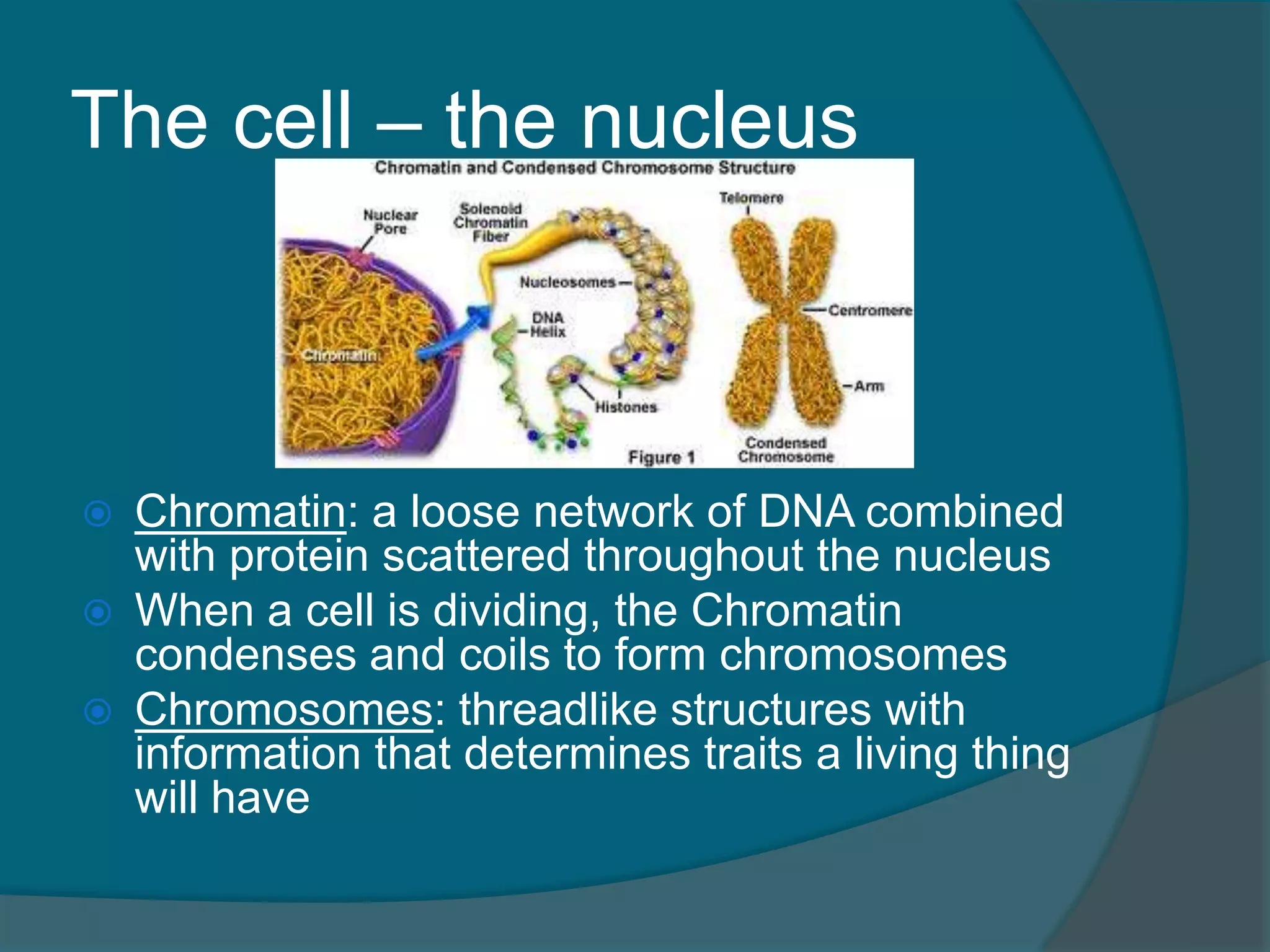
The cell –the nucleus
Chromatin: a loose network of DNA combined
with protein scattered throughout the nucleus
When a cell is dividing, the Chromatin
condenses and coils to form chromosomes
Chromosomes: threadlike structures with
information that determines traits a living thing
will have
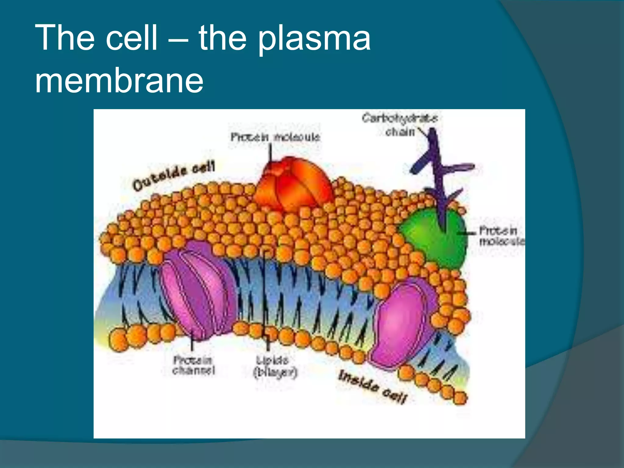
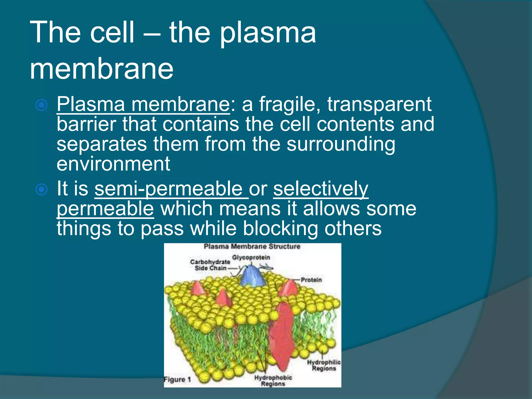
The cell –the plasma
membrane
Plasma membrane: a fragile, transparent
barrier that contains the cell contents and
separates them from the surrounding
environment
It is semi-permeable or selectively
permeable which means it allows some
things to pass while blocking others
20.
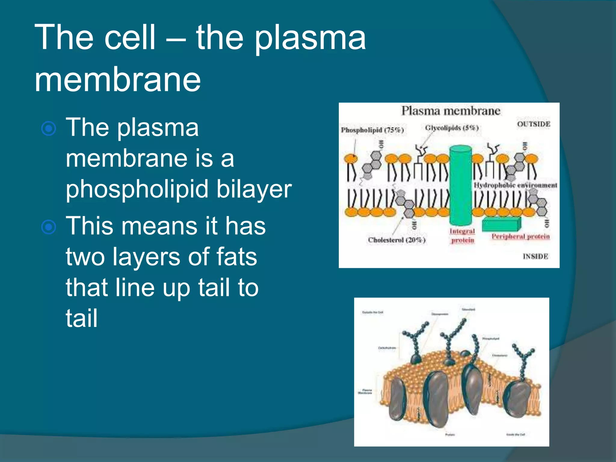
The cell –the plasma
membrane
The plasma
membrane is a
phospholipid bilayer
This means it has
two layers of fats
that line up tail to
tail
21.
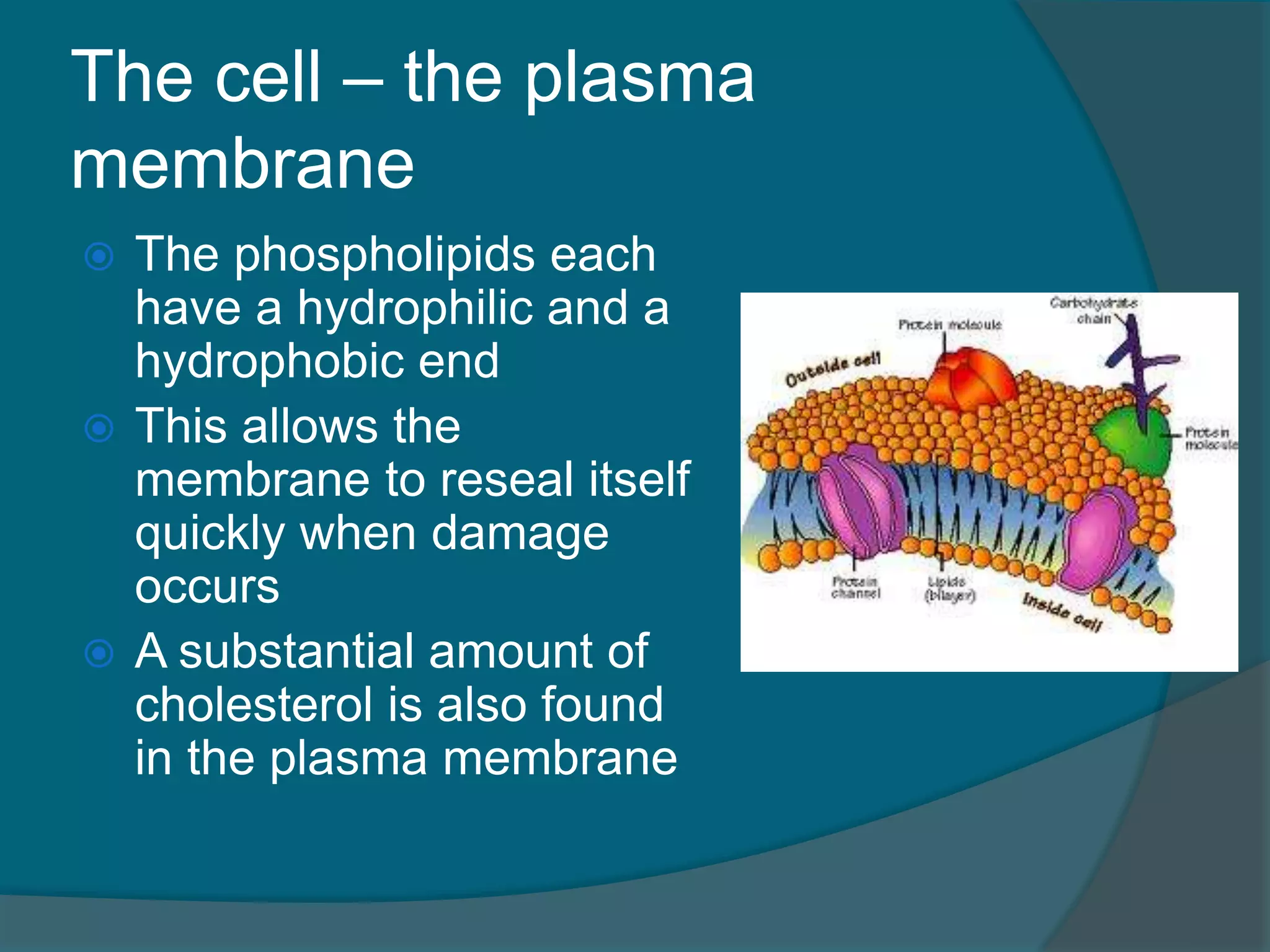
The cell –the plasma
membrane
The phospholipids each
have a hydrophilic and a
hydrophobic end
This allows the
membrane to reseal itself
quickly when damage
occurs
A substantial amount of
cholesterol is also found
in the plasma membrane
22.
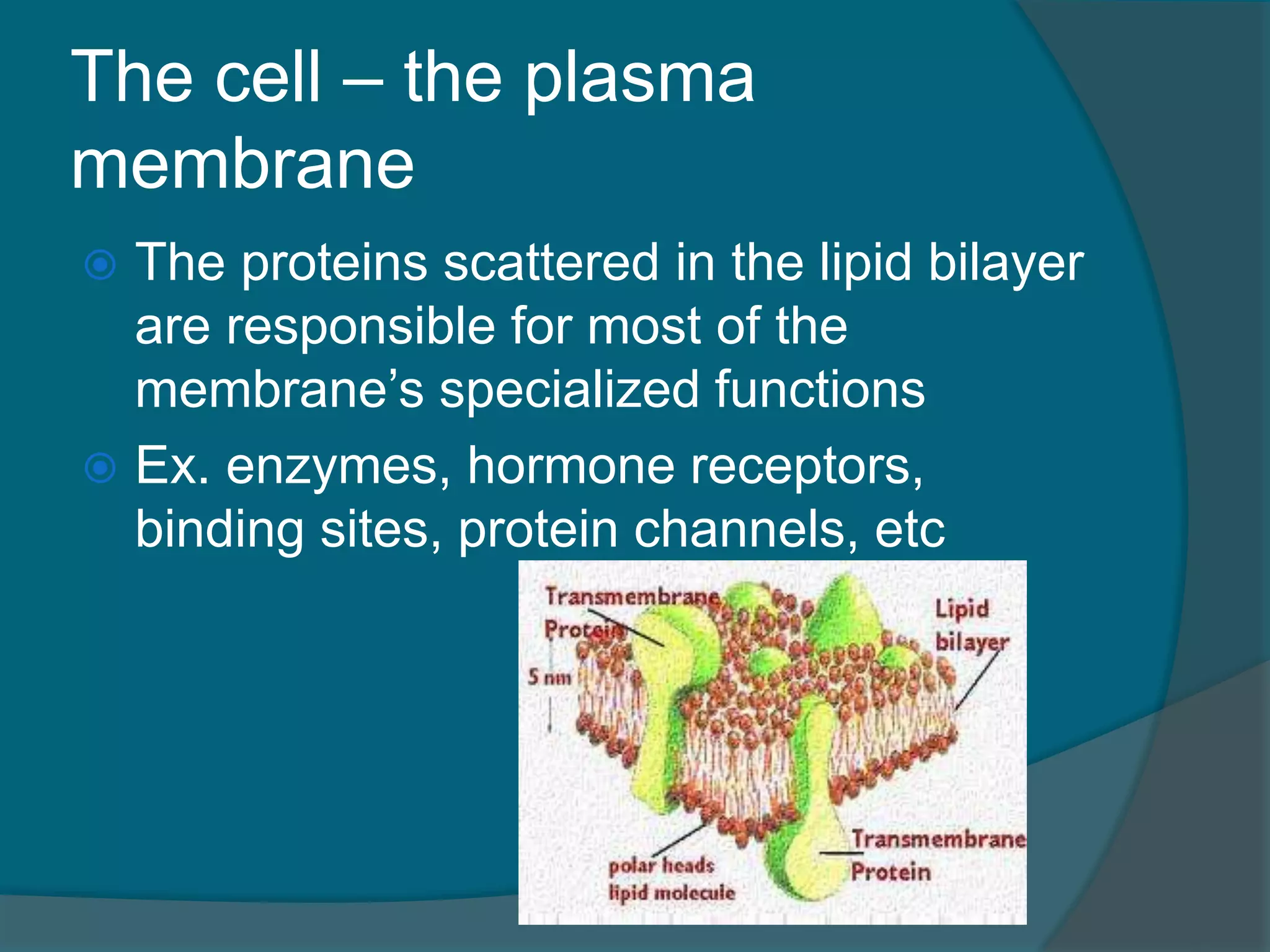
The cell –the plasma
membrane
The proteins scattered in the lipid bilayer
are responsible for most of the
membrane’s specialized functions
Ex. enzymes, hormone receptors,
binding sites, protein channels, etc
23.
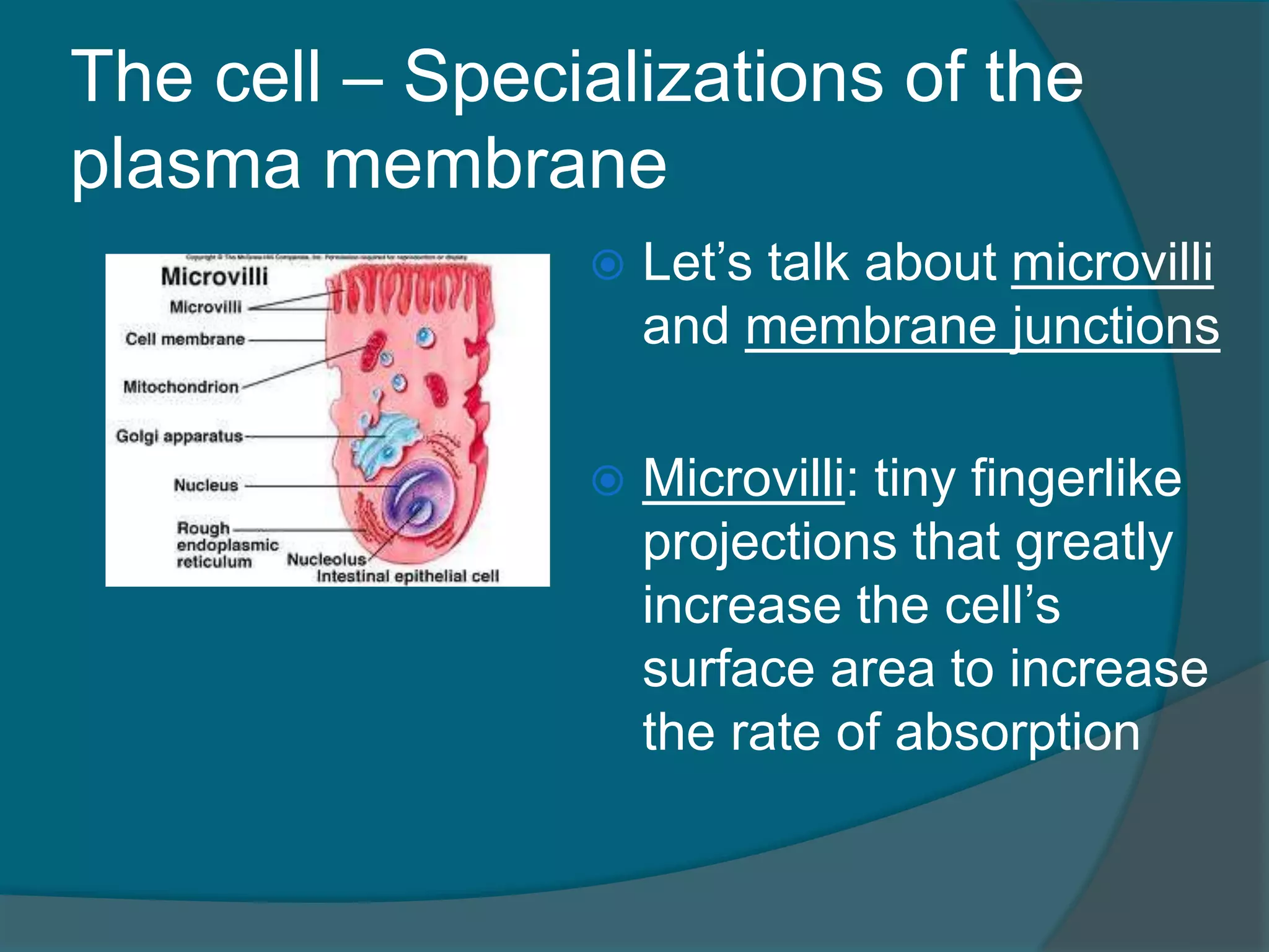
The cell –Specializations of the
plasma membrane
Let’s talk about microvilli
and membrane junctions
Microvilli: tiny fingerlike
projections that greatly
increase the cell’s
surface area to increase
the rate of absorption
24.
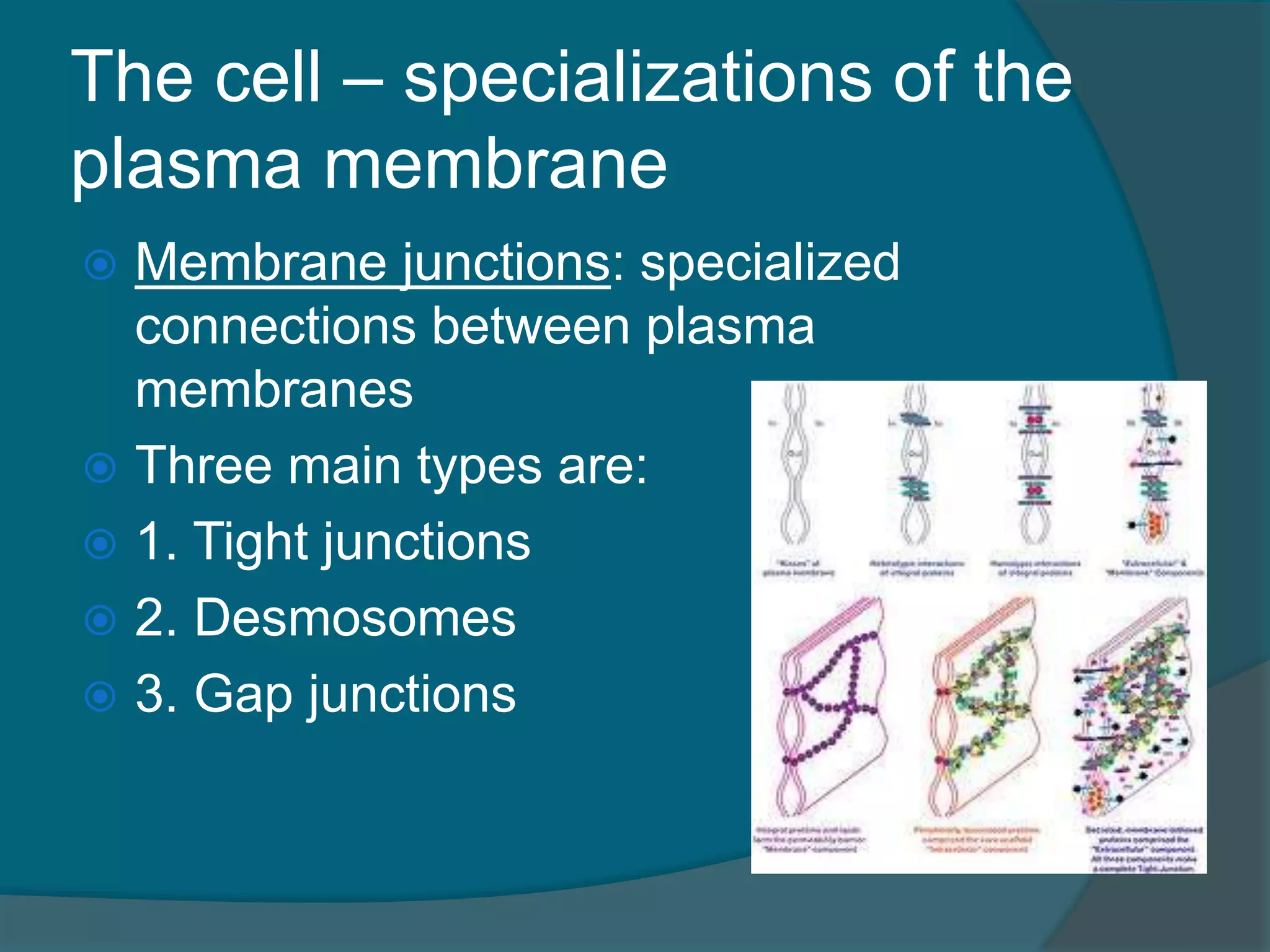
The cell –specializations of the
plasma membrane
Membrane junctions: specialized
connections between plasma
membranes
Three main types are:
1. Tight junctions
2. Desmosomes
3. Gap junctions
25.
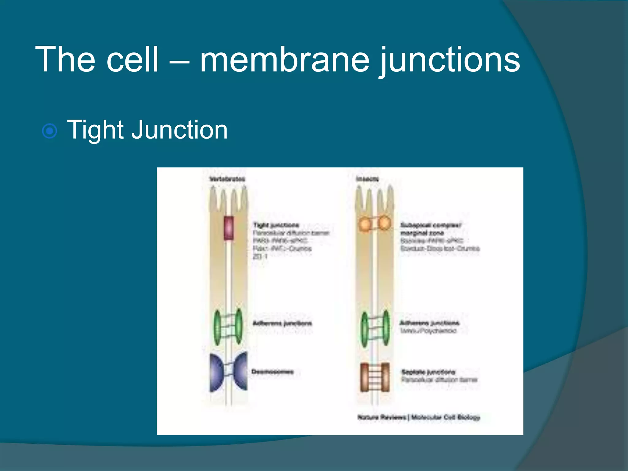
The cell –membrane junctions
1. Tight junctions: impermeable
junctions that bind cells together into
leakproof sheets that prevent
substances from passing through the
extracellular space between cells
Plasma membranes fuse together like a
zipper
Ex. in the small intestine, these junctions
prevent digestive enzymes from seeping
into the bloodstream
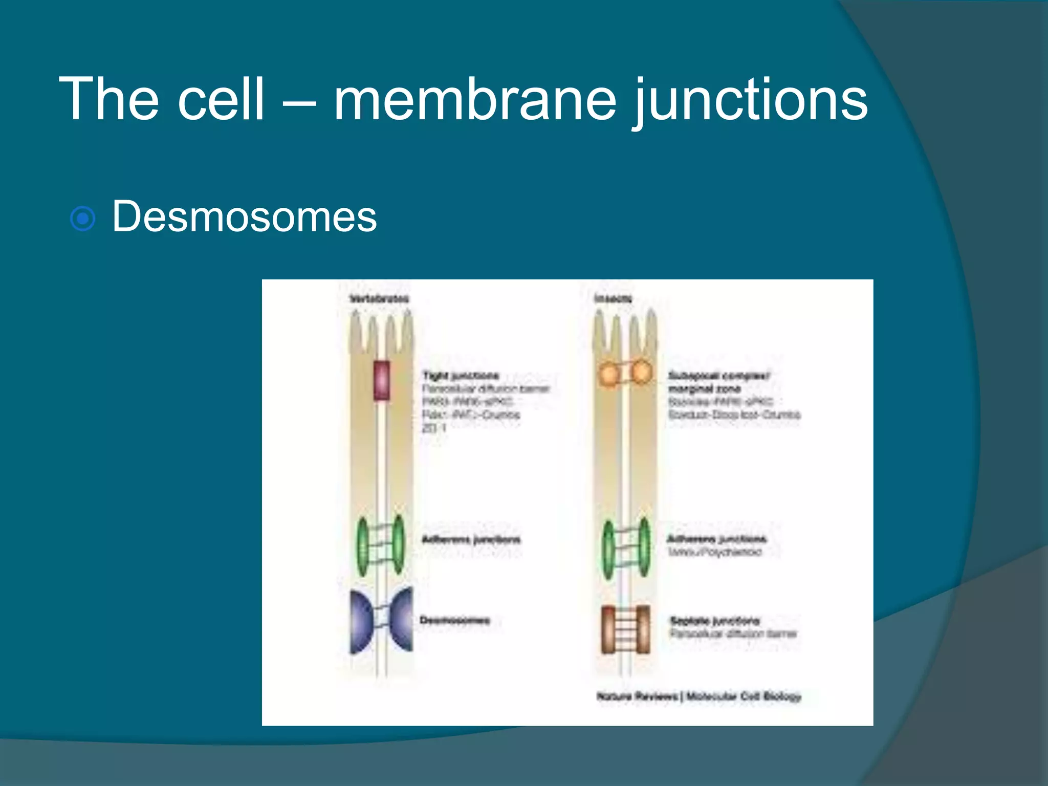
The cell –membrane junctions
2. Desmosomes: anchoring junctions that
prevent cells subjected to mechanical
stress from being pulled apart
Structurally these junctions are buttonlike
thickenings of adjacent plasma membranes
(plaques), connected by fine protein
filaments
Thicker protein filaments extend from the
plaques inside the cells to the plaques on
the cells’ opposite side, forming an internal
system of strong wires
Ex. skin cells
The cell –membrane junctions
3. Gap junctions: common to heart cells
and embryonic cells, these junctions
function mainly to allow communication
Chemical molecules (nutrients, ions, etc)
pass directly from one cell to another
through the gap
In gap junctions, the neighboring cells are
connected by connexons: hollow cylinders
composed of proteins that span the entire
width of the adjoining membranes
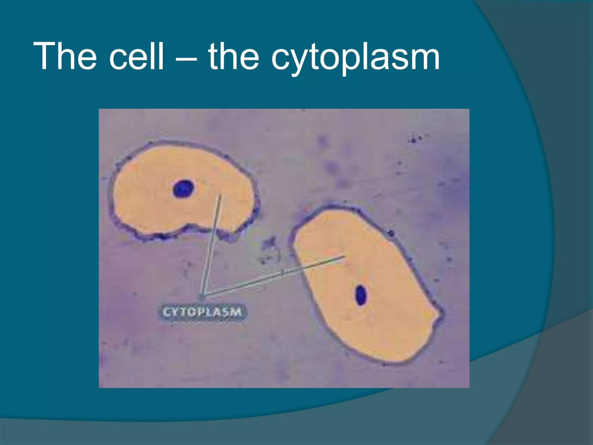
The cell –the cytoplasm
Cytoplasm: the cellular material outside
the nucleus and inside the plasma
membrane
It is where most chemical reactions
occur inside the cell
Made of three major elements:
1. the cytosol
2. the organelles
3. inclusions
32.
The cell –the cytoplasm
The cytosol is the semitransparent fluid
that suspends the other elements
The organelles or “tiny organs” are the
machinery of the cell
Inclusions are chemical substances that
may or may not be present, depend on
the cell type
Include stored nutrient, lipids, glycogen,
mucus, various crystallized products, etc
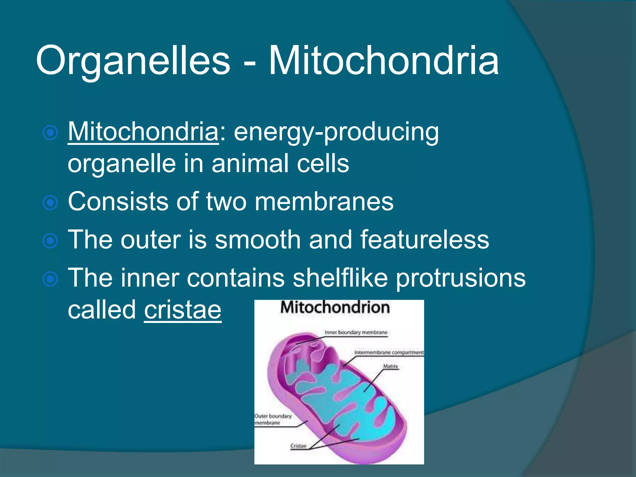
Organelles - Mitochondria
Mitochondria: energy-producing
organelle in animal cells
Consists of two membranes
The outer is smooth and featureless
The inner contains shelflike protrusions
called cristae
35.
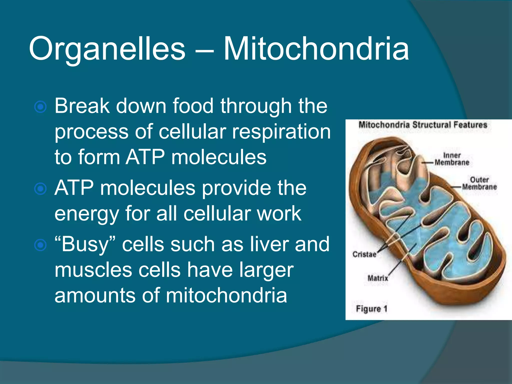
Organelles – Mitochondria
Break down food through the
process of cellular respiration
to form ATP molecules
ATP molecules provide the
energy for all cellular work
“Busy” cells such as liver and
muscles cells have larger
amounts of mitochondria
36.
Organelles - Ribosomes
Ribosomes: tiny, bilobed, dark bodies
made of proteins and RNA
Site of protein synthesis in the cell
Two types:
Free – free floating in the cell
Bound/Attached – attached to the
Endoplasmic Reticulum
37.
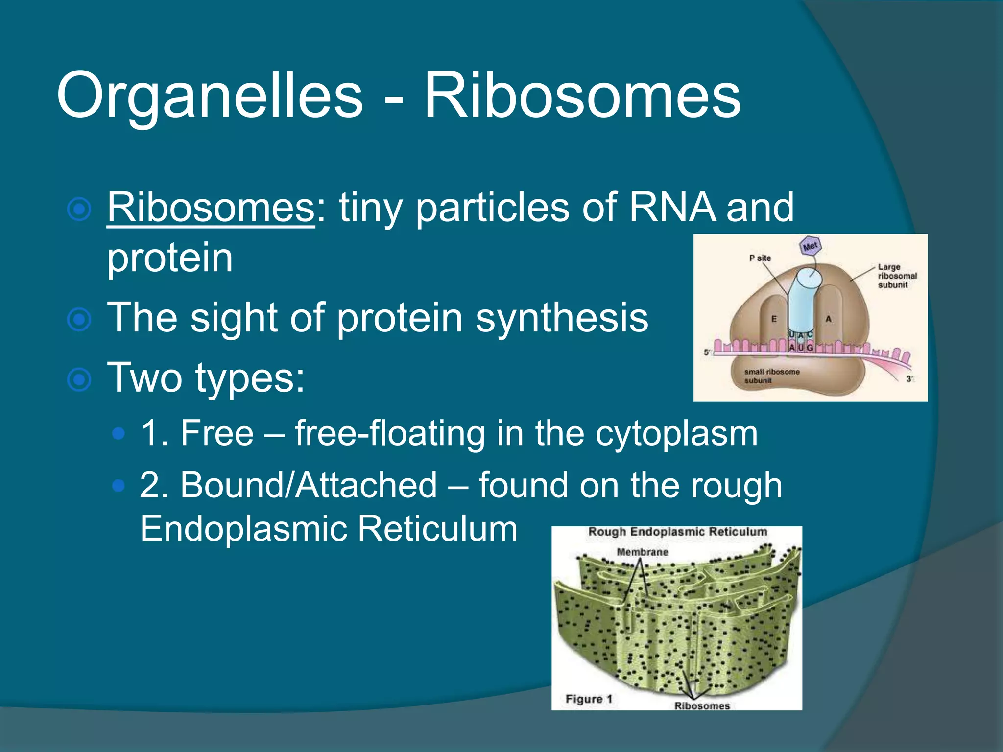
Organelles - Ribosomes
Ribosomes: tiny particles of RNA and
protein
The sight of protein synthesis
Two types:
1. Free – free-floating in the cytoplasm
2. Bound/Attached – found on the rough
Endoplasmic Reticulum
38.
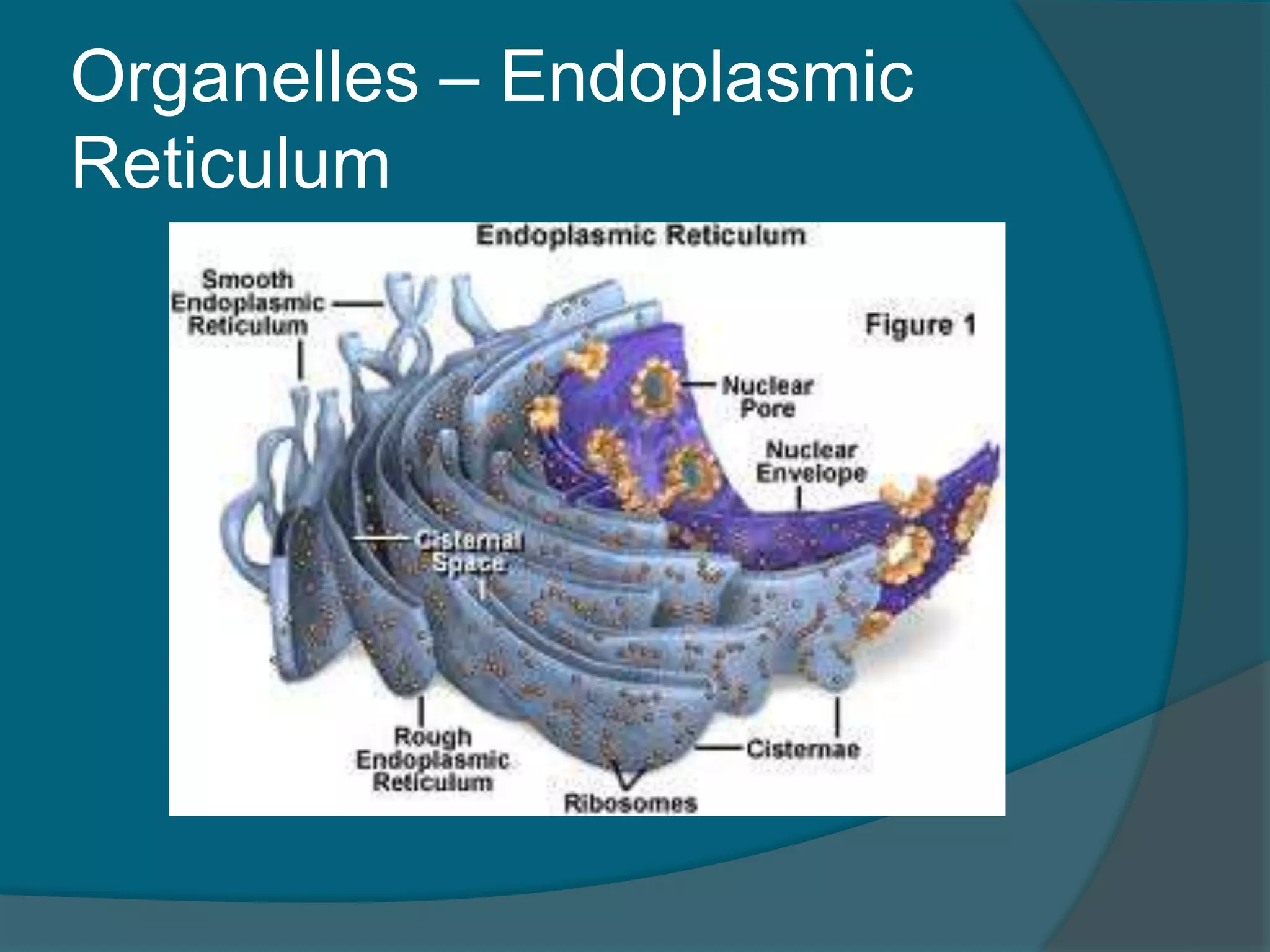
Organelles – Endoplasmic
Reticulum
Endoplasmic Reticulum: a system of
fluid-filled sacs and membranes located
near the nucleus that packages and
exports protein, lipids and other small
molecules.
Accounts for about half of a cell’s
membrane
39.
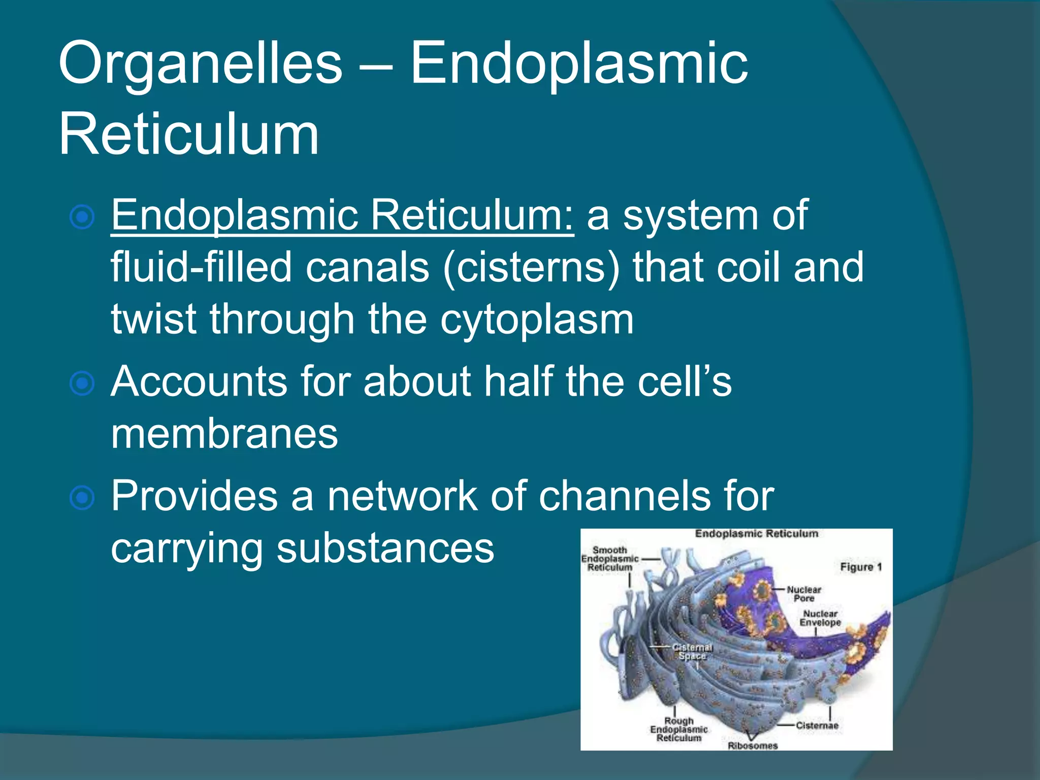
Organelles – Endoplasmic
Reticulum
Endoplasmic Reticulum: a system of
fluid-filled canals (cisterns) that coil and
twist through the cytoplasm
Accounts for about half the cell’s
membranes
Provides a network of channels for
carrying substances
40.
Organelles – Endoplasmic
Reticulum
Two forms of the ER:
1. Rough ER: studded with ribosomes
All of the building materials of cellular
membranes are formed either in or on it:
Proteins are packaged and sent out in
transport vesicles
Greater number in organs that require more
proteins,
○ Ex. pancreas
41.
Organelles – Endoplasmic
Reticulum
2. Smooth ER: plays no role in protein
synthesis
Functions in lipid metabolism and
detoxification
Therefore there are many smooth ER in
liver cells
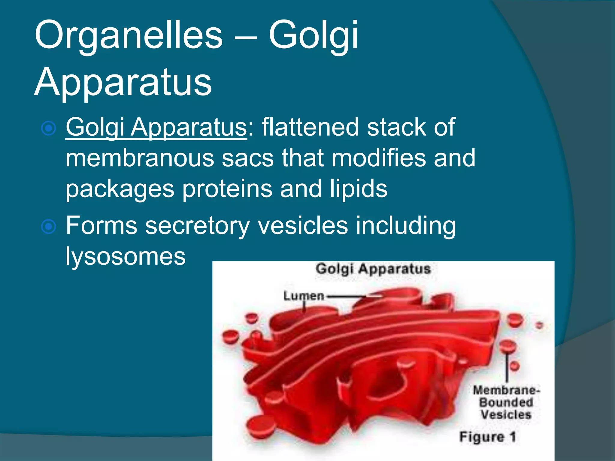
Organelles – Golgi
Apparatus
Golgi Apparatus: flattened stack of
membranous sacs that modifies and
packages proteins and lipids
Forms secretory vesicles including
lysosomes
44.
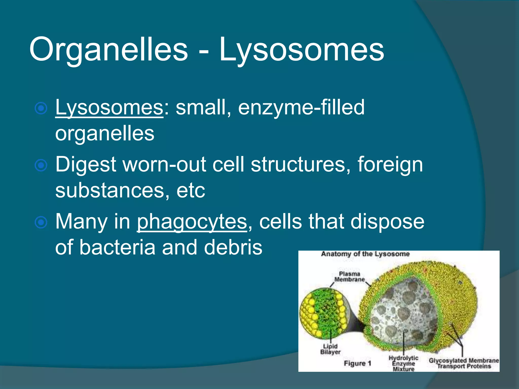
Organelles - Lysosomes
Lysosomes: small, enzyme-filled
organelles
Digest worn-out cell structures, foreign
substances, etc
Many in phagocytes, cells that dispose
of bacteria and debris
45.
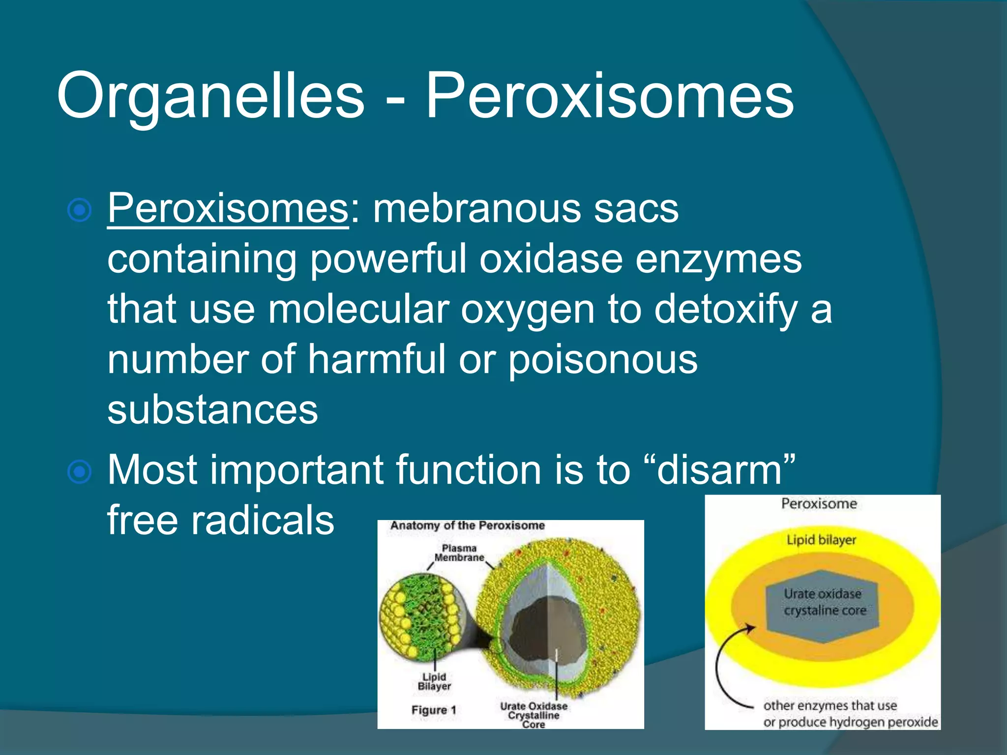
Organelles - Peroxisomes
Peroxisomes: mebranous sacs
containing powerful oxidase enzymes
that use molecular oxygen to detoxify a
number of harmful or poisonous
substances
Most important function is to “disarm”
free radicals
46.
Organelles - Peroxisomes
Free radicals: highly reactive chemicals
with unpaired electrons that can
scramble the structure of proteins and
nucleic acids
Free radicals are usually produced by
cellular respiration but if they
accumulate they have devastating
effects on the cell
47.
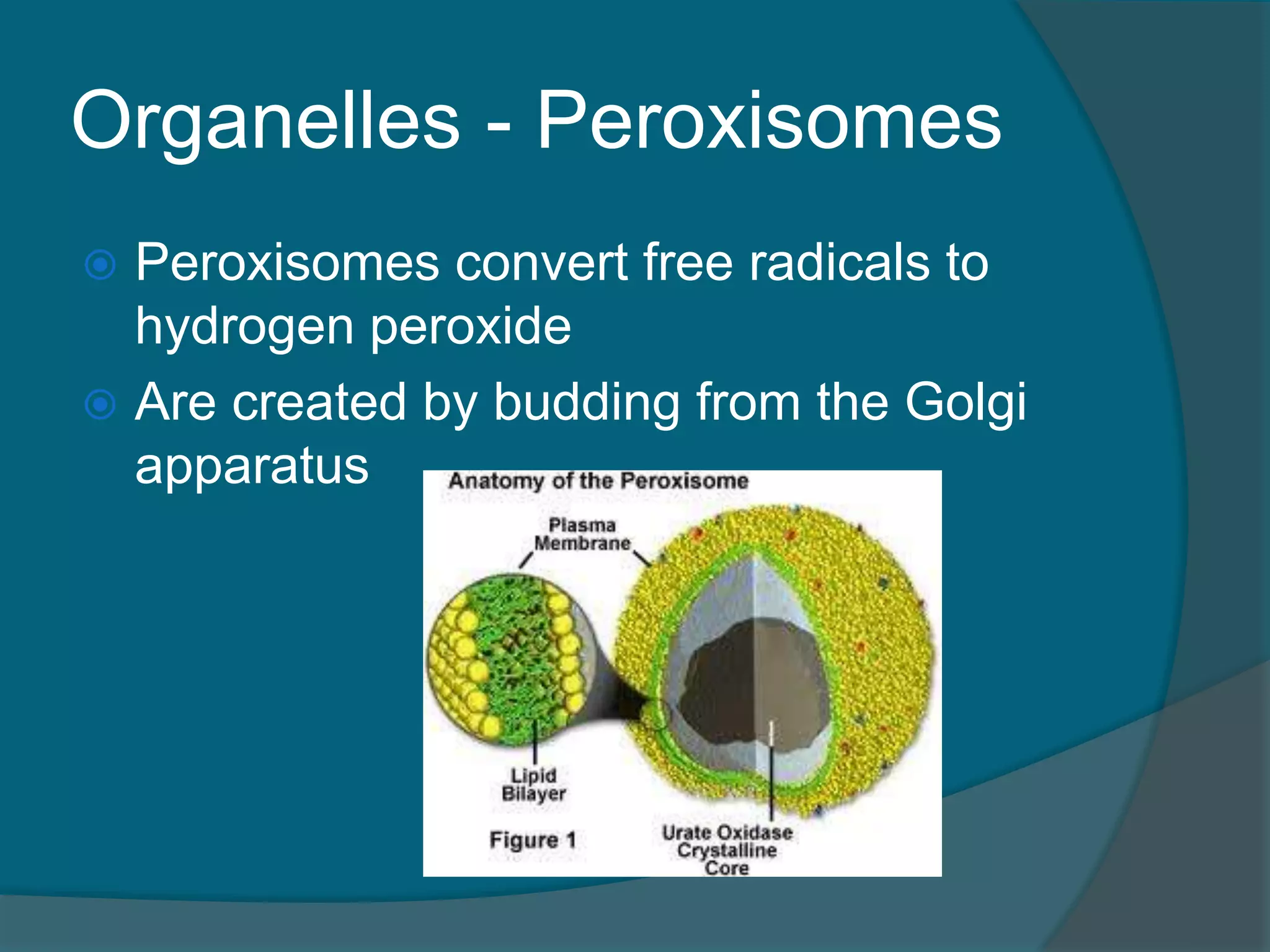
Organelles - Peroxisomes
Peroxisomes convert free radicals to
hydrogen peroxide
Are created by budding from the Golgi
apparatus
48.
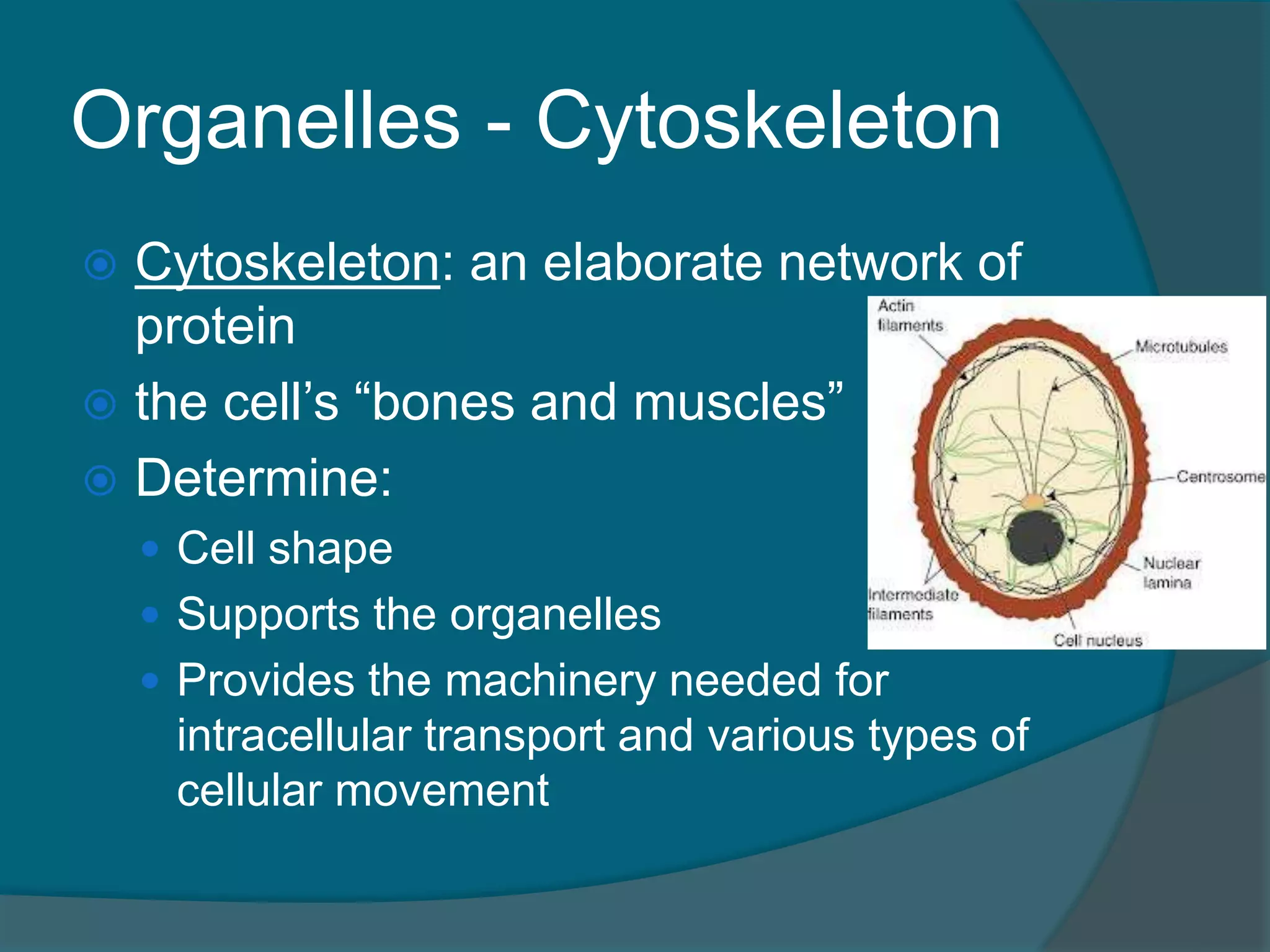
Organelles - Cytoskeleton
Cytoskeleton: an elaborate network of
protein
the cell’s “bones and muscles”
Determine:
Cell shape
Supports the organelles
Provides the machinery needed for
intracellular transport and various types of
cellular movement
49.
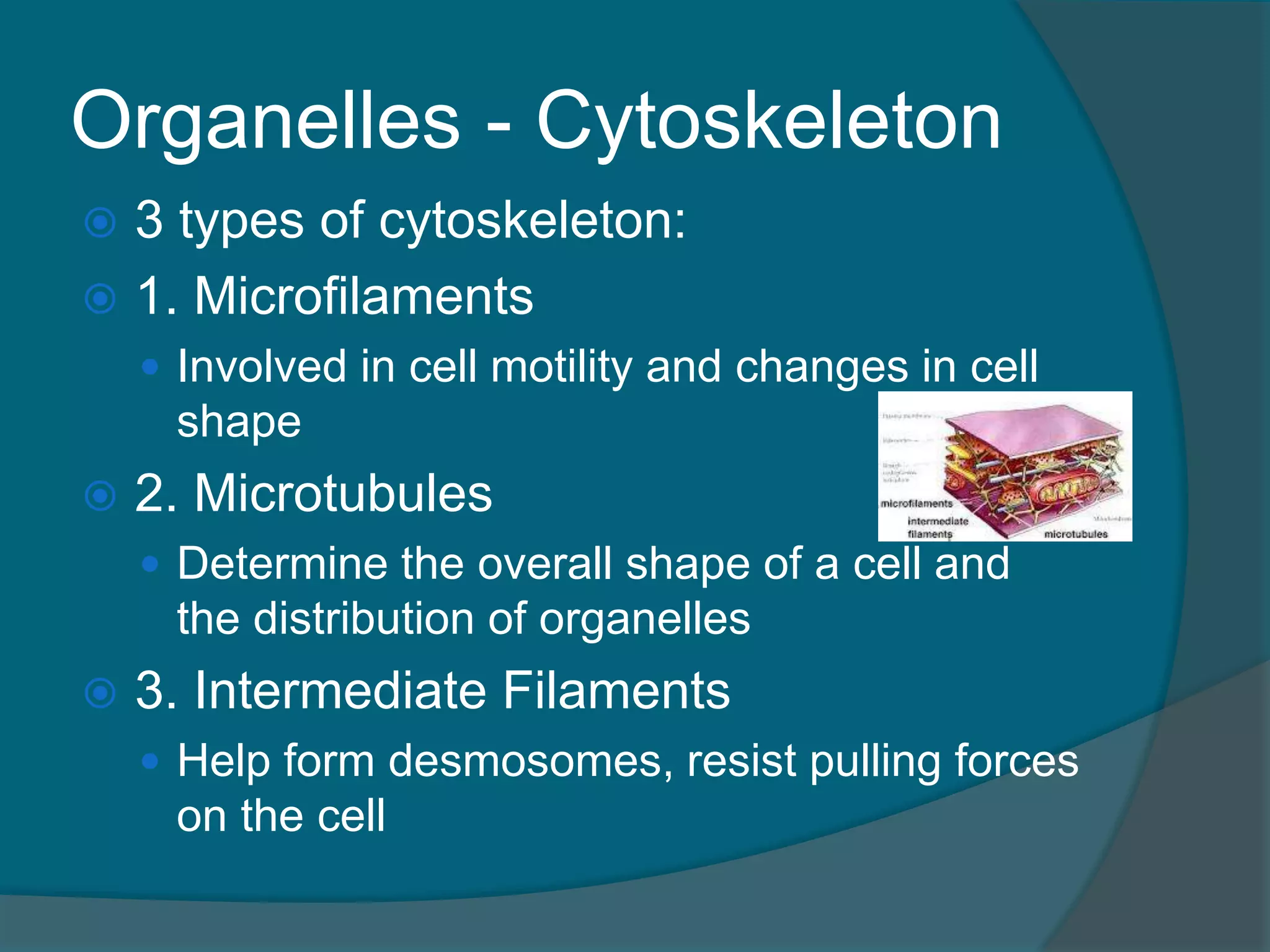
Organelles - Cytoskeleton
3 types of cytoskeleton:
1. Microfilaments
Involved in cell motility and changes in cell
shape
2. Microtubules
Determine the overall shape of a cell and
the distribution of organelles
3. Intermediate Filaments
Help form desmosomes, resist pulling forces
on the cell
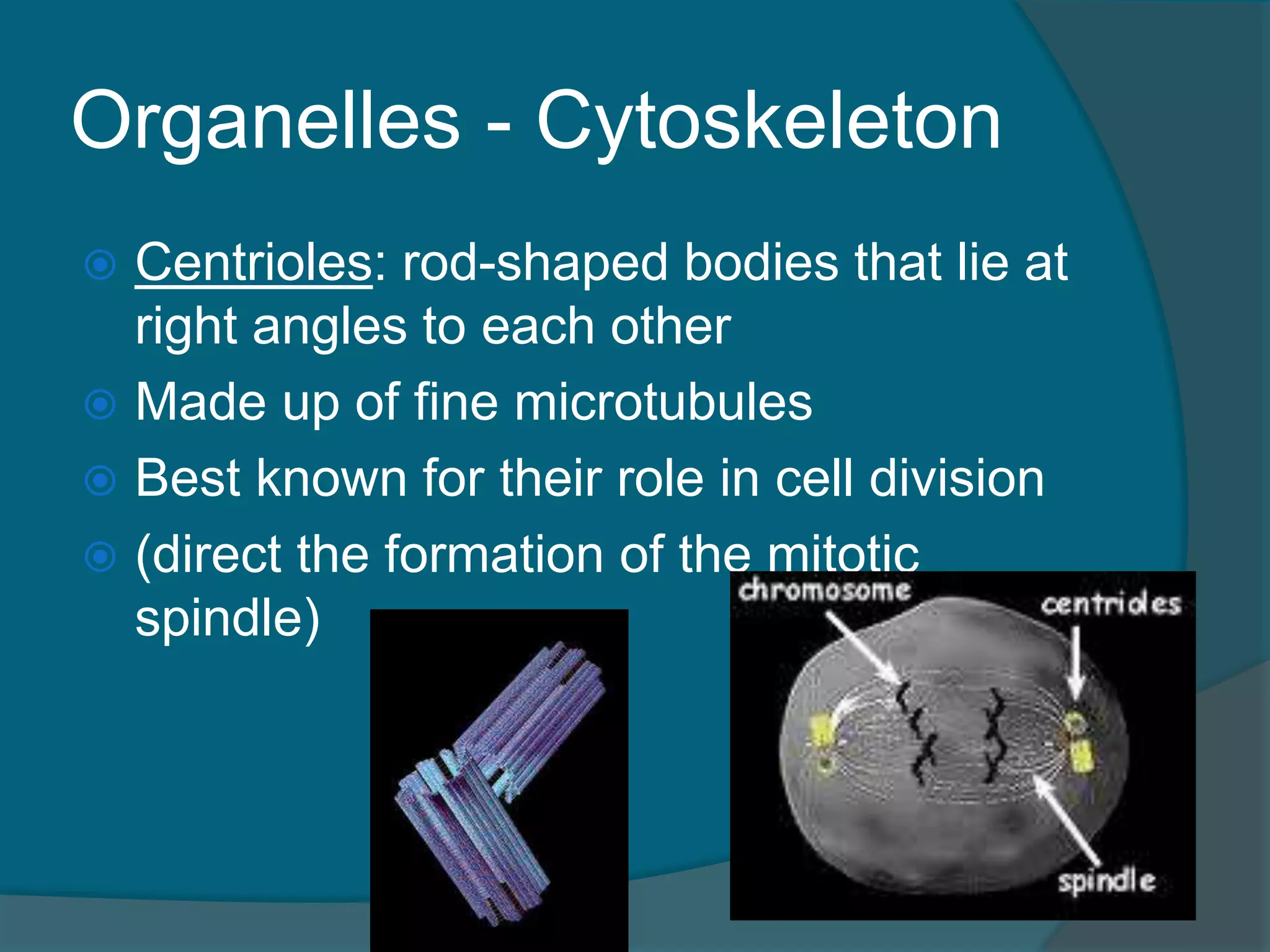
Organelles - Cytoskeleton
Centrioles: rod-shaped bodies that lie at
right angles to each other
Made up of fine microtubules
Best known for their role in cell division
(direct the formation of the mitotic
spindle)
52.
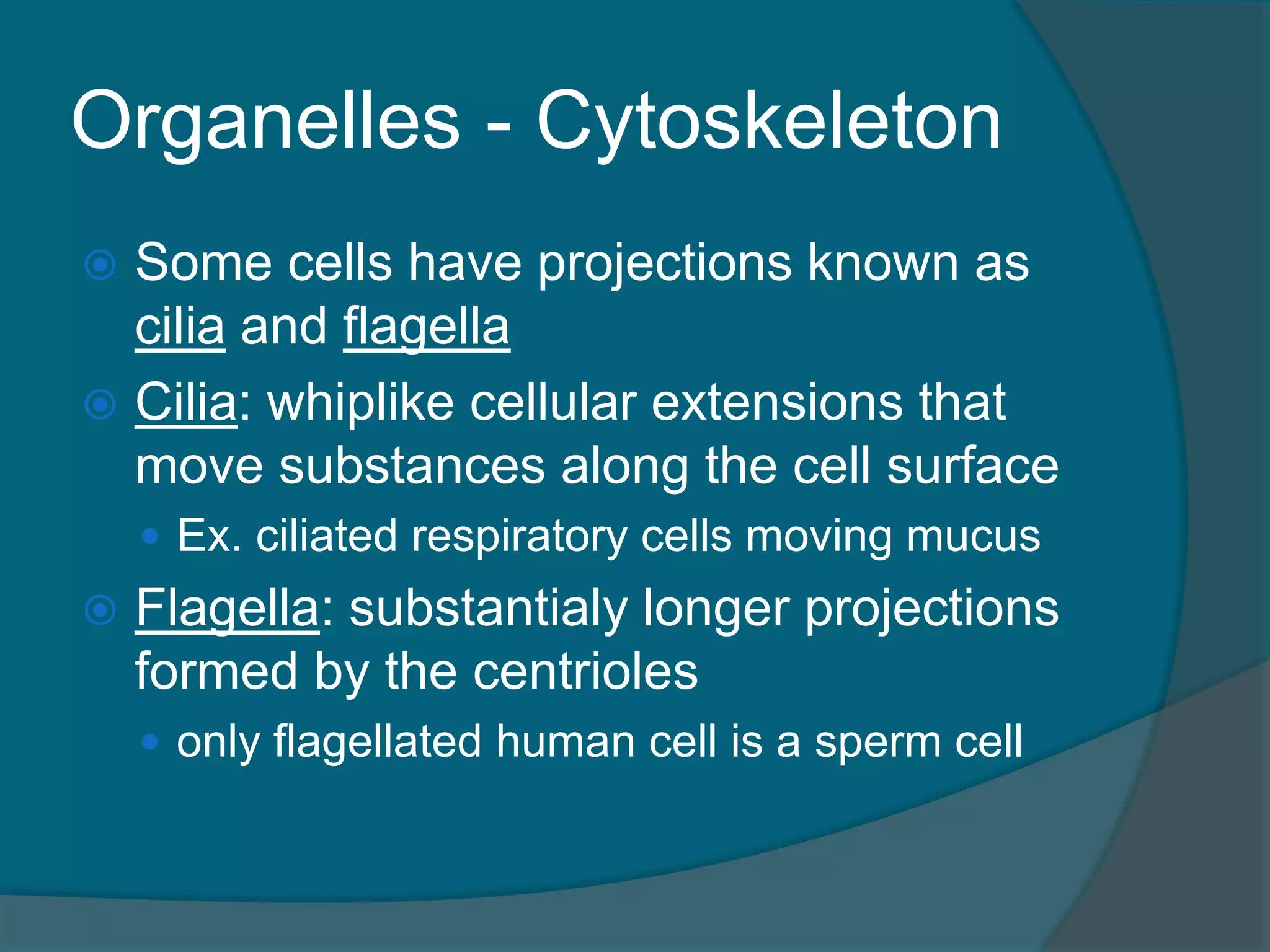
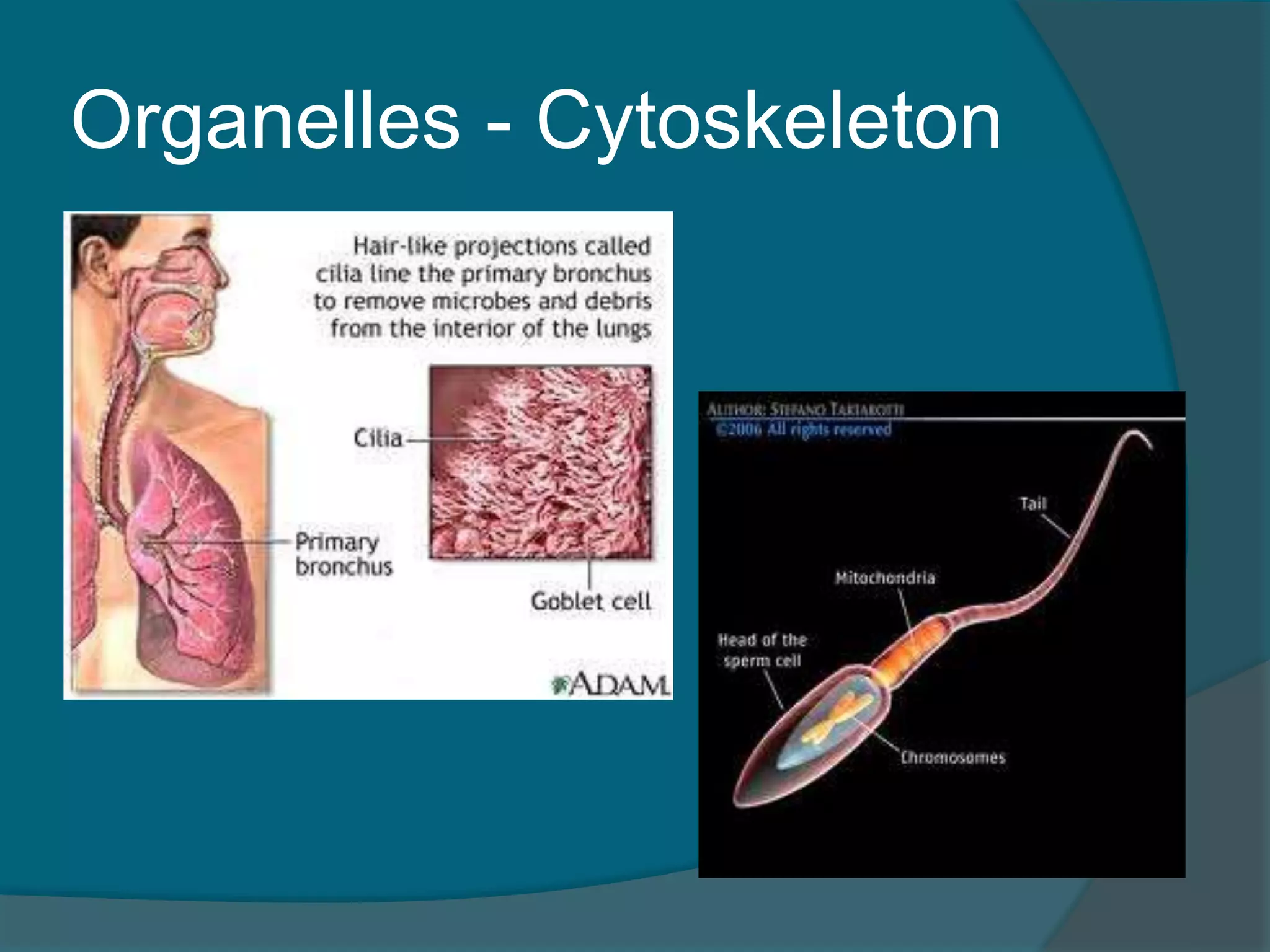
Organelles - Cytoskeleton
Some cells have projections known as
cilia and flagella
Cilia: whiplike cellular extensions that
move substances along the cell surface
Ex. ciliated respiratory cells moving mucus
Flagella: substantialy longer projections
formed by the centrioles
only flagellated human cell is a sperm cell
Cell Diversity
1.Cells that connect body parts
2. Cell that covers and lines body organs
3. Cells that move organs and body parts
4. Cells that stores nutrients
5. Cells that fight disease
6. Cells that gather information and
controls body functions
7. Cells of reproduction
56.
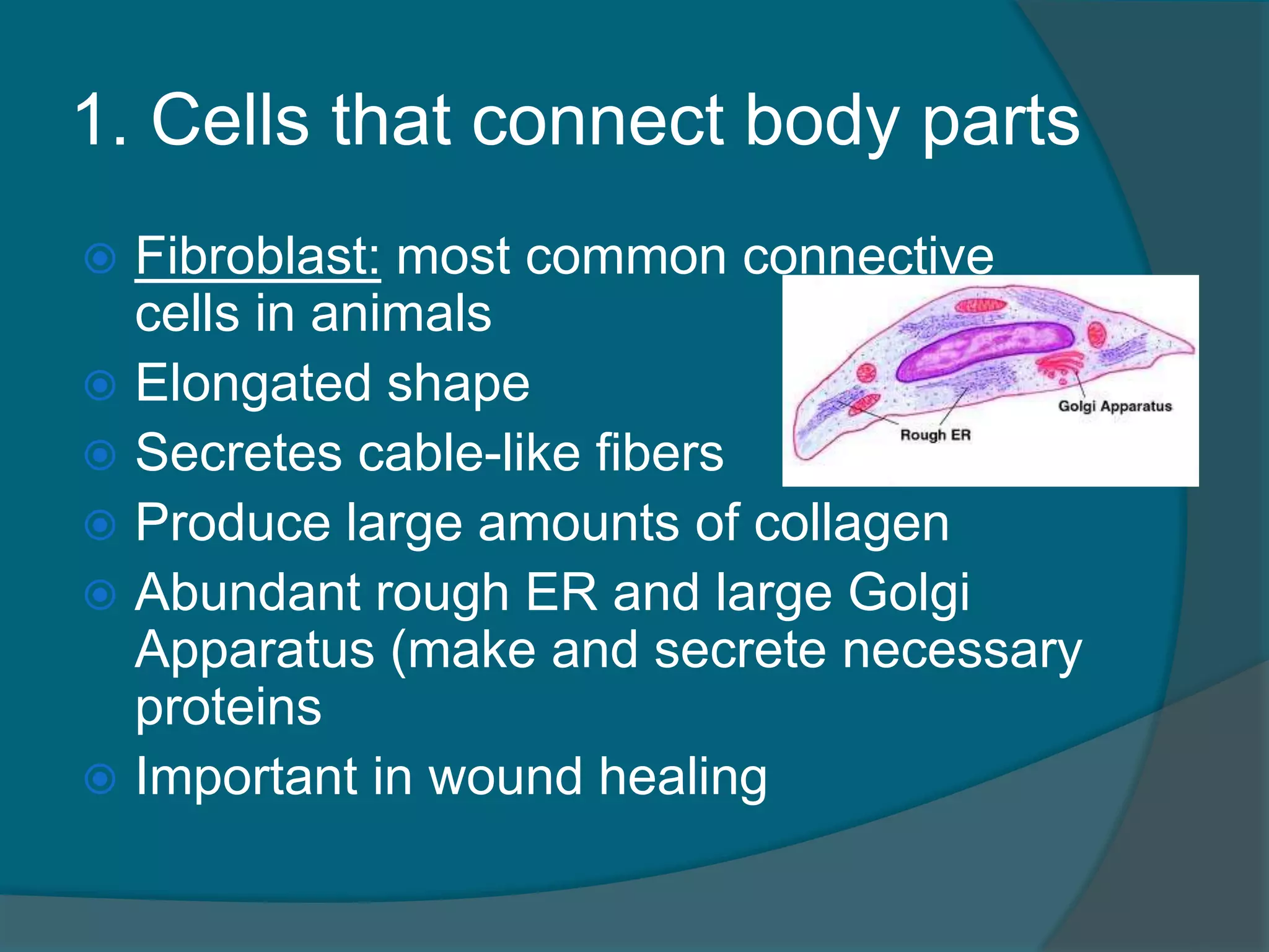
1. Cells thatconnect body parts
Fibroblast: most common connective
cells in animals
Elongated shape
Secretes cable-like fibers
Produce large amounts of collagen
Abundant rough ER and large Golgi
Apparatus (make and secrete necessary
proteins
Important in wound healing
57.
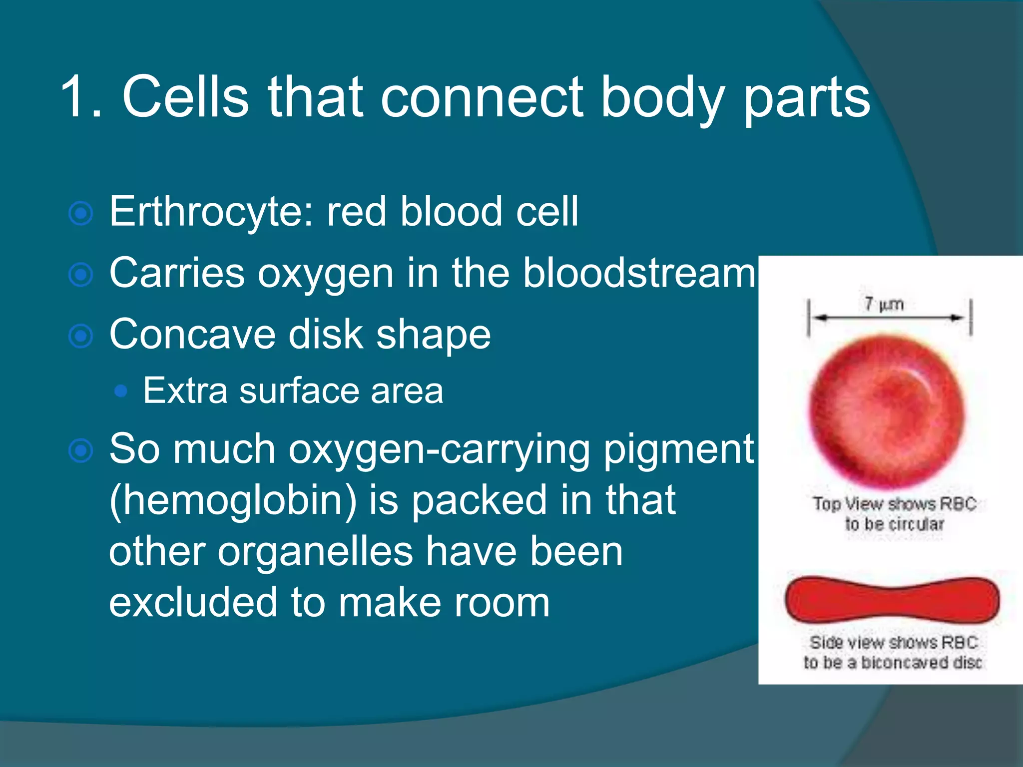
1. Cells thatconnect body parts
Erthrocyte: red blood cell
Carries oxygen in the bloodstream
Concave disk shape
Extra surface area
So much oxygen-carrying pigment
(hemoglobin) is packed in that
other organelles have been
excluded to make room
58.
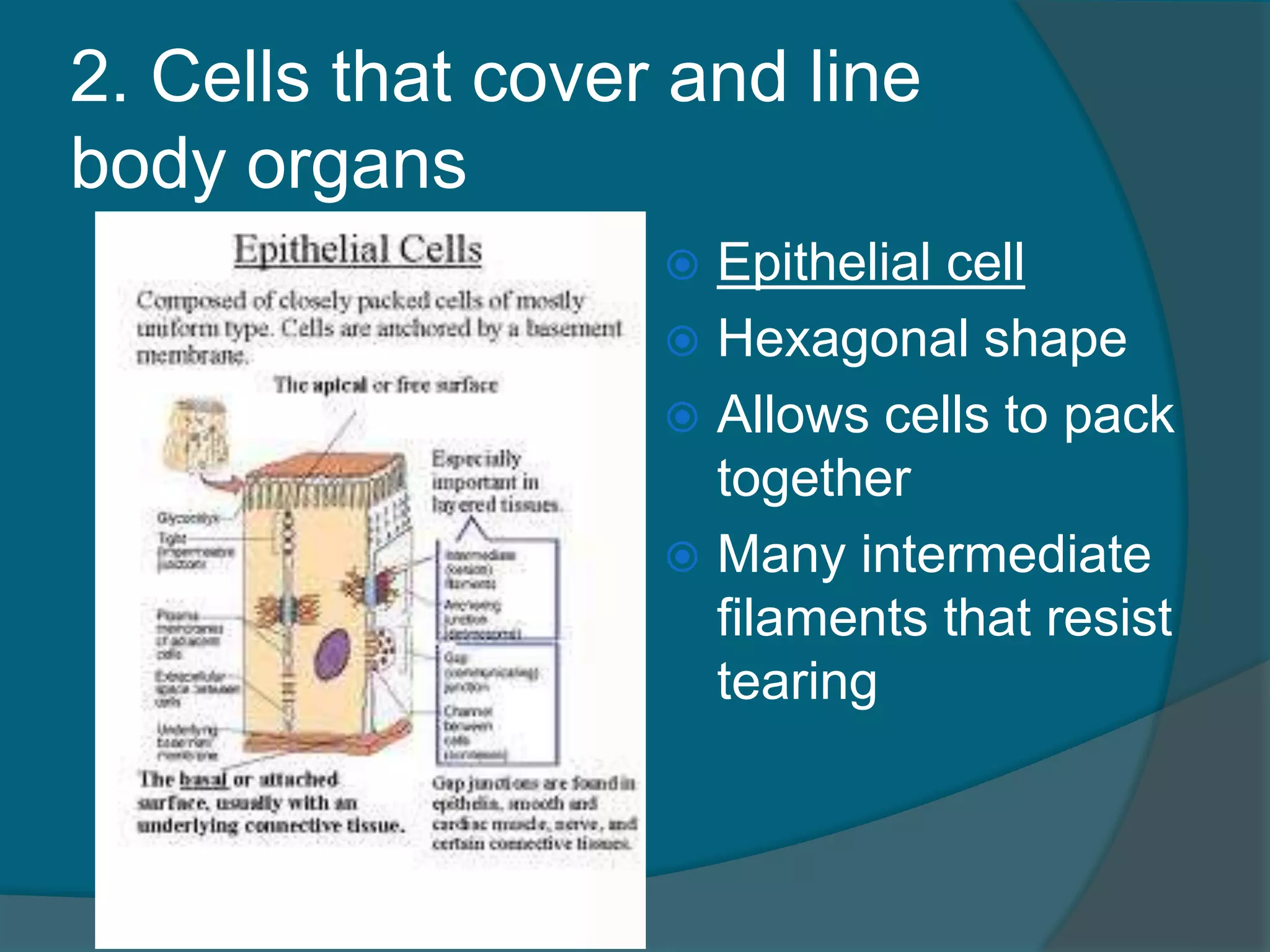
2. Cells thatcover and line
body organs
Epithelial cell
Hexagonal shape
Allows cells to pack
together
Many intermediate
filaments that resist
tearing
59.
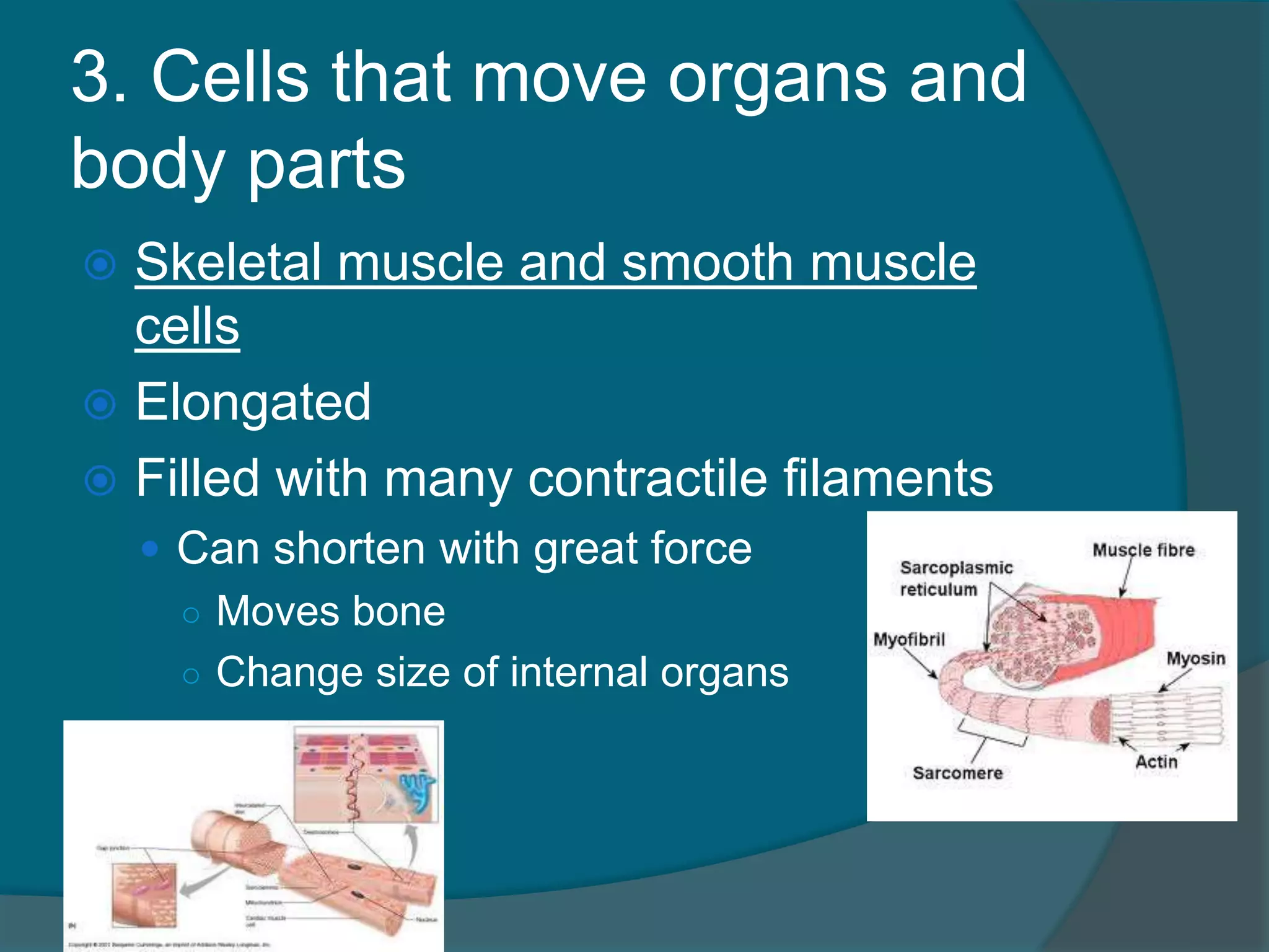
3. Cells thatmove organs and
body parts
Skeletal muscle and smooth muscle
cells
Elongated
Filled with many contractile filaments
Can shorten with great force
○ Moves bone
○ Change size of internal organs
60.
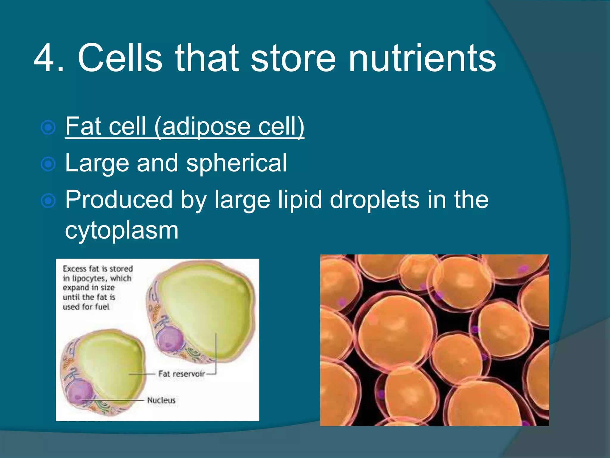
4. Cells thatstore nutrients
Fat cell (adipose cell)
Large and spherical
Produced by large lipid droplets in the
cytoplasm
61.
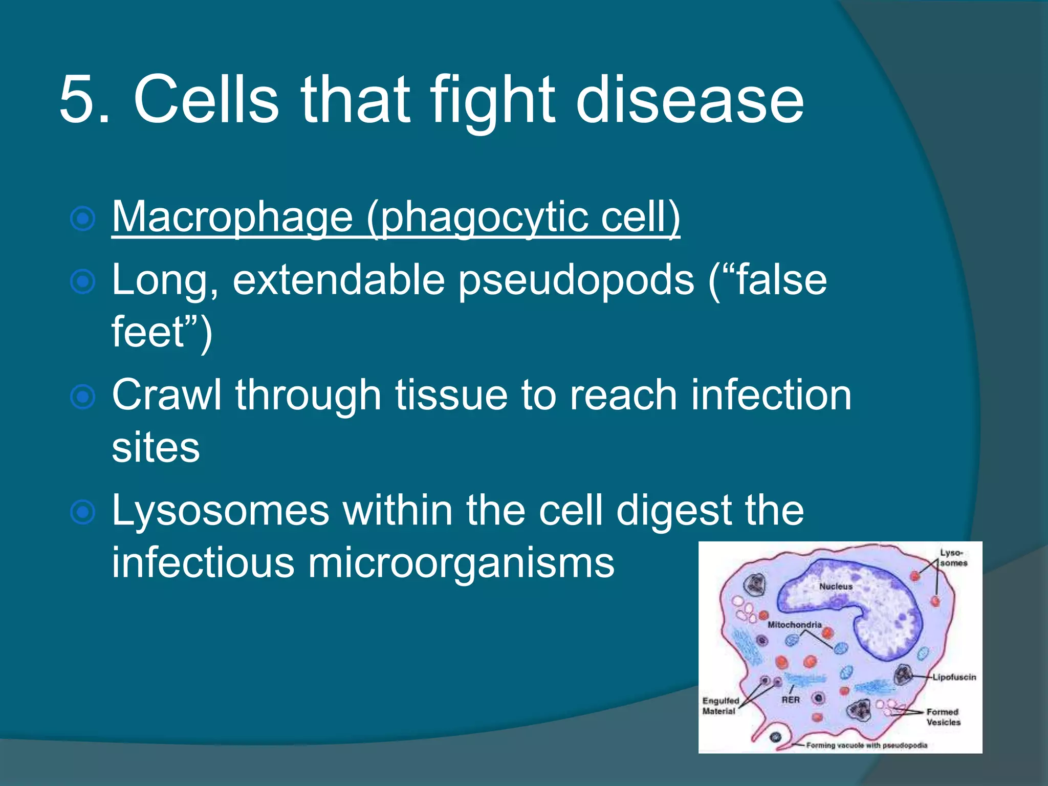
5. Cells thatfight disease
Macrophage (phagocytic cell)
Long, extendable pseudopods (“false
feet”)
Crawl through tissue to reach infection
sites
Lysosomes within the cell digest the
infectious microorganisms
62.
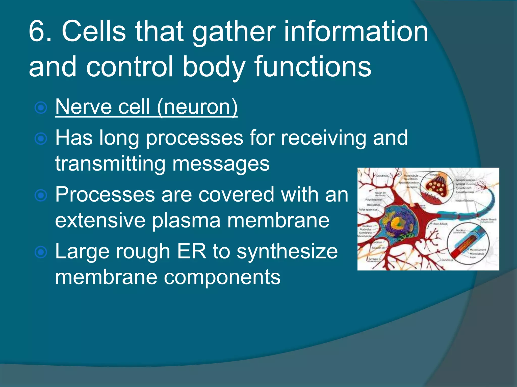
6. Cells thatgather information
and control body functions
Nerve cell (neuron)
Has long processes for receiving and
transmitting messages
Processes are covered with an
extensive plasma membrane
Large rough ER to synthesize
membrane components
63.
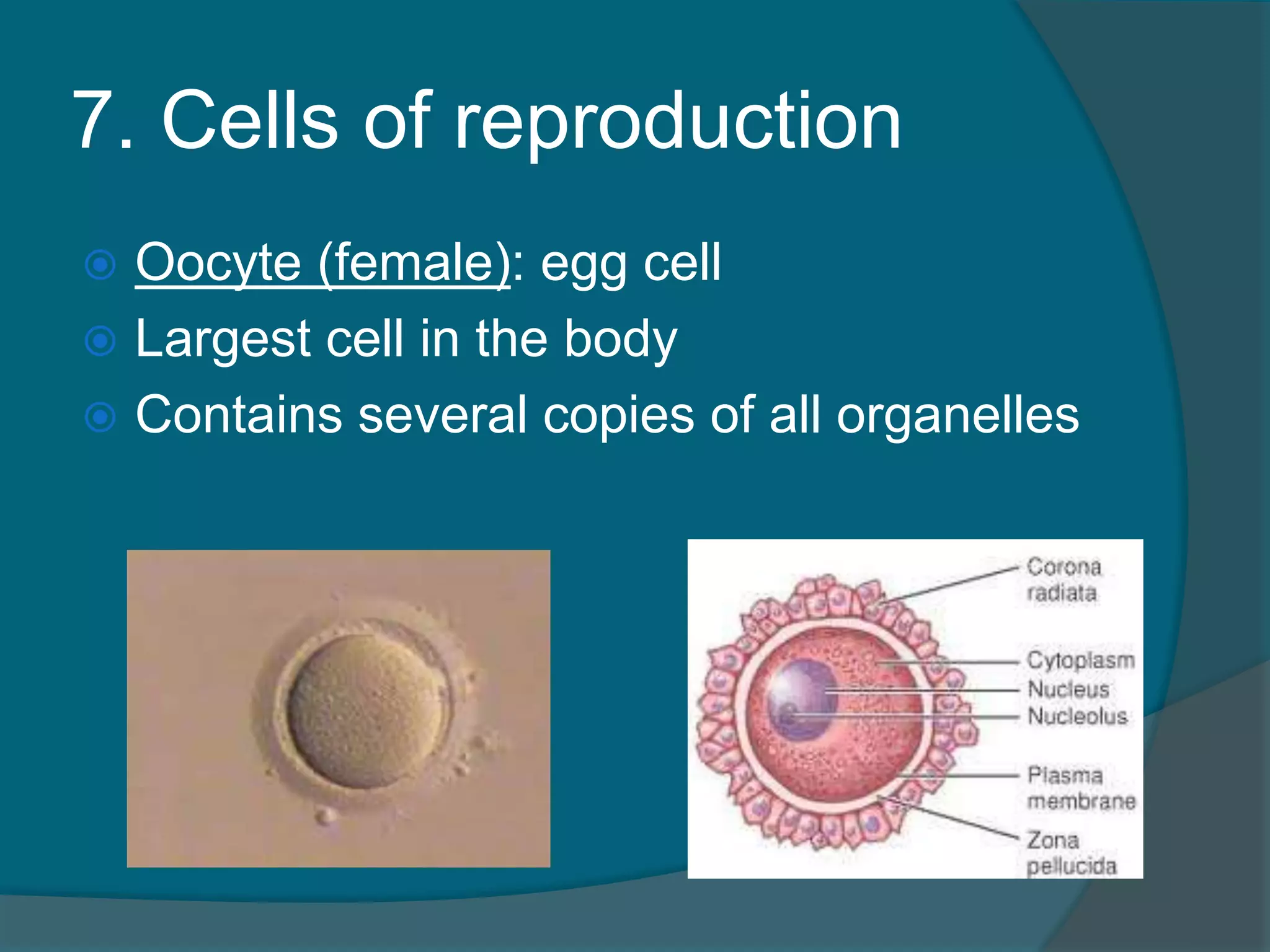
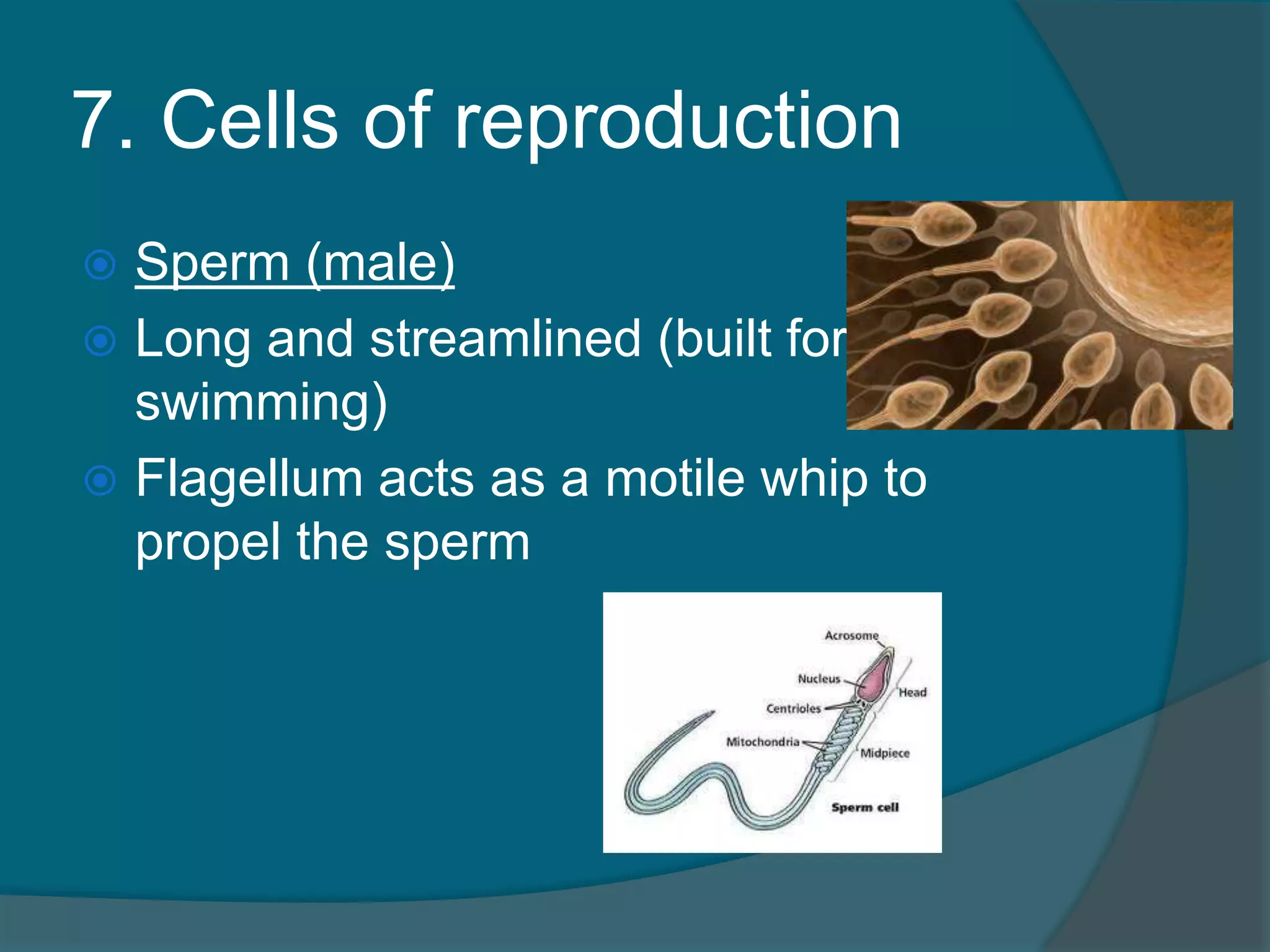
7. Cells ofreproduction
Oocyte (female): egg cell
Largest cell in the body
Contains several copies of all organelles
64.
7. Cells ofreproduction
Sperm (male)
Long and streamlined (built for
swimming)
Flagellum acts as a motile whip to
propel the sperm
66.
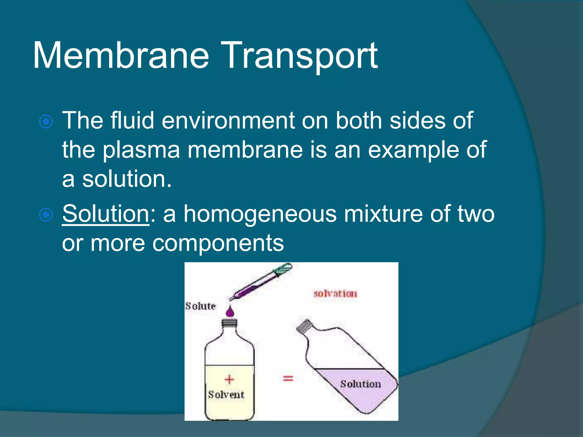
Membrane Transport
Thefluid environment on both sides of
the plasma membrane is an example of
a solution.
Solution: a homogeneous mixture of two
or more components
67.
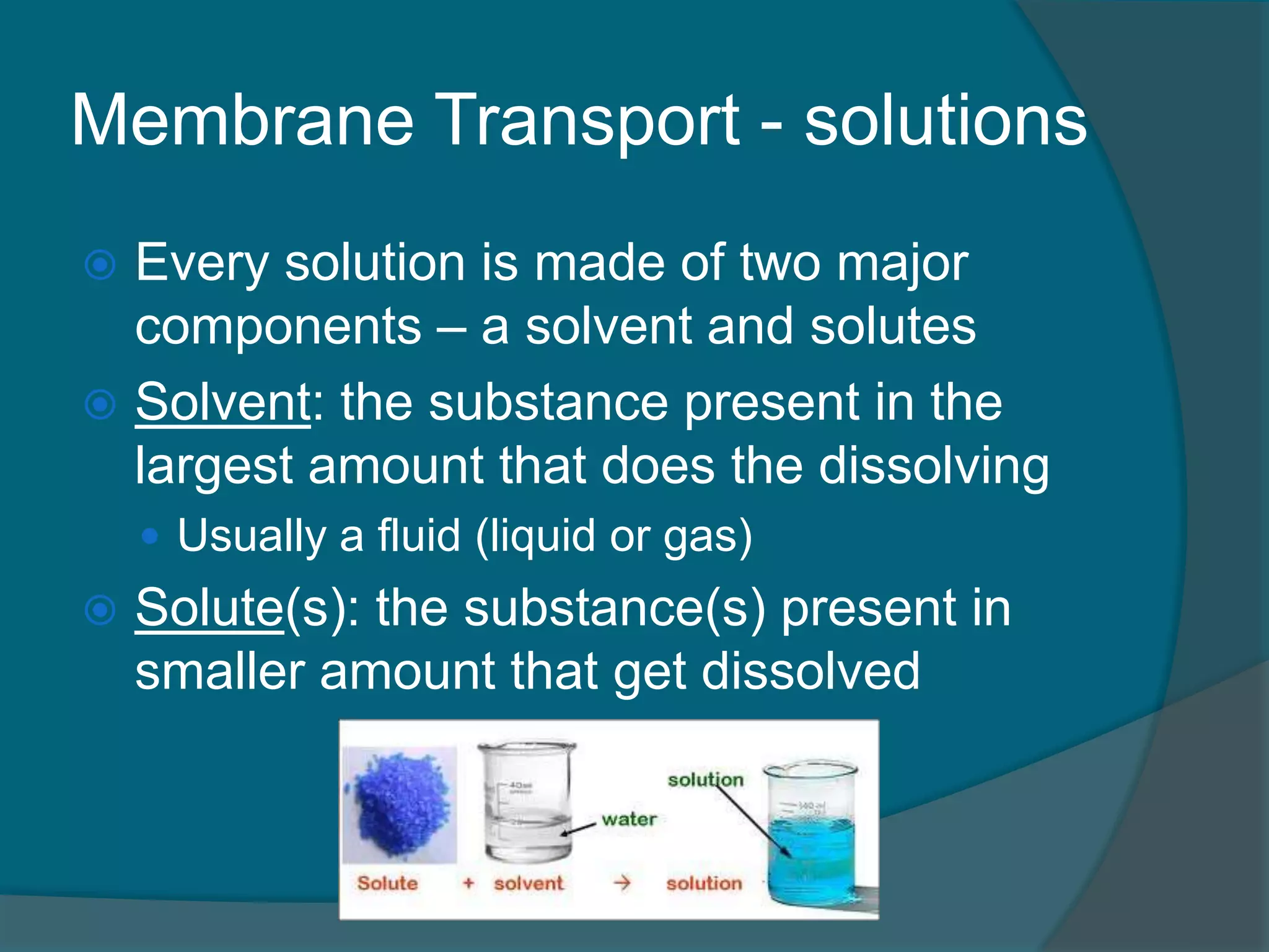
Membrane Transport -solutions
Every solution is made of two major
components – a solvent and solutes
Solvent: the substance present in the
largest amount that does the dissolving
Usually a fluid (liquid or gas)
Solute(s): the substance(s) present in
smaller amount that get dissolved
68.
Membrane Transport
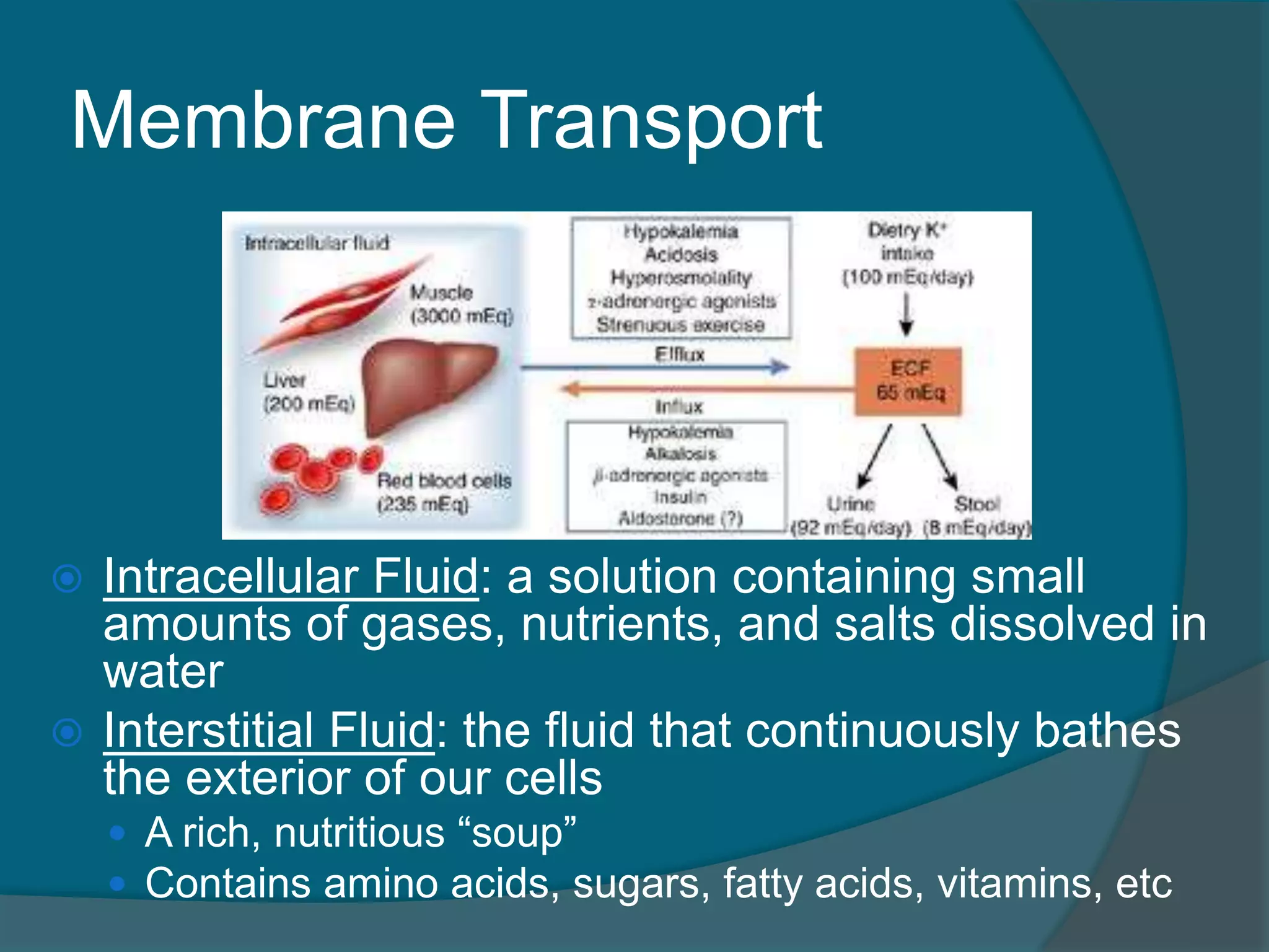
IntracellularFluid: a solution containing small
amounts of gases, nutrients, and salts dissolved in
water
Interstitial Fluid: the fluid that continuously bathes
the exterior of our cells
A rich, nutritious “soup”
Contains amino acids, sugars, fatty acids, vitamins, etc
69.
Membrane Transport
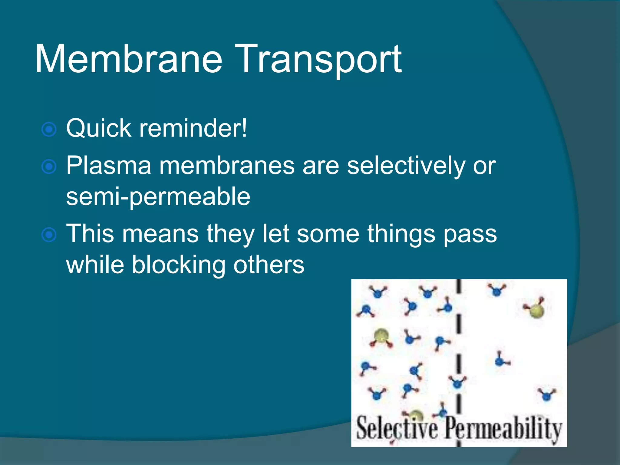
Quickreminder!
Plasma membranes are selectively or
semi-permeable
This means they let some things pass
while blocking others
70.
Membrane Transport
Movementof substances through the
plasma membrane happens two ways
1. Passive Transport
2. Active Transport
71.
Passive Transport
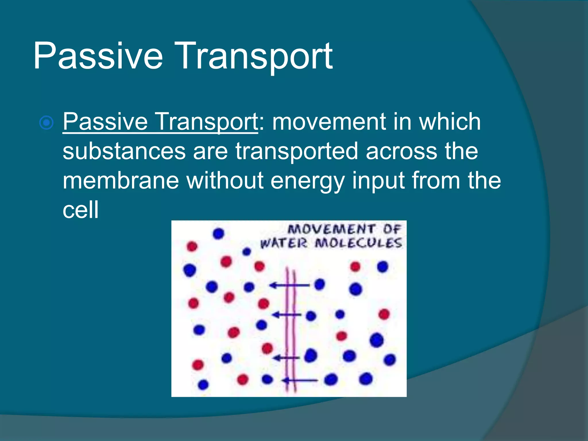
PassiveTransport: movement in which
substances are transported across the
membrane without energy input from the
cell
72.
Passive Transport
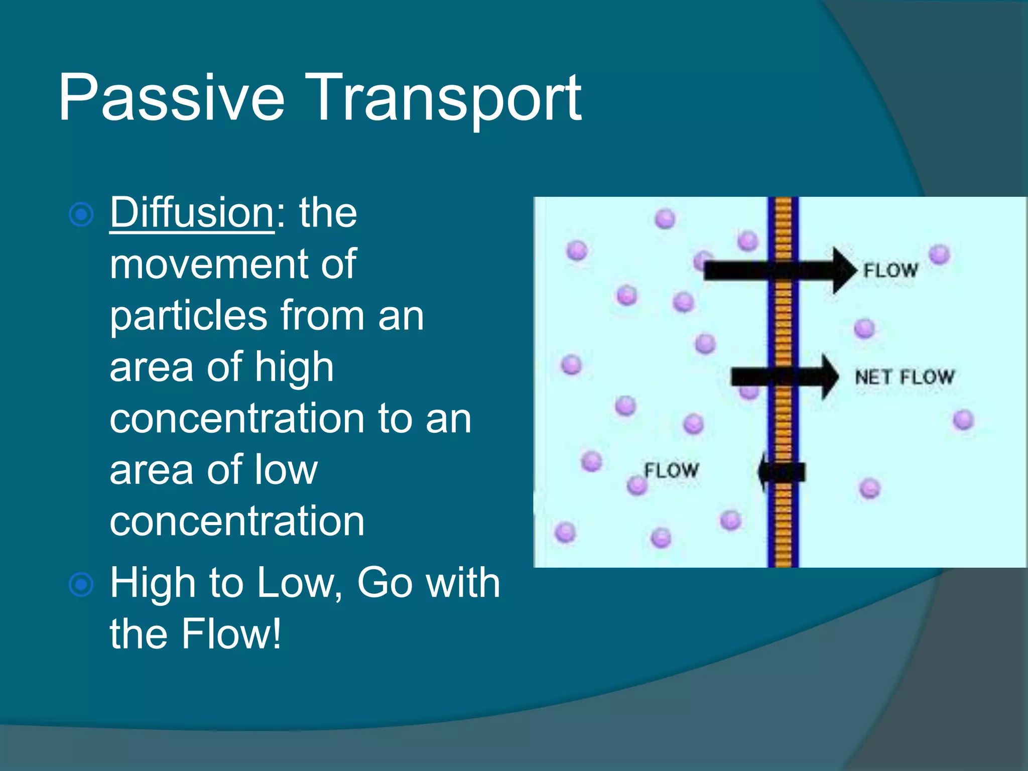
Diffusion:the
movement of
particles from an
area of high
concentration to an
area of low
concentration
High to Low, Go with
the Flow!
73.
Passive Transport
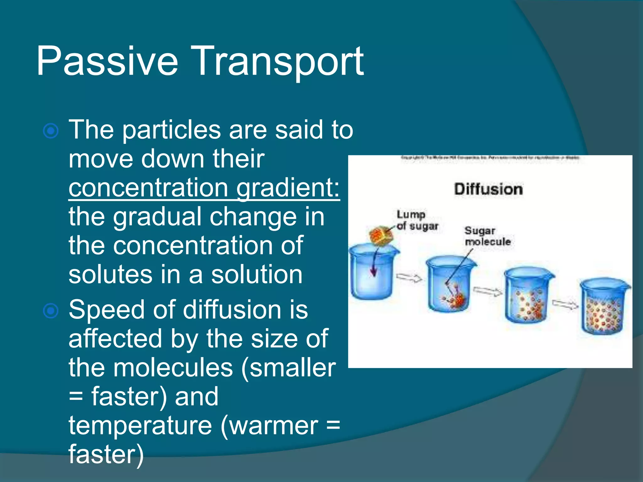
Theparticles are said to
move down their
concentration gradient:
the gradual change in
the concentration of
solutes in a solution
Speed of diffusion is
affected by the size of
the molecules (smaller
= faster) and
temperature (warmer =
faster)
74.
Passive Transport
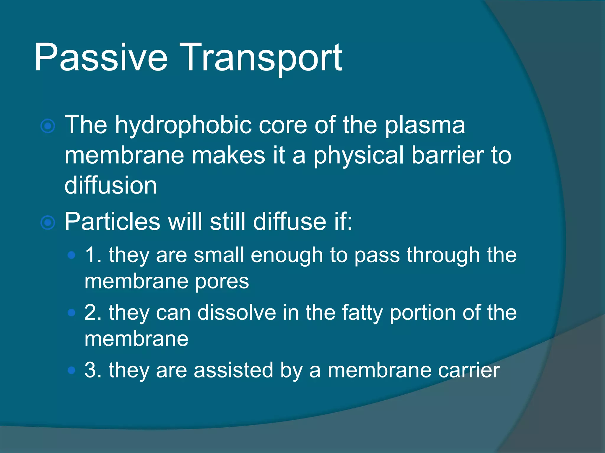
Thehydrophobic core of the plasma
membrane makes it a physical barrier to
diffusion
Particles will still diffuse if:
1. they are small enough to pass through the
membrane pores
2. they can dissolve in the fatty portion of the
membrane
3. they are assisted by a membrane carrier
75.
Passive Transport
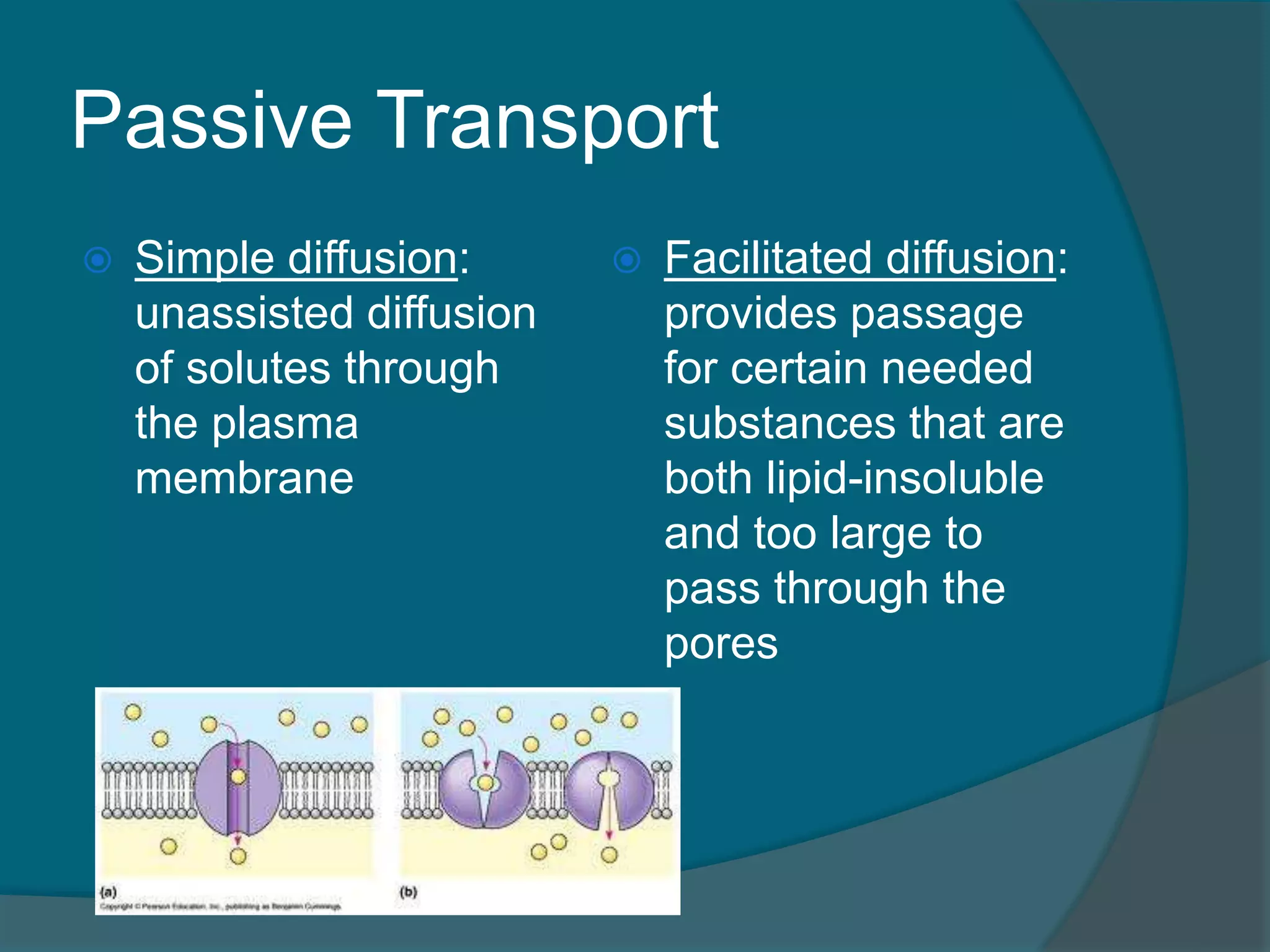
Simplediffusion:
unassisted diffusion
of solutes through
the plasma
membrane
Facilitated diffusion:
provides passage
for certain needed
substances that are
both lipid-insoluble
and too large to
pass through the
pores
76.
Passive Transport
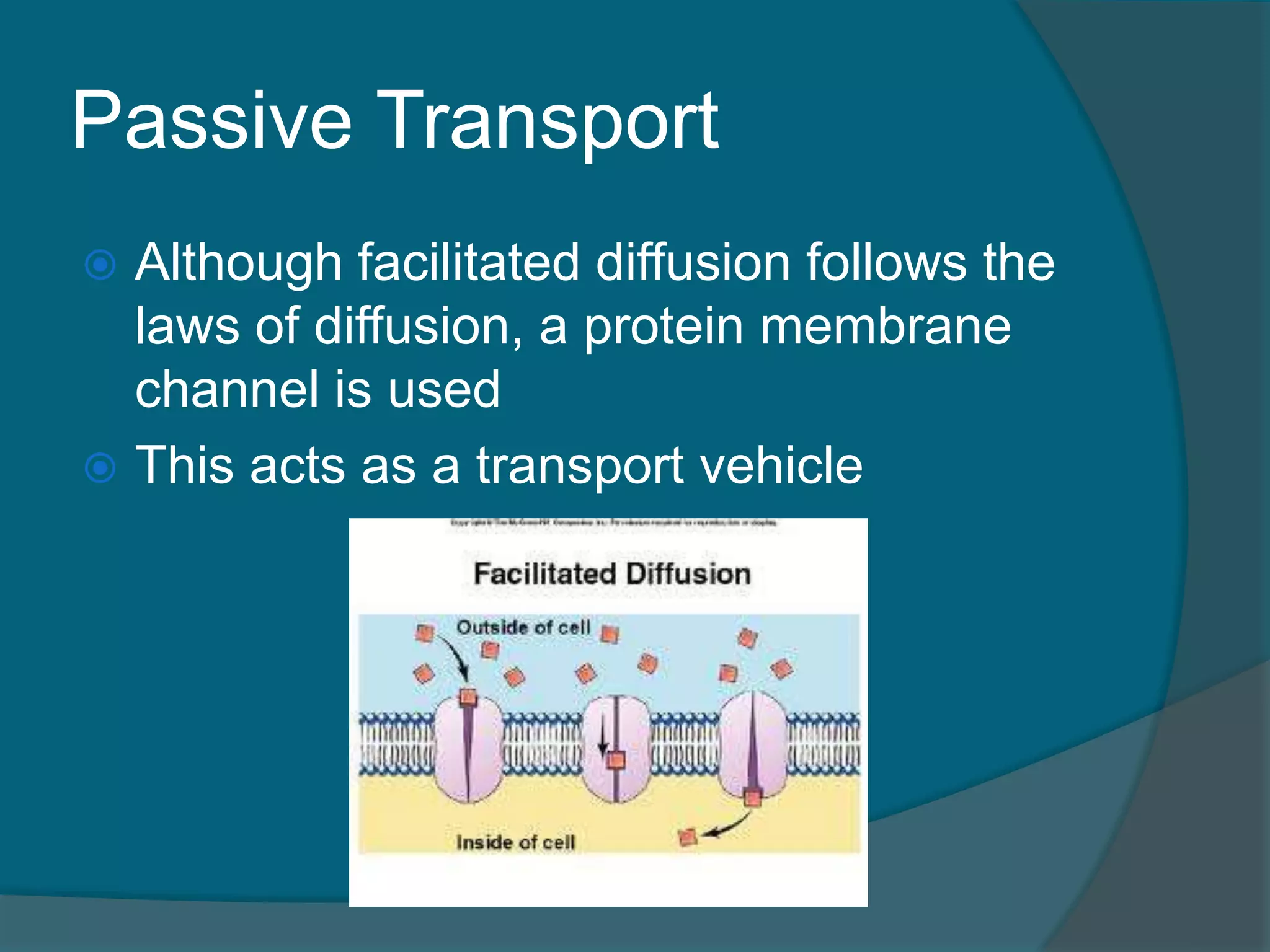
Althoughfacilitated diffusion follows the
laws of diffusion, a protein membrane
channel is used
This acts as a transport vehicle
77.
Passive Transport
Substancesthat
pass into and out
of cells by diffusion
save energy
Includes the
movement of key
molecules like
water, glucose,
oxygen and carbon
dioxide
78.
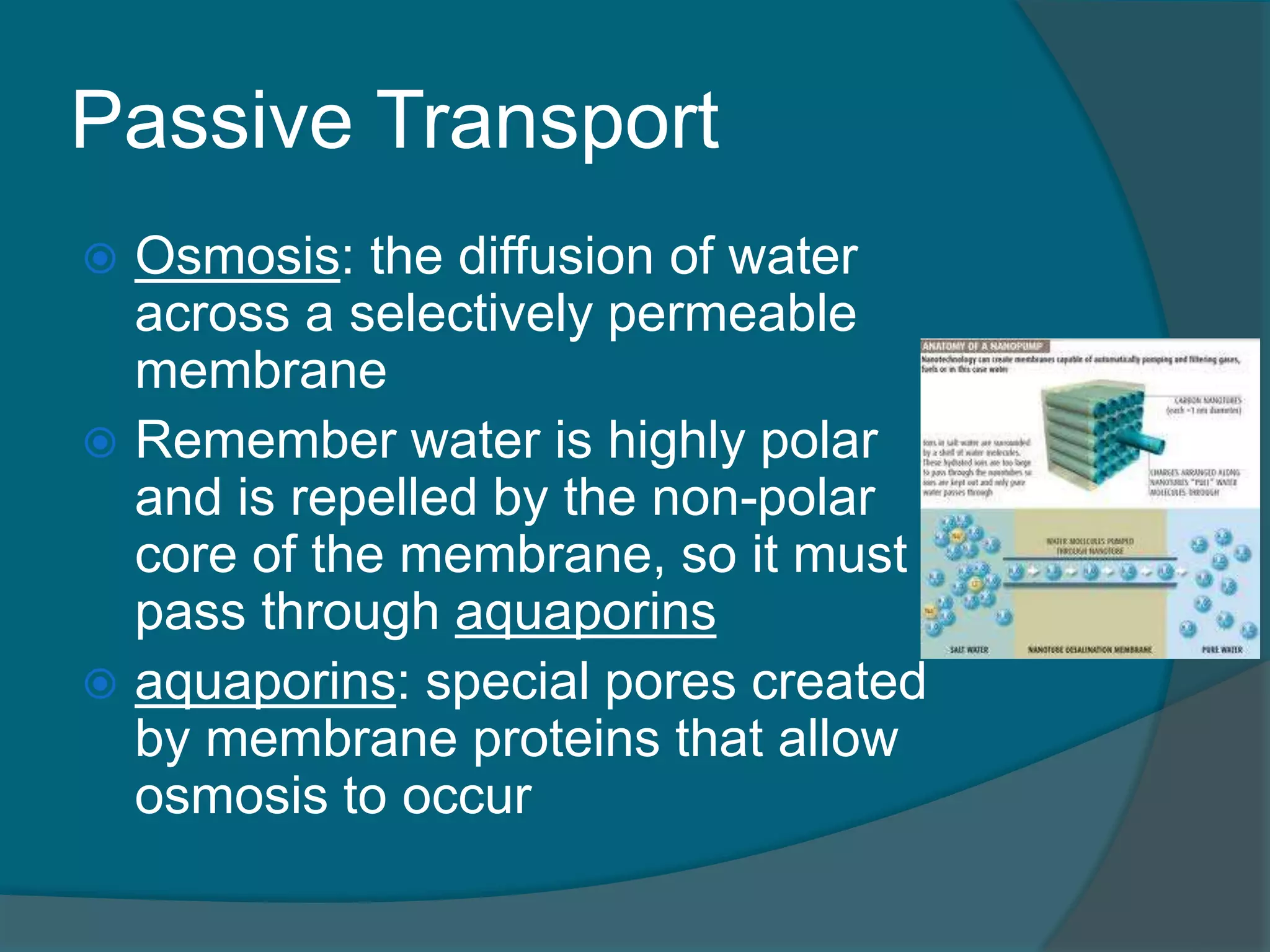
Passive Transport
Osmosis:the diffusion of water
across a selectively permeable
membrane
Remember water is highly polar
and is repelled by the non-polar
core of the membrane, so it must
pass through aquaporins
aquaporins: special pores created
by membrane proteins that allow
osmosis to occur
79.
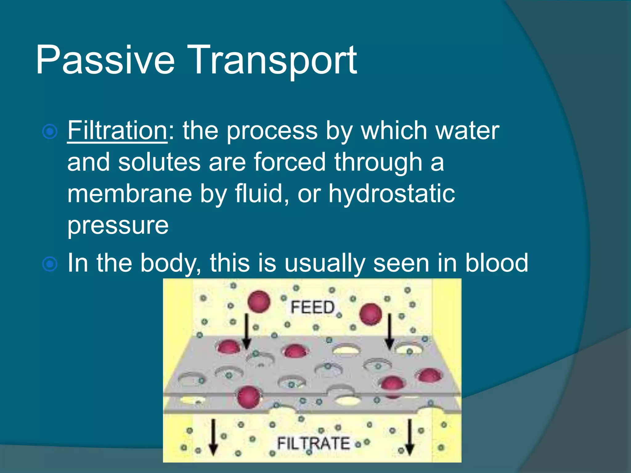
Passive Transport
Filtration:the process by which water
and solutes are forced through a
membrane by fluid, or hydrostatic
pressure
In the body, this is usually seen in blood
80.
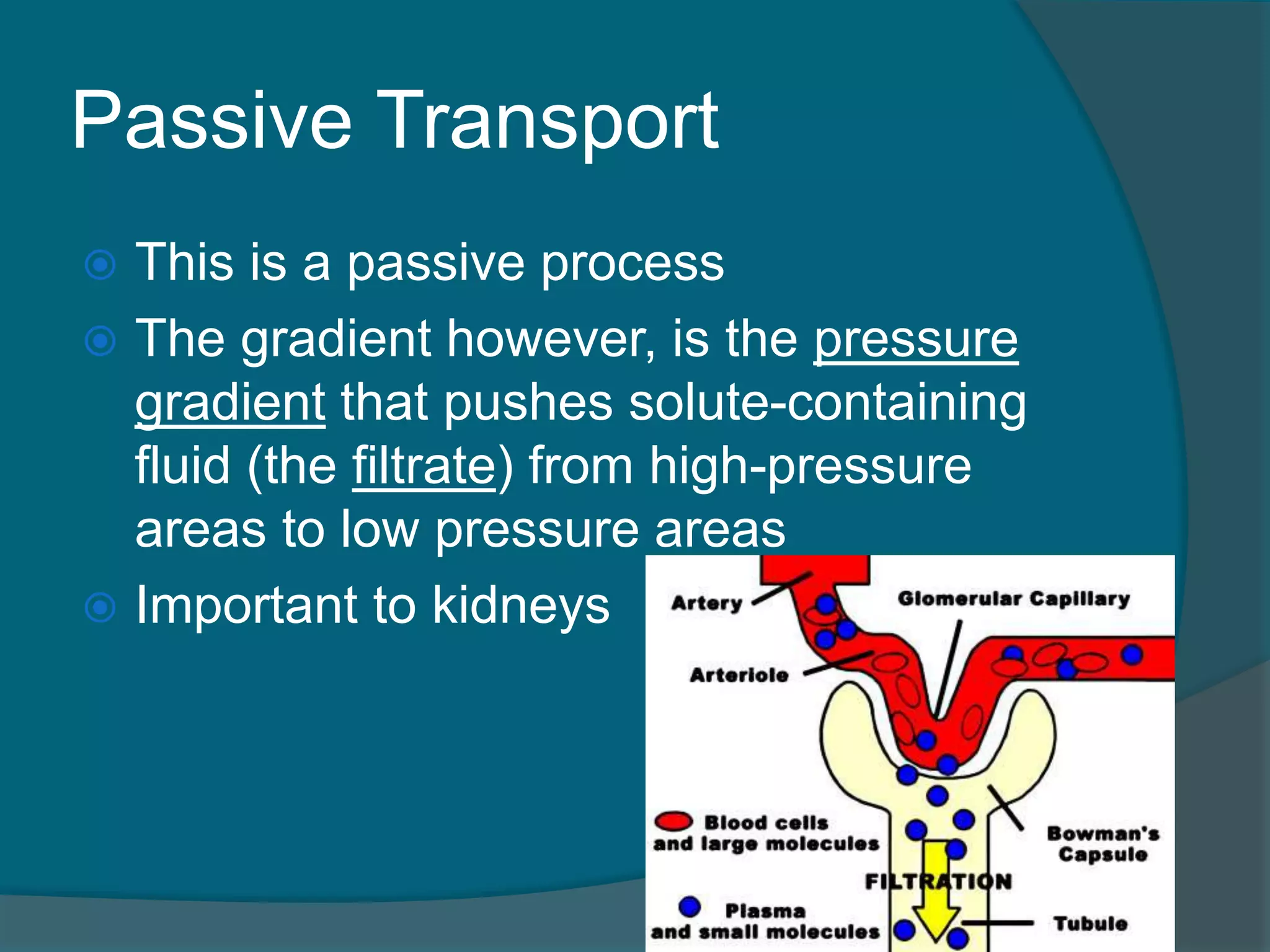
Passive Transport
Thisis a passive process
The gradient however, is the pressure
gradient that pushes solute-containing
fluid (the filtrate) from high-pressure
areas to low pressure areas
Important to kidneys
81.
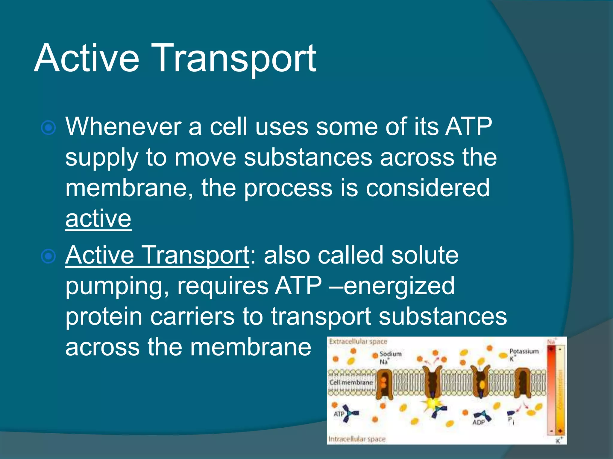
Active Transport
Whenevera cell uses some of its ATP
supply to move substances across the
membrane, the process is considered
active
Active Transport: also called solute
pumping, requires ATP –energized
protein carriers to transport substances
across the membrane
82.
Active Transport
TheATP-energized protein carriers used
in active transport are called solute
pumps
Amino acids, some sugars, and most
ions are transported across the
membrane in this way
And in most cases, they travel against
the concentration gradient
This is opposite to the direction in which
substances would normally flow
83.
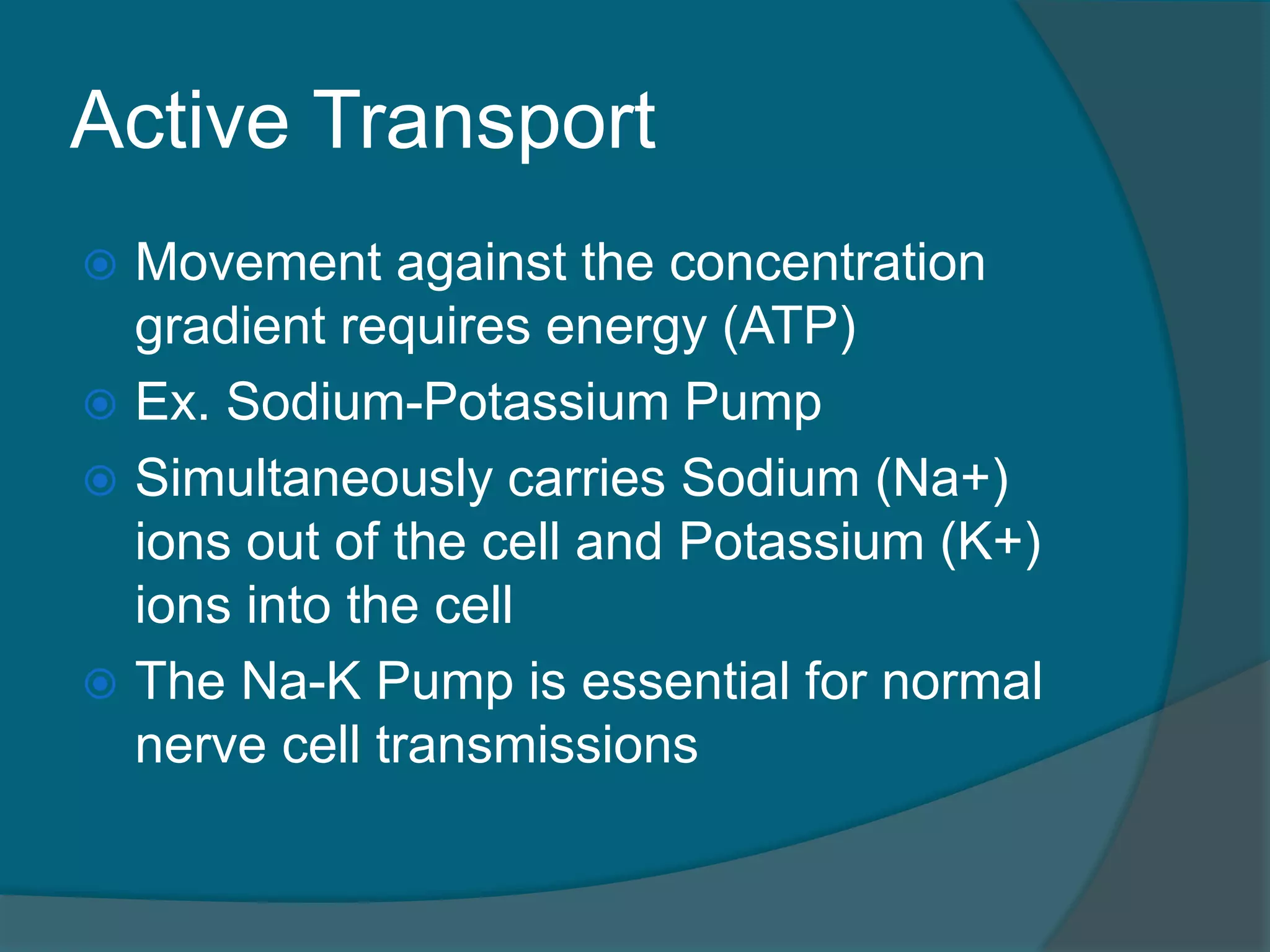
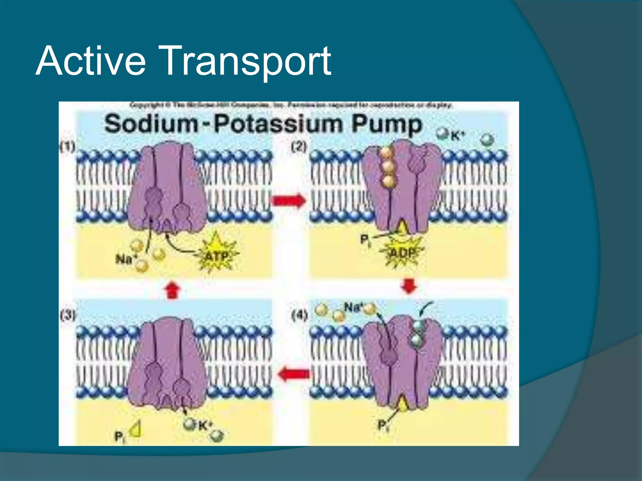
Active Transport
Movementagainst the concentration
gradient requires energy (ATP)
Ex. Sodium-Potassium Pump
Simultaneously carries Sodium (Na+)
ions out of the cell and Potassium (K+)
ions into the cell
The Na-K Pump is essential for normal
nerve cell transmissions
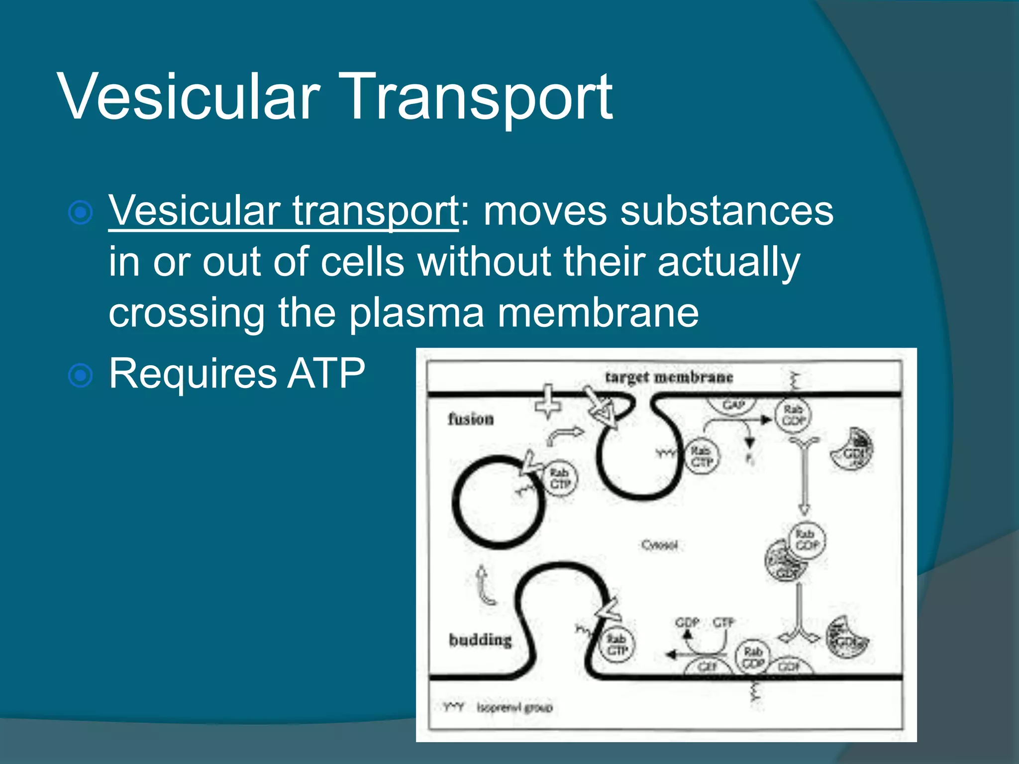
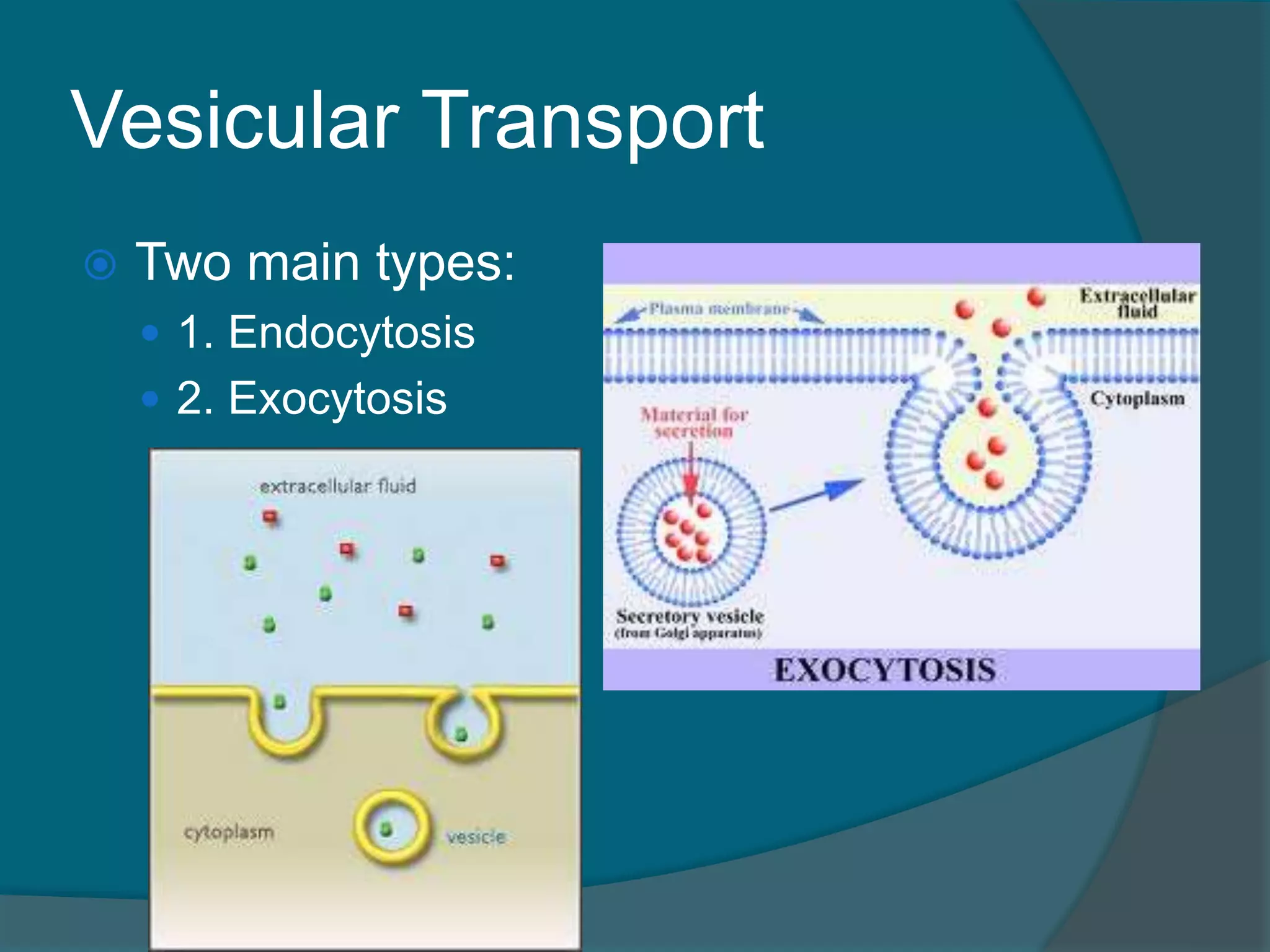
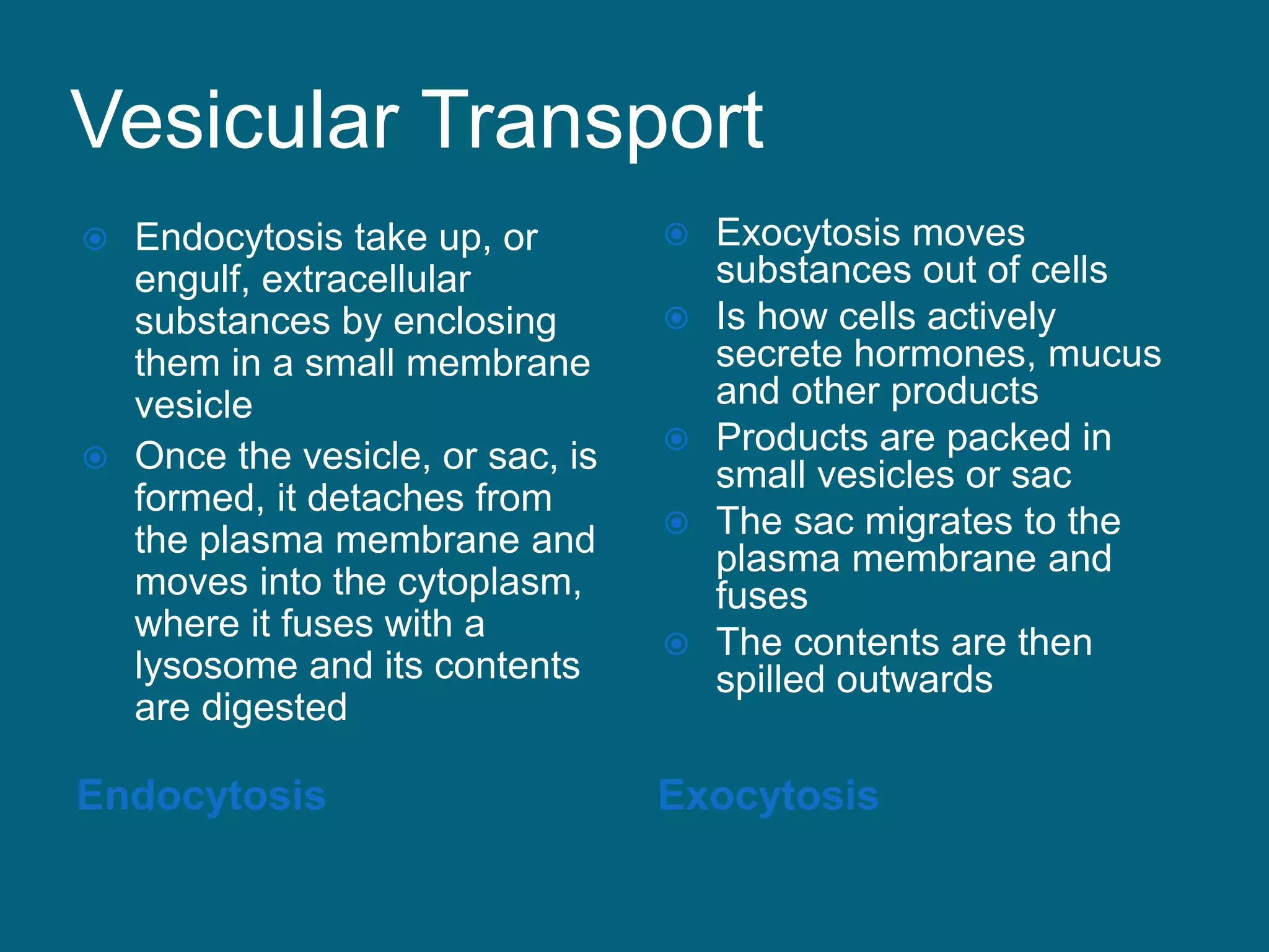
Vesicular Transport
Endocytosis Exocytosis
Endocytosis take up, or
engulf, extracellular
substances by enclosing
them in a small membrane
vesicle
Once the vesicle, or sac, is
formed, it detaches from
the plasma membrane and
moves into the cytoplasm,
where it fuses with a
lysosome and its contents
are digested
Exocytosis moves
substances out of cells
Is how cells actively
secrete hormones, mucus
and other products
Products are packed in
small vesicles or sac
The sac migrates to the
plasma membrane and
fuses
The contents are then
spilled outwards
88.
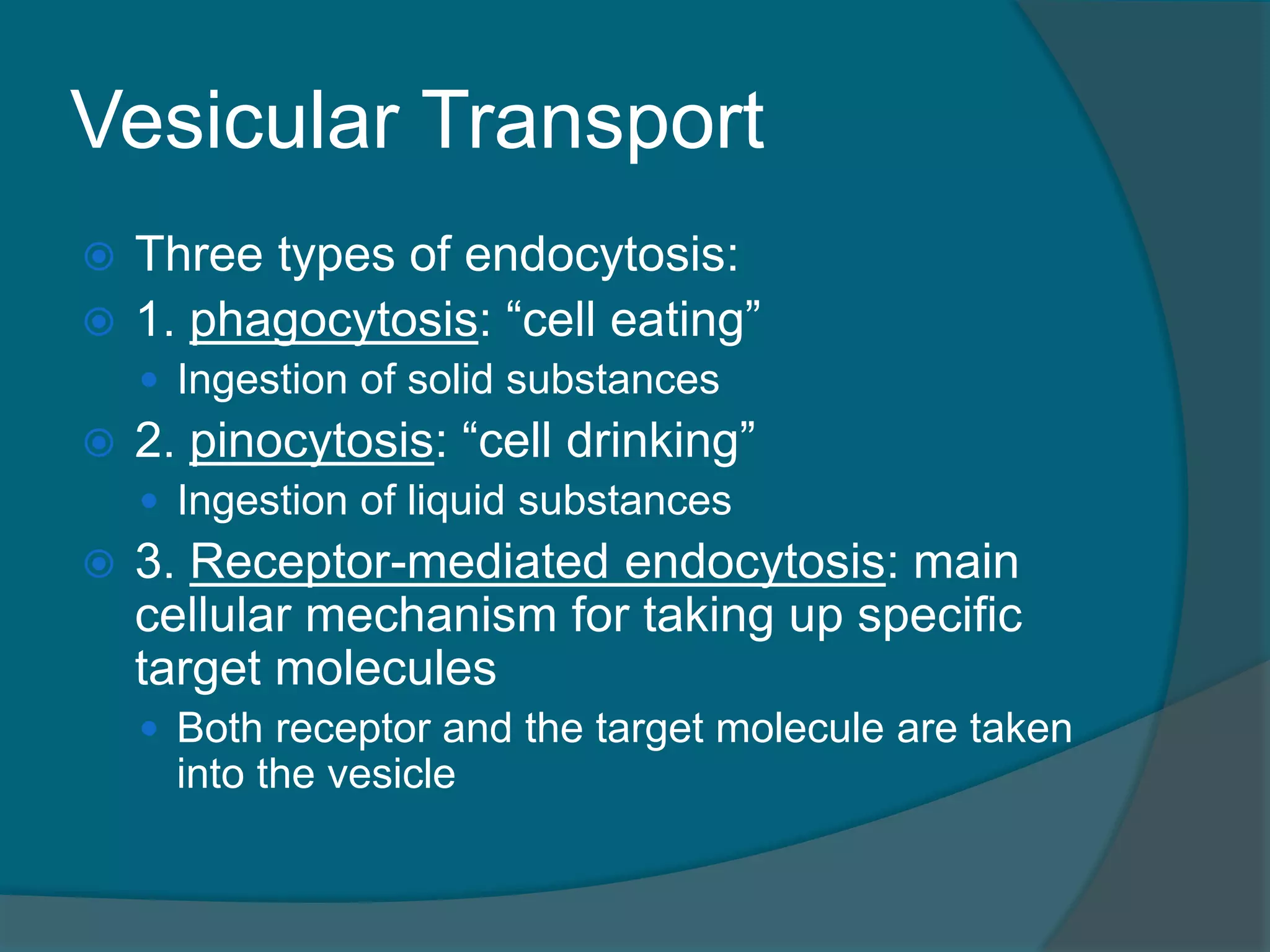
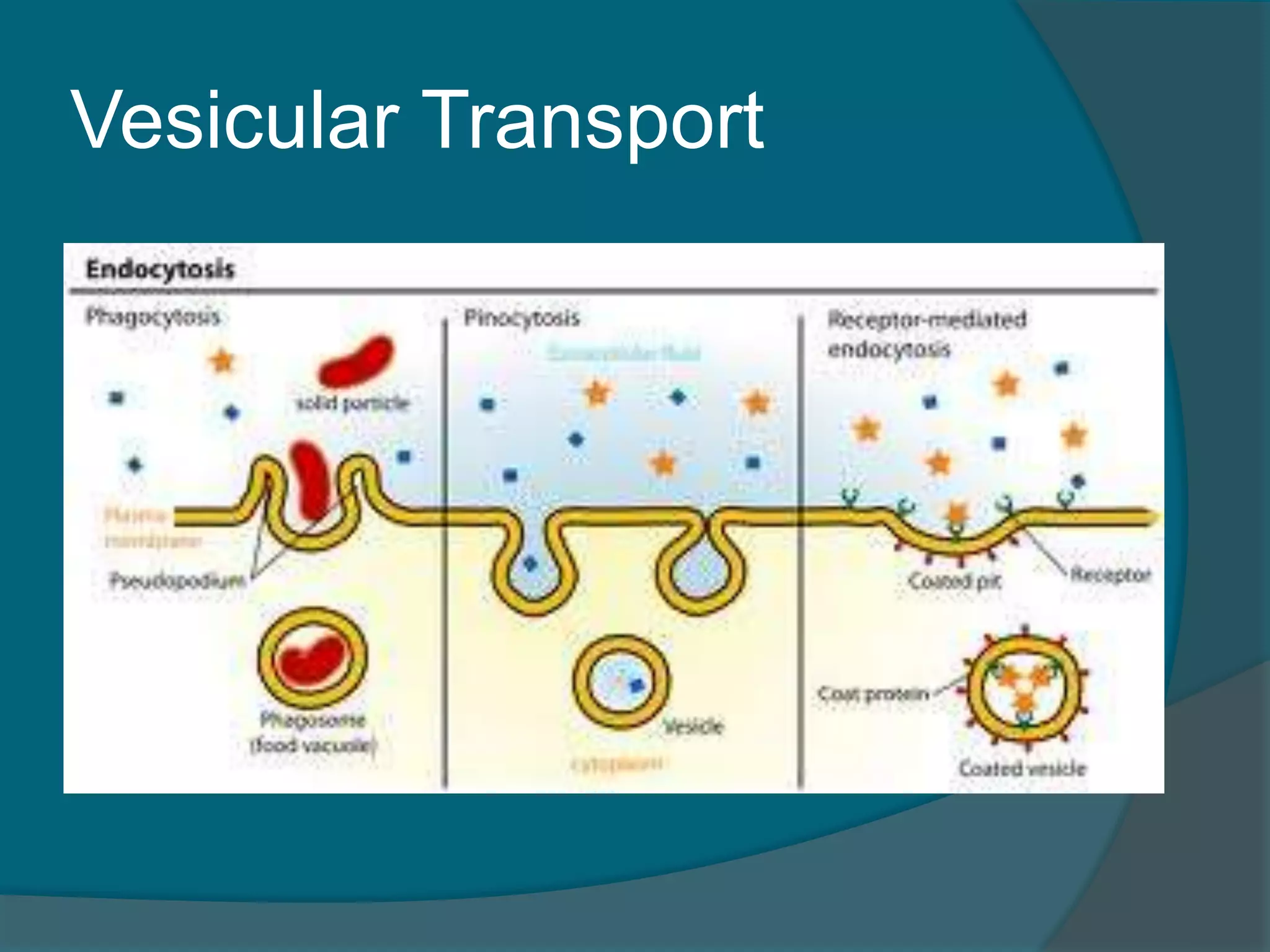
Vesicular Transport
Threetypes of endocytosis:
1. phagocytosis: “cell eating”
Ingestion of solid substances
2. pinocytosis: “cell drinking”
Ingestion of liquid substances
3. Receptor-mediated endocytosis: main
cellular mechanism for taking up specific
target molecules
Both receptor and the target molecule are taken
into the vesicle
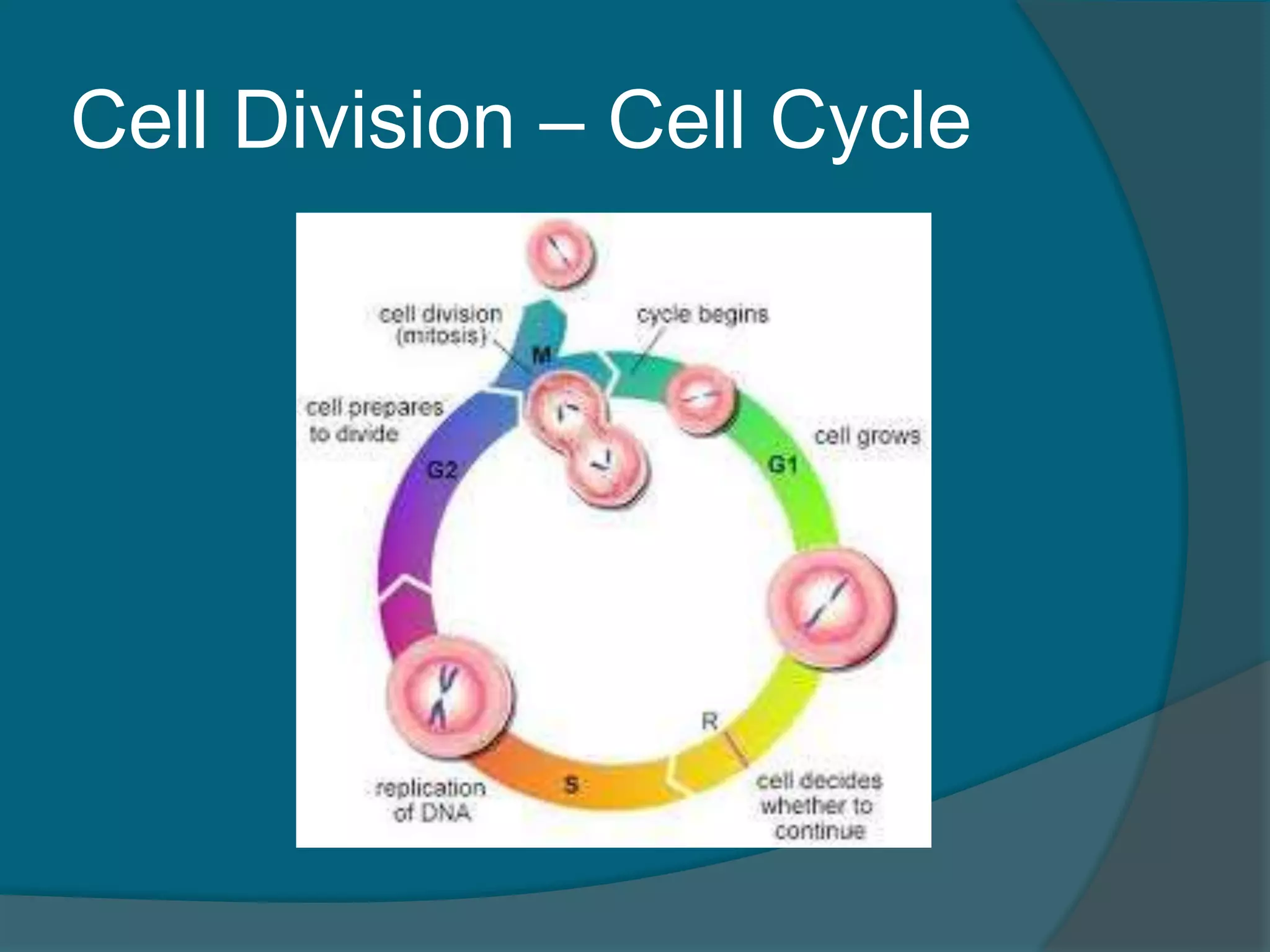
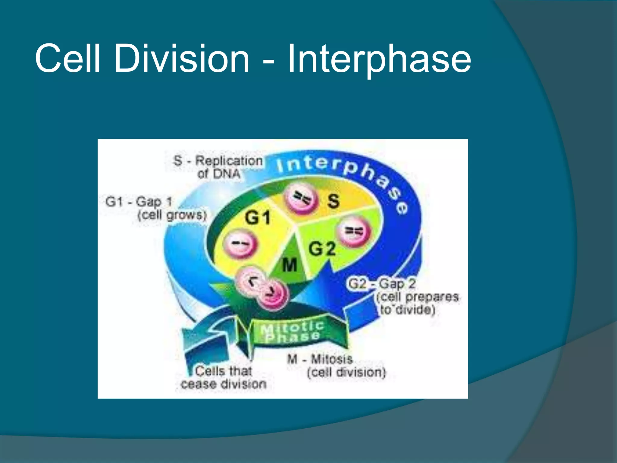
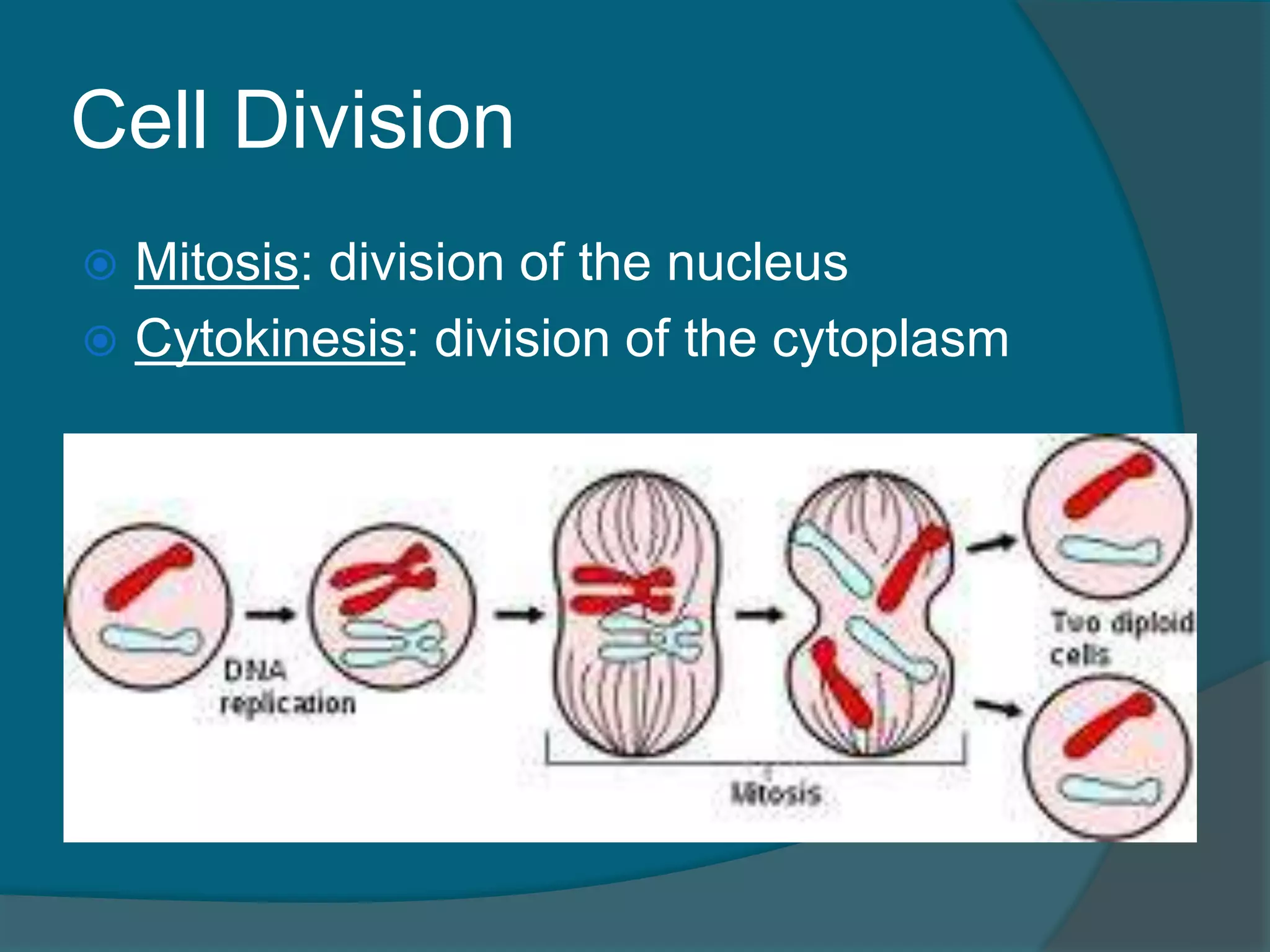
Cell Division
Thecell life cycle is the series of
changes a cell goes through from the
time it is formed until it divides
The cycle has two major periods:
1. Interphase, in which the cell grows
and carries on it usual metabolic
activities
2. Cell Division, time when the cell
reproduces itself
Cell Division -Interphase
Interphase has three major stages
1. G1 – Growth 1
Cell increases in size
2. S – Synthesis
DNA and organelles are replicated
3. G2 – Growth 2
Continued cell growth before division
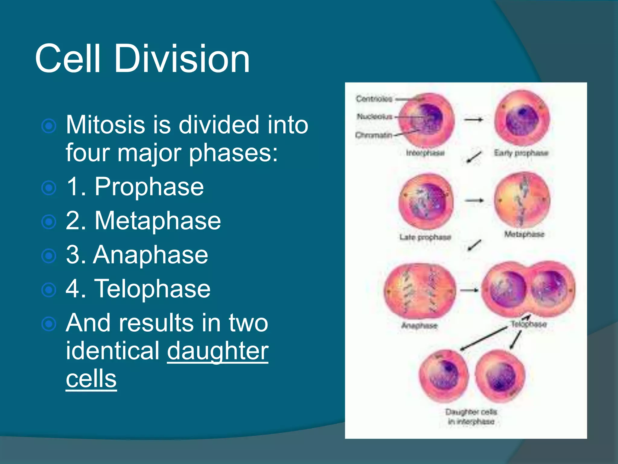
Cell Division
Mitosisis divided into
four major phases:
1. Prophase
2. Metaphase
3. Anaphase
4. Telophase
And results in two
identical daughter
cells
97.
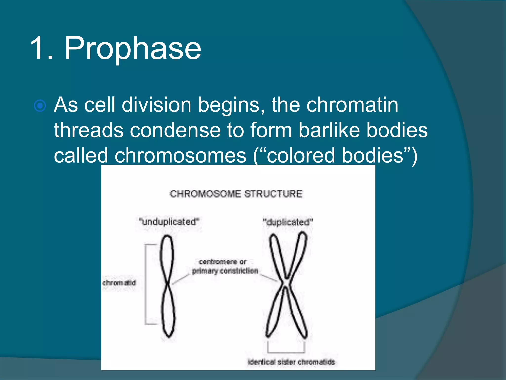
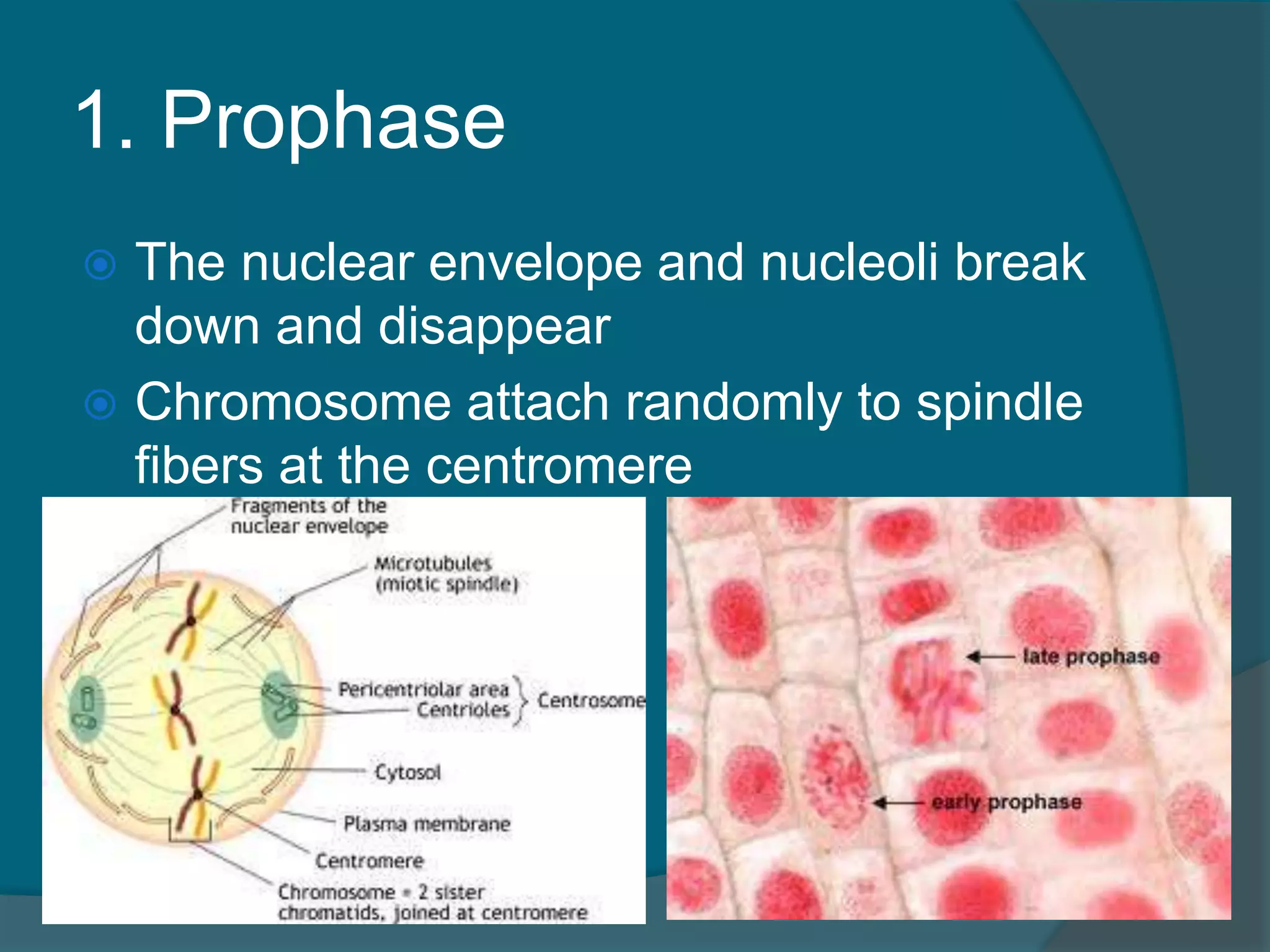
1. Prophase
Ascell division begins, the chromatin
threads condense to form barlike bodies
called chromosomes (“colored bodies”)
98.
1. Prophase
Thecentrioles separate from each other
and begin to move to opposite sides
(“poles”) of the cell
The direct the assembly of the mitotic
spindle
The mitotic spindle provides the
structure for attachment and movement
of the chromosomes for the duration of
mitosis
99.
1. Prophase
Thenuclear envelope and nucleoli break
down and disappear
Chromosome attach randomly to spindle
fibers at the centromere
100.
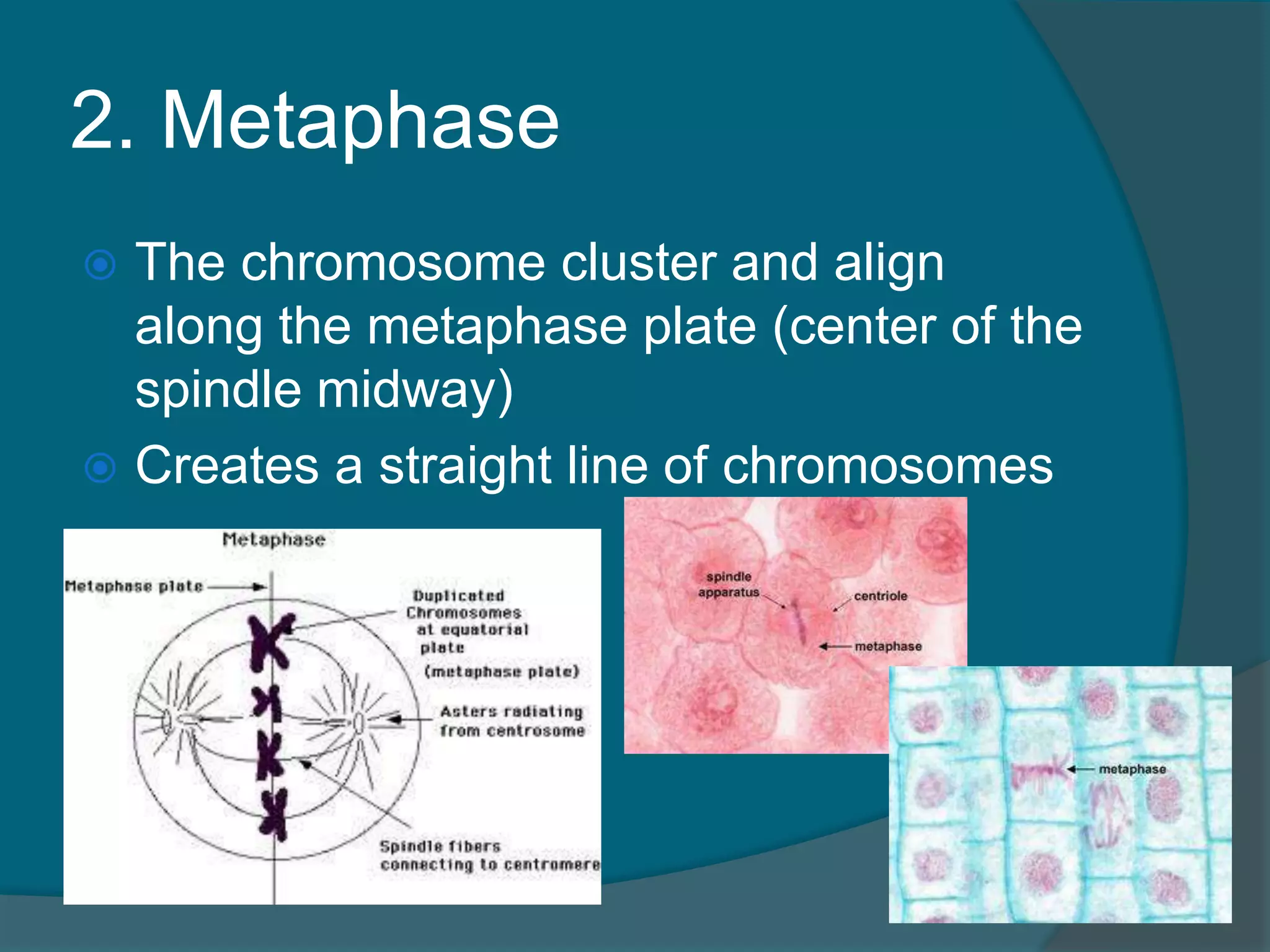
2. Metaphase
Thechromosome cluster and align
along the metaphase plate (center of the
spindle midway)
Creates a straight line of chromosomes
101.
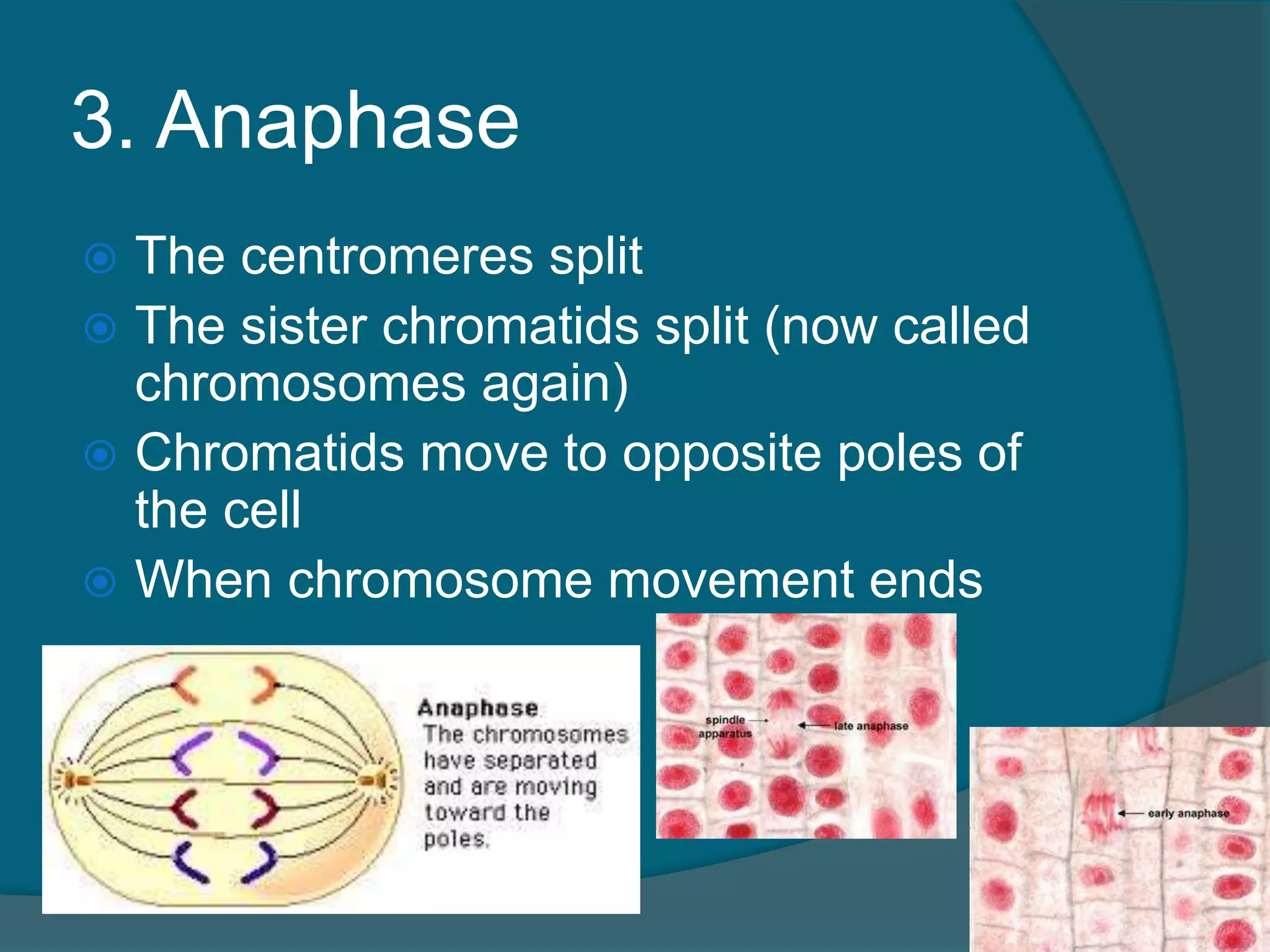
3. Anaphase
Thecentromeres split
The sister chromatids split (now called
chromosomes again)
Chromatids move to opposite poles of
the cell
When chromosome movement ends
102.
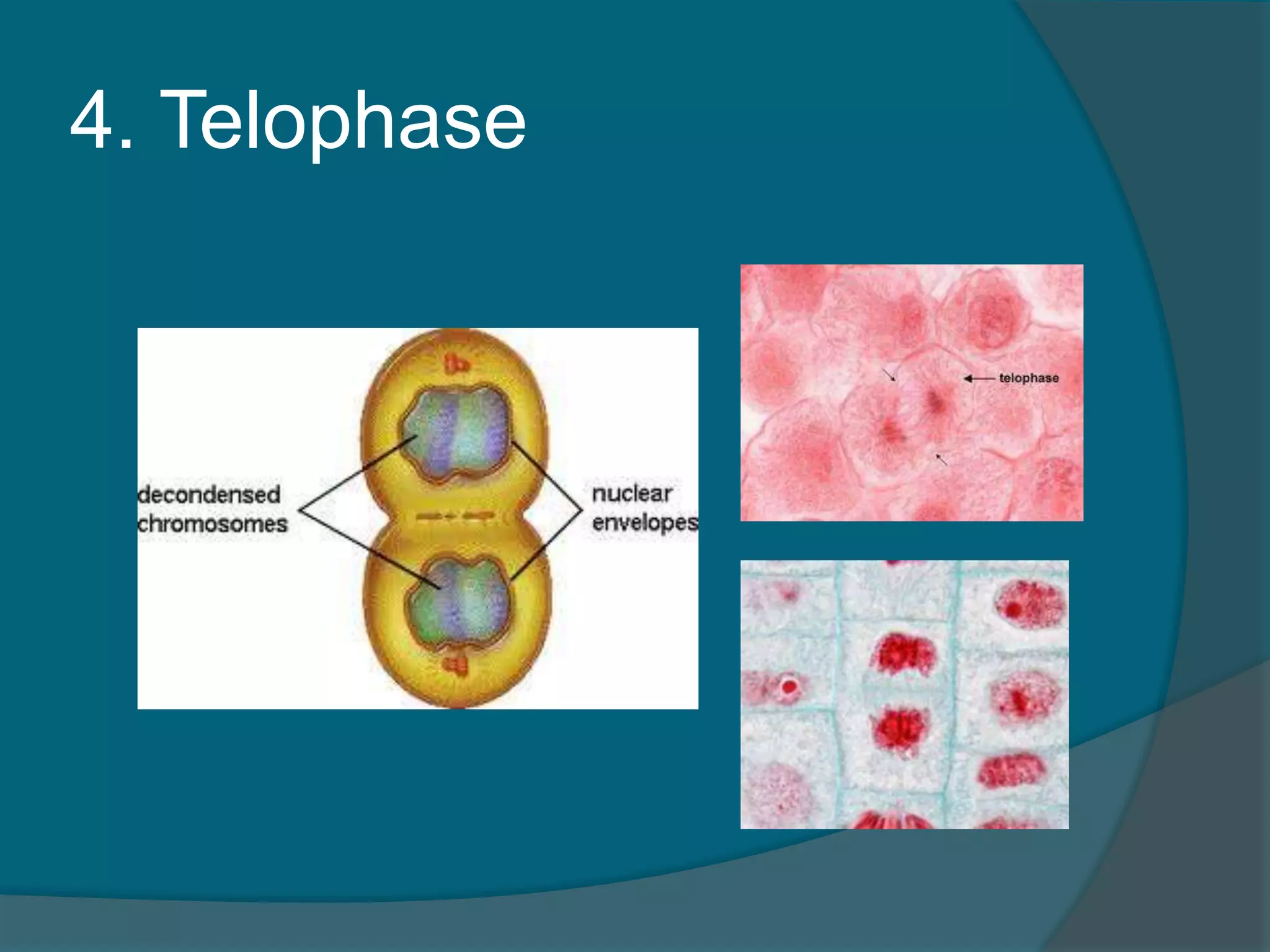
4. Telophase
Essentiallyprophase in reverse
Chromosomes uncoil and become
chromatin again
Spindle fibers break down and
disappear
Nuclear envelopes reform and nucleoli
reappear around each group of
chromatin
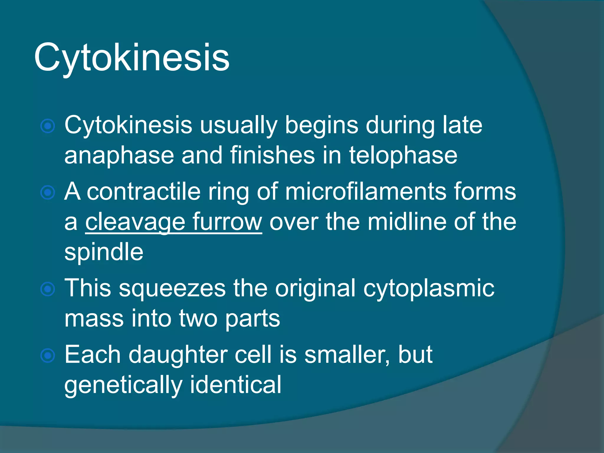
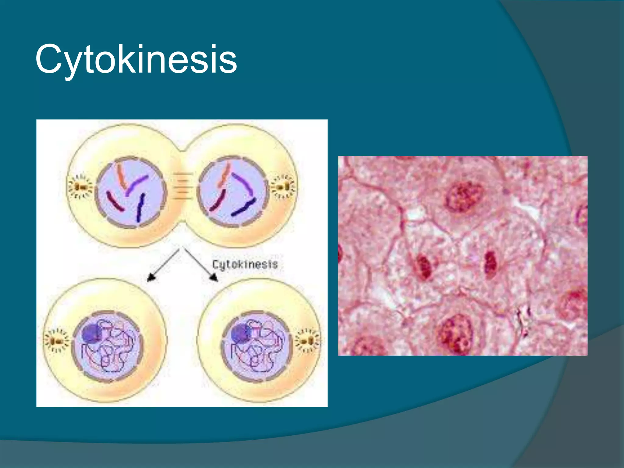
Cytokinesis
Cytokinesis usuallybegins during late
anaphase and finishes in telophase
A contractile ring of microfilaments forms
a cleavage furrow over the midline of the
spindle
This squeezes the original cytoplasmic
mass into two parts
Each daughter cell is smaller, but
genetically identical
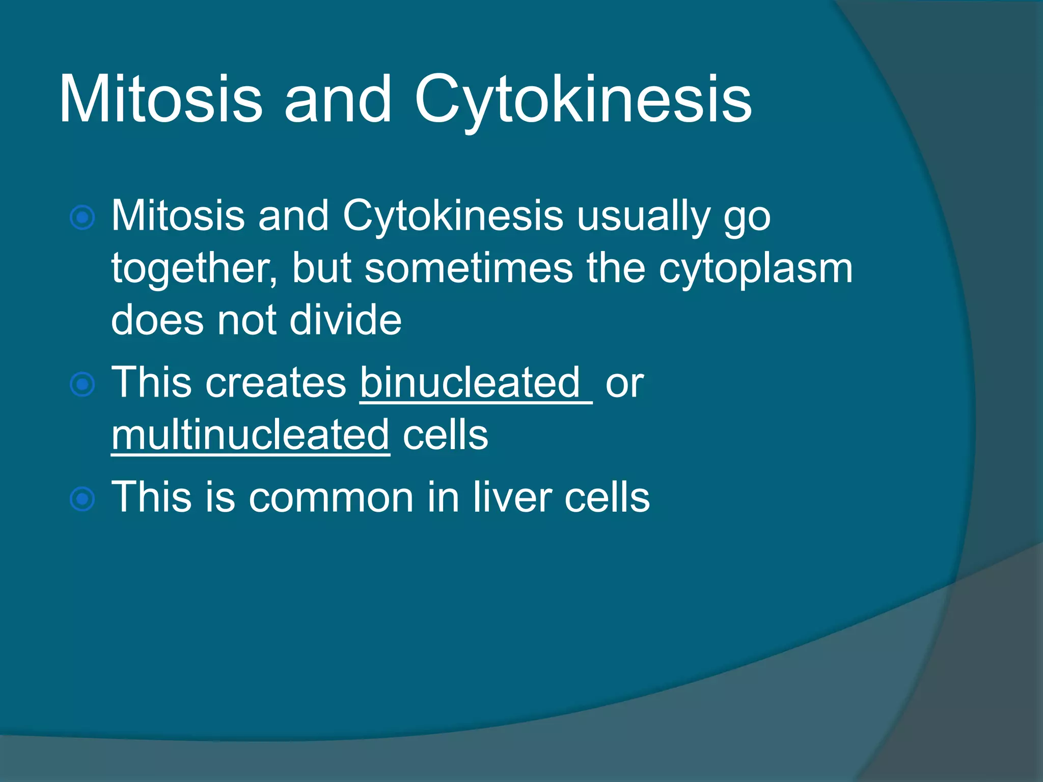
Mitosis and Cytokinesis
Mitosis and Cytokinesis usually go
together, but sometimes the cytoplasm
does not divide
This creates binucleated or
multinucleated cells
This is common in liver cells
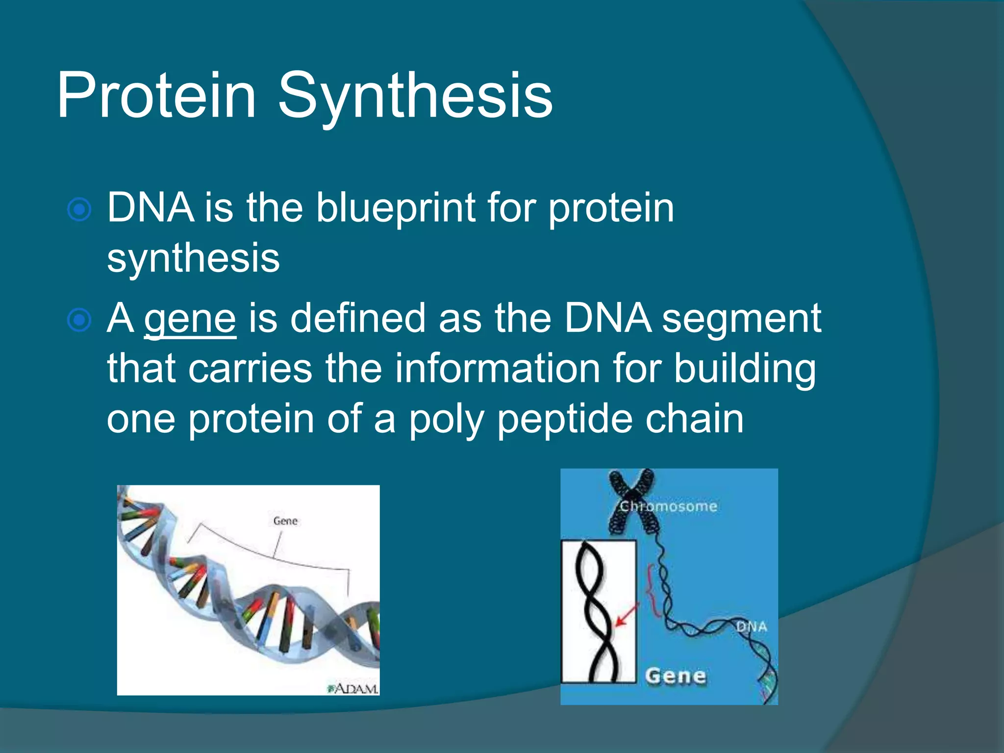
Protein Synthesis
DNAis the blueprint for protein
synthesis
A gene is defined as the DNA segment
that carries the information for building
one protein of a poly peptide chain
109.
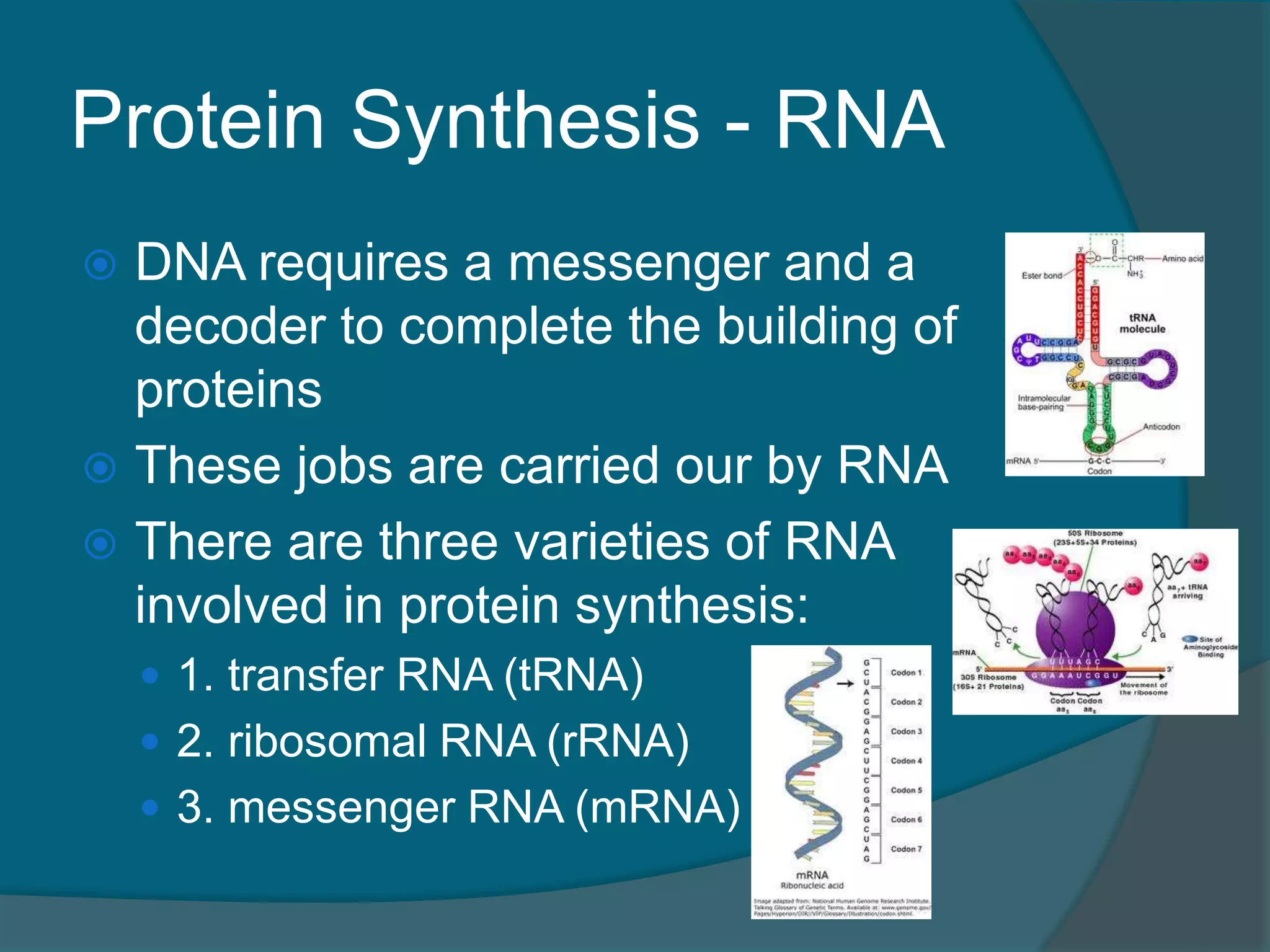
Protein Synthesis -RNA
DNA requires a messenger and a
decoder to complete the building of
proteins
These jobs are carried our by RNA
There are three varieties of RNA
involved in protein synthesis:
1. transfer RNA (tRNA)
2. ribosomal RNA (rRNA)
3. messenger RNA (mRNA)
110.
Protein Synthesis
ProteinSynthesis occurs in two major
phases:
1. Transcription – when complementary
mRNA is made at the DNA gene
2. Translation – when the information carried
in the mRNA molecules is “decoded” and
used to assemble proteins
111.
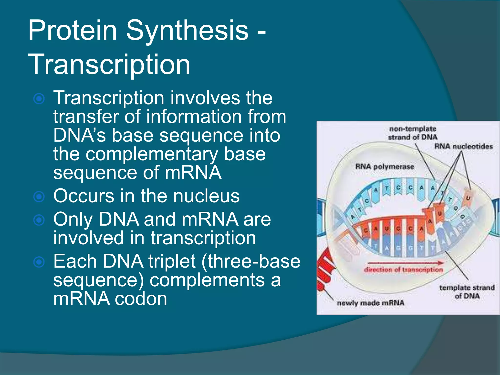
Protein Synthesis -
Transcription
Transcription involves the
transfer of information from
DNA’s base sequence into
the complementary base
sequence of mRNA
Occurs in the nucleus
Only DNA and mRNA are
involved in transcription
Each DNA triplet (three-base
sequence) complements a
mRNA codon
112.
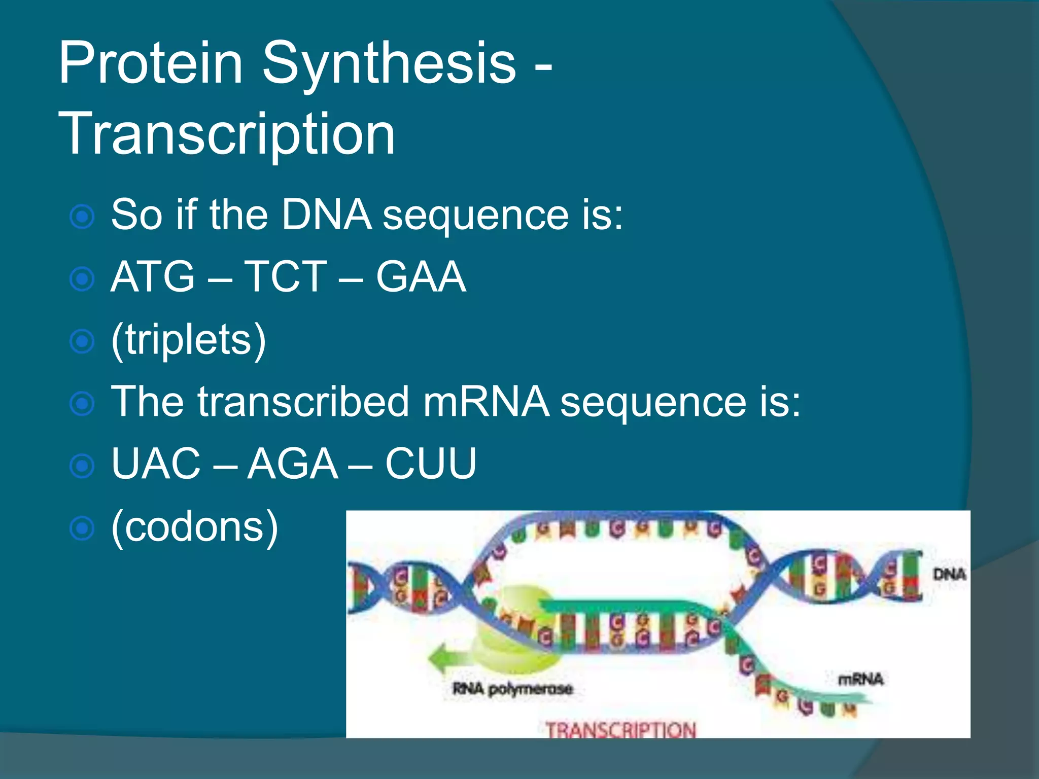
Protein Synthesis -
Transcription
So if the DNA sequence is:
ATG – TCT – GAA
(triplets)
The transcribed mRNA sequence is:
UAC – AGA – CUU
(codons)
113.
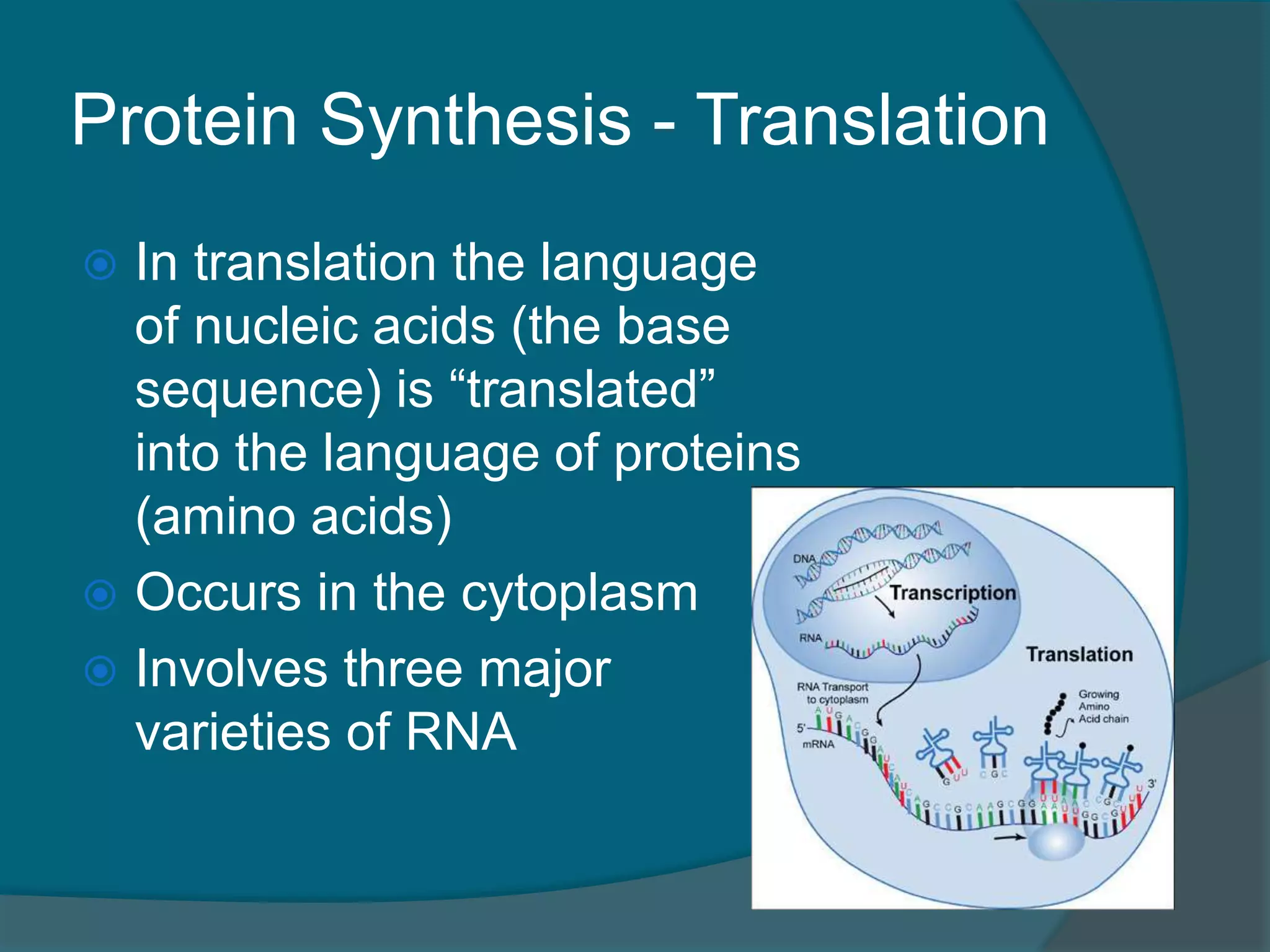
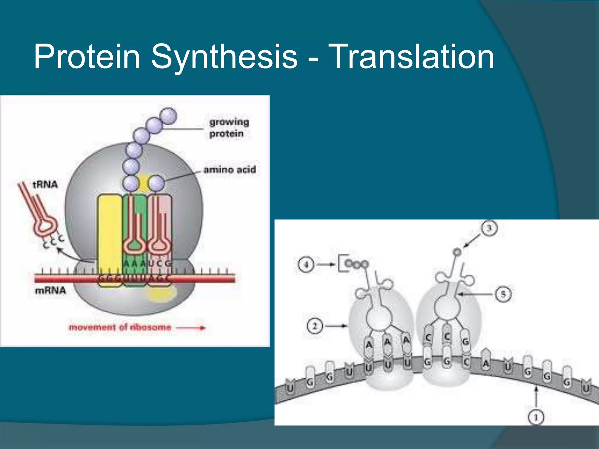
Protein Synthesis -Translation
In translation the language
of nucleic acids (the base
sequence) is “translated”
into the language of proteins
(amino acids)
Occurs in the cytoplasm
Involves three major
varieties of RNA
114.
Protein Synthesis -Translation
Once the mRNA attaches to the
ribosome, tRNA comes into the picture
Each tRNA carries or “transfers” an
amino acid to the ribosome
They match a three-base anticodon
with the codon of the mRNA as it reads
through the ribsome
Protein Synthesis -Translation
Once the first tRNA has moved itself into
the correct position, the ribosome moves
the mRNA strand along, bringing the next
codon into position to be read by the tRNA
As each amino acid is brought in, they are
joined together by enzymes
As the amino acids join, each tRNA is
released
When the last codon, or “stop” codon is
read, the protein is released
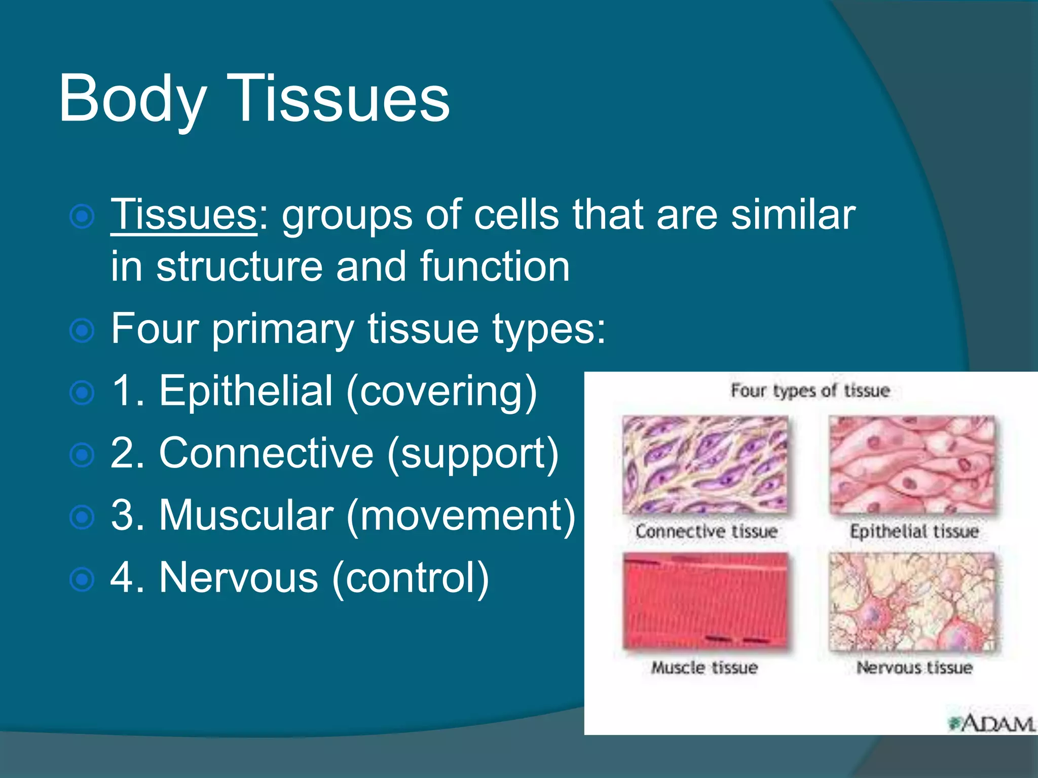
Body Tissues
Tissues:groups of cells that are similar
in structure and function
Four primary tissue types:
1. Epithelial (covering)
2. Connective (support)
3. Muscular (movement)
4. Nervous (control)
120.
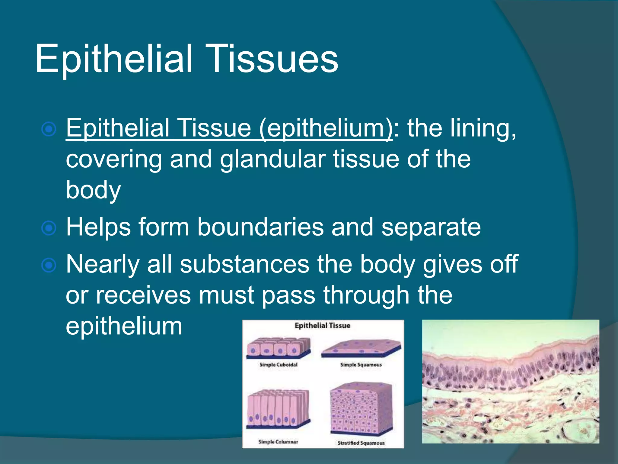
Epithelial Tissues
EpithelialTissue (epithelium): the lining,
covering and glandular tissue of the
body
Helps form boundaries and separate
Nearly all substances the body gives off
or receives must pass through the
epithelium
Epithelial Tissues -
Characteristics
1. Fit closely together (except glandular cells)
Bound together by many desmosomes and tight
junctions
2. One free edge or surface
Apical surface
3. Lower surface rests on a basement
membrane
4. No blood supply of their own
Avascular
Depend on diffusion from the capillaries
5. Regenerate themselves, if well nourished
Epithelial Tissues -
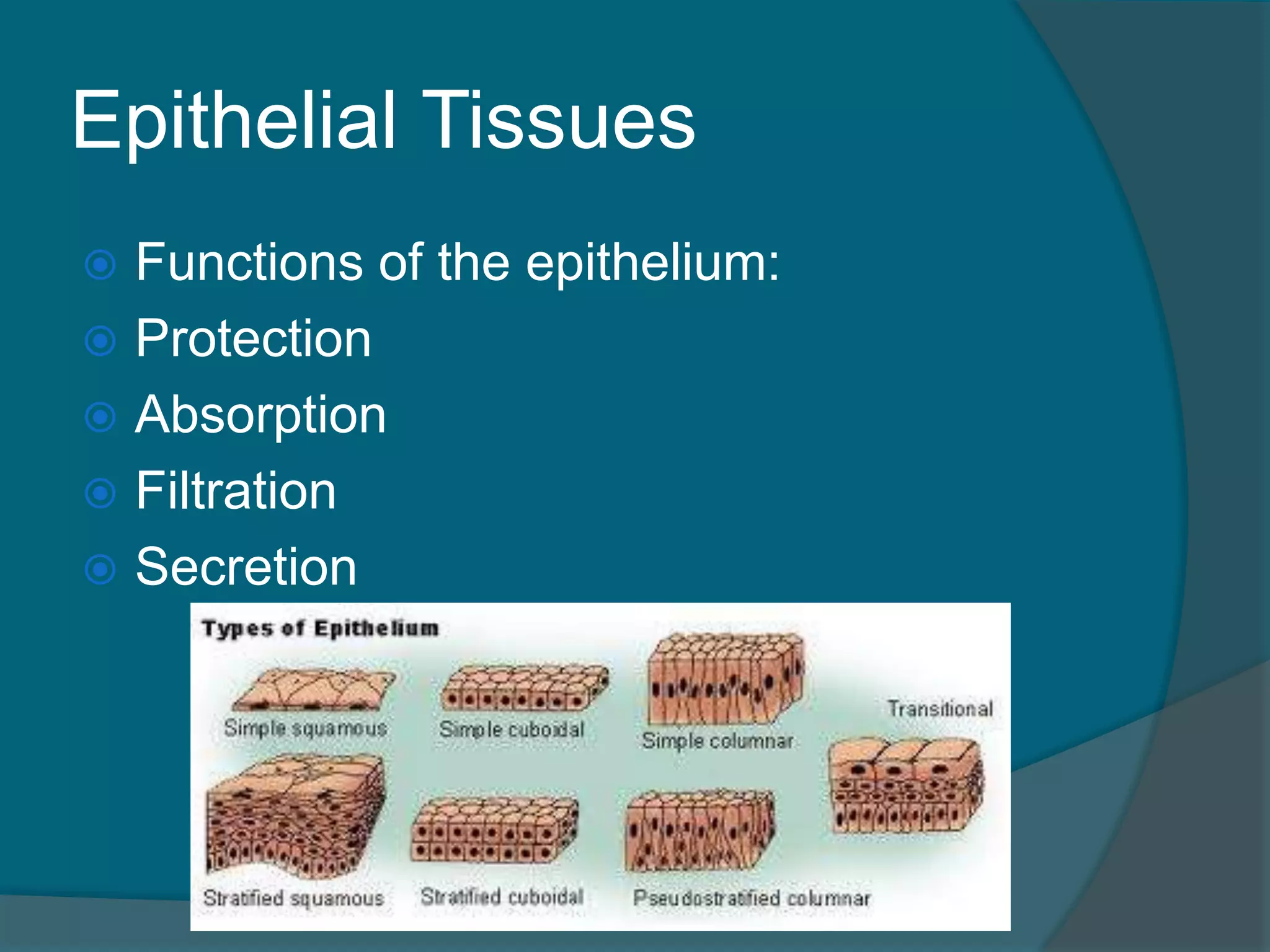
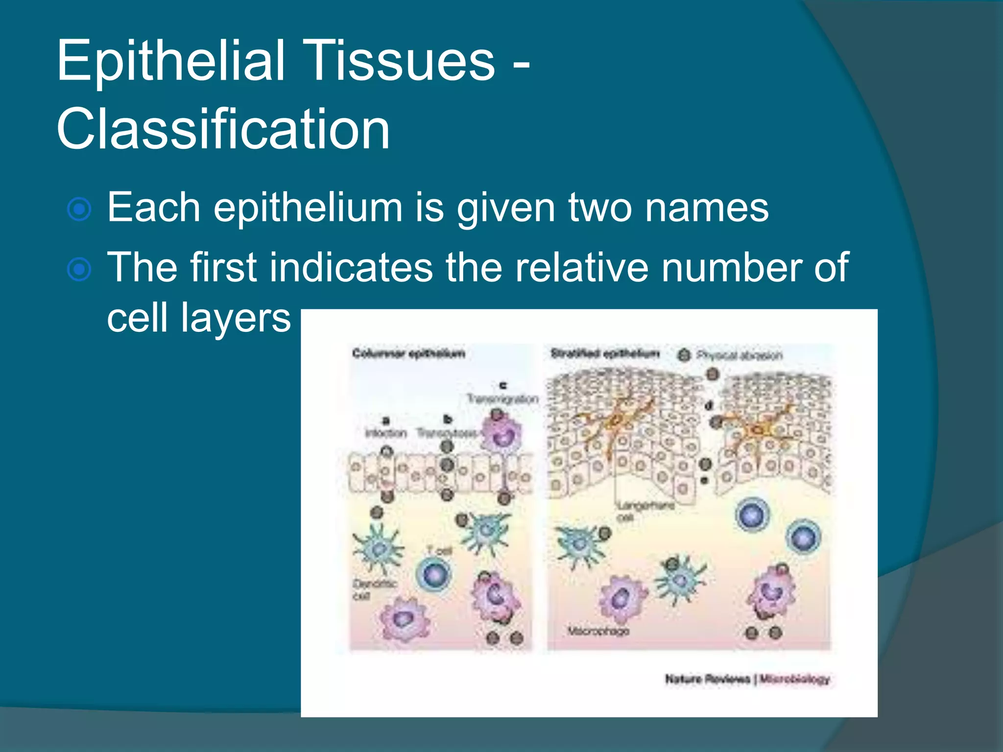
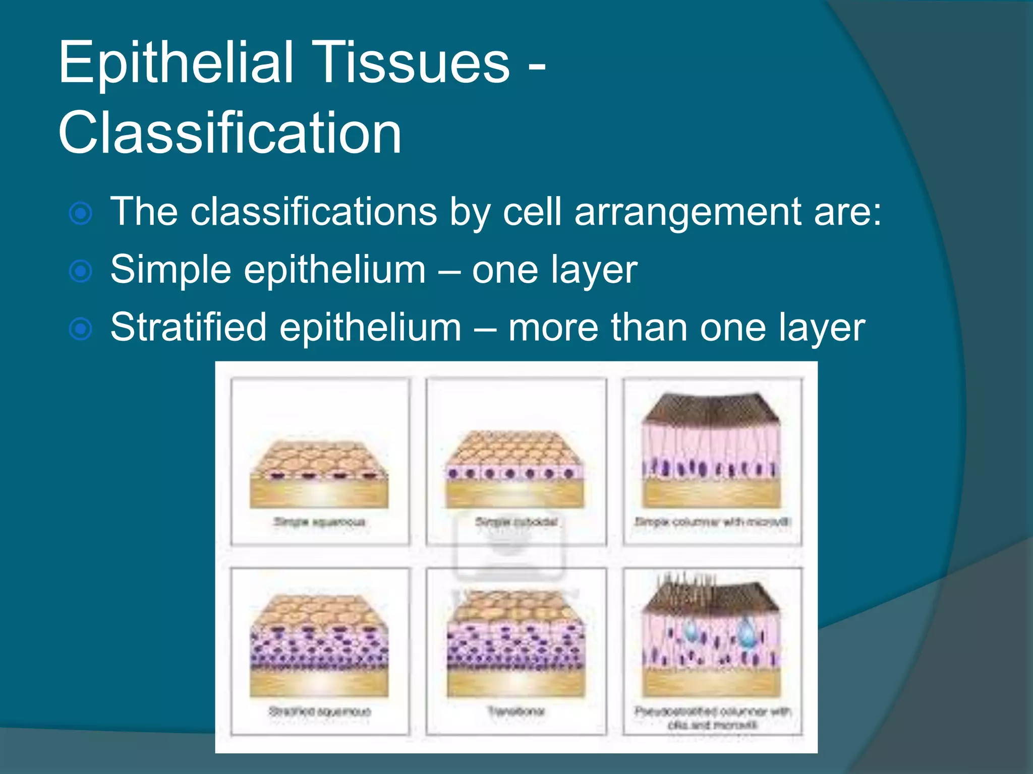
Classification
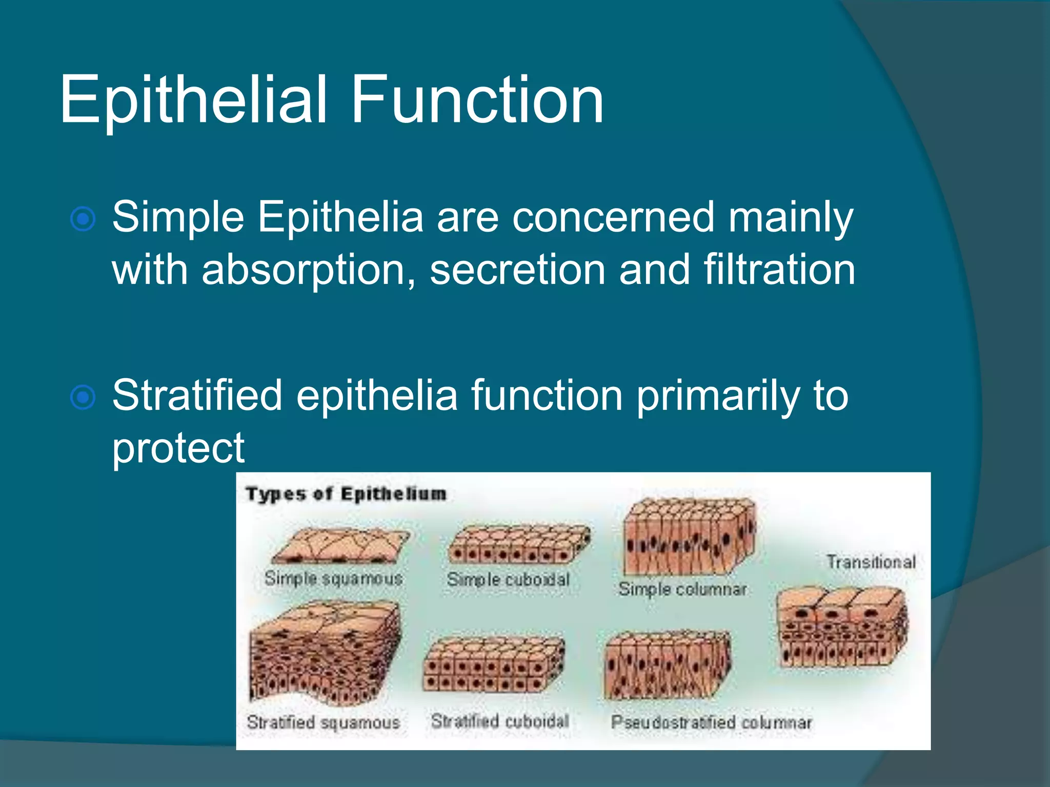
The classifications by cell arrangement are:
Simple epithelium – one layer
Stratified epithelium – more than one layer
125.
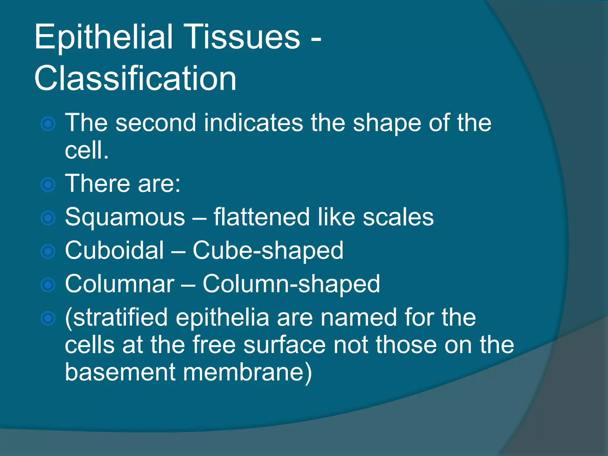
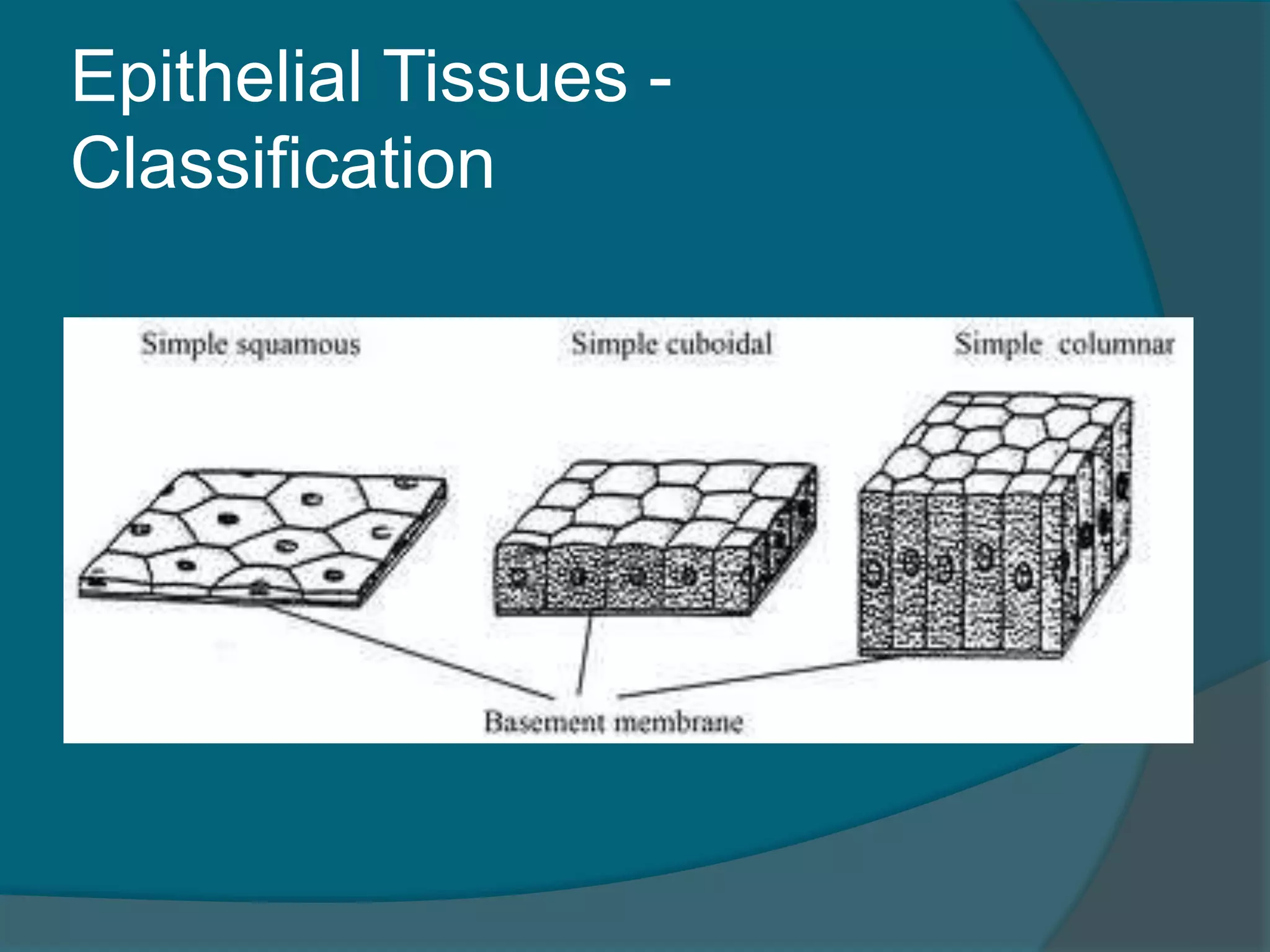
Epithelial Tissues -
Classification
The second indicates the shape of the
cell.
There are:
Squamous – flattened like scales
Cuboidal – Cube-shaped
Columnar – Column-shaped
(stratified epithelia are named for the
cells at the free surface not those on the
basement membrane)
Epithelial Function
SimpleEpithelia are concerned mainly
with absorption, secretion and filtration
Stratified epithelia function primarily to
protect
128.
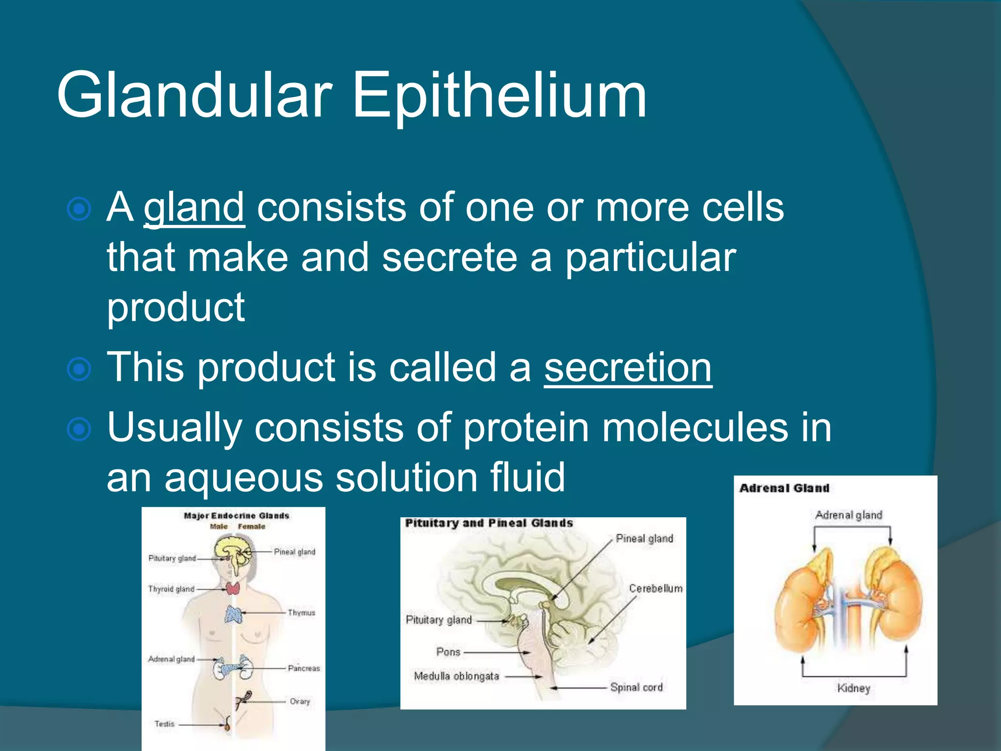
Glandular Epithelium
Agland consists of one or more cells
that make and secrete a particular
product
This product is called a secretion
Usually consists of protein molecules in
an aqueous solution fluid
129.
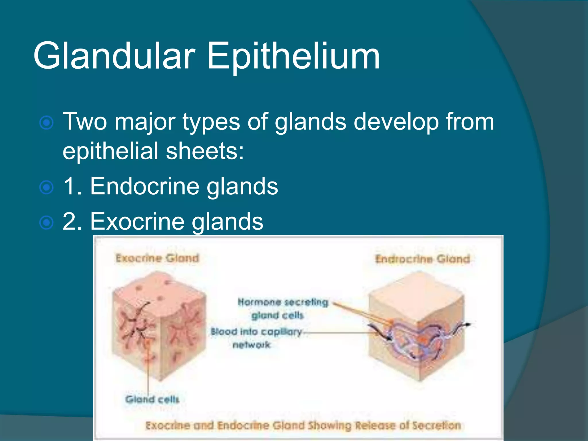
Glandular Epithelium
Twomajor types of glands develop from
epithelial sheets:
1. Endocrine glands
2. Exocrine glands
130.
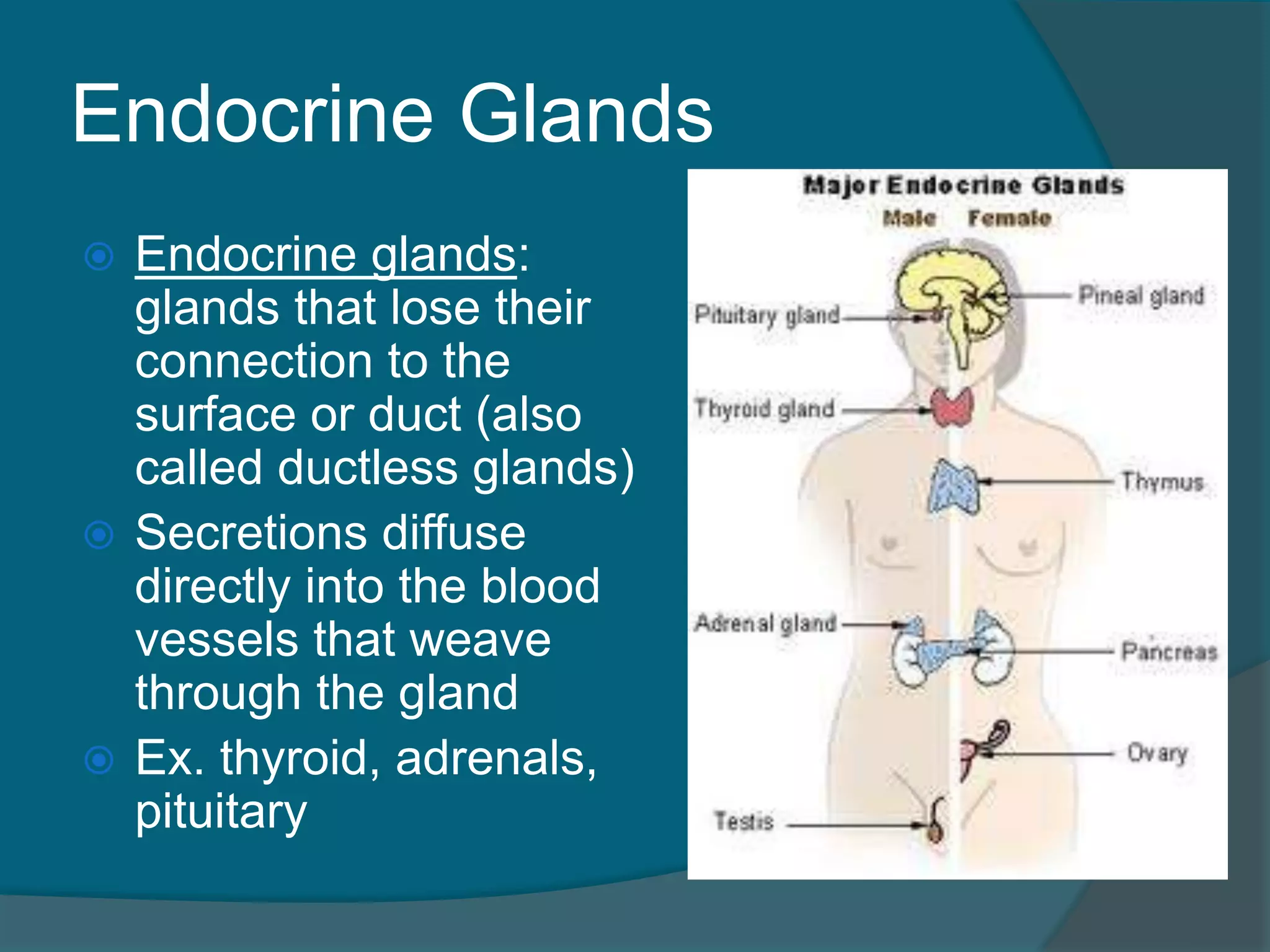
Endocrine Glands
Endocrineglands:
glands that lose their
connection to the
surface or duct (also
called ductless glands)
Secretions diffuse
directly into the blood
vessels that weave
through the gland
Ex. thyroid, adrenals,
pituitary
131.
Exocrine Glands
Exocrineglands:
gland that retain
their ducts
Secretions empty
through the ducts to
the epithelial
surface
Ex. sweat and oil
glands, liver,
pancreas
132.
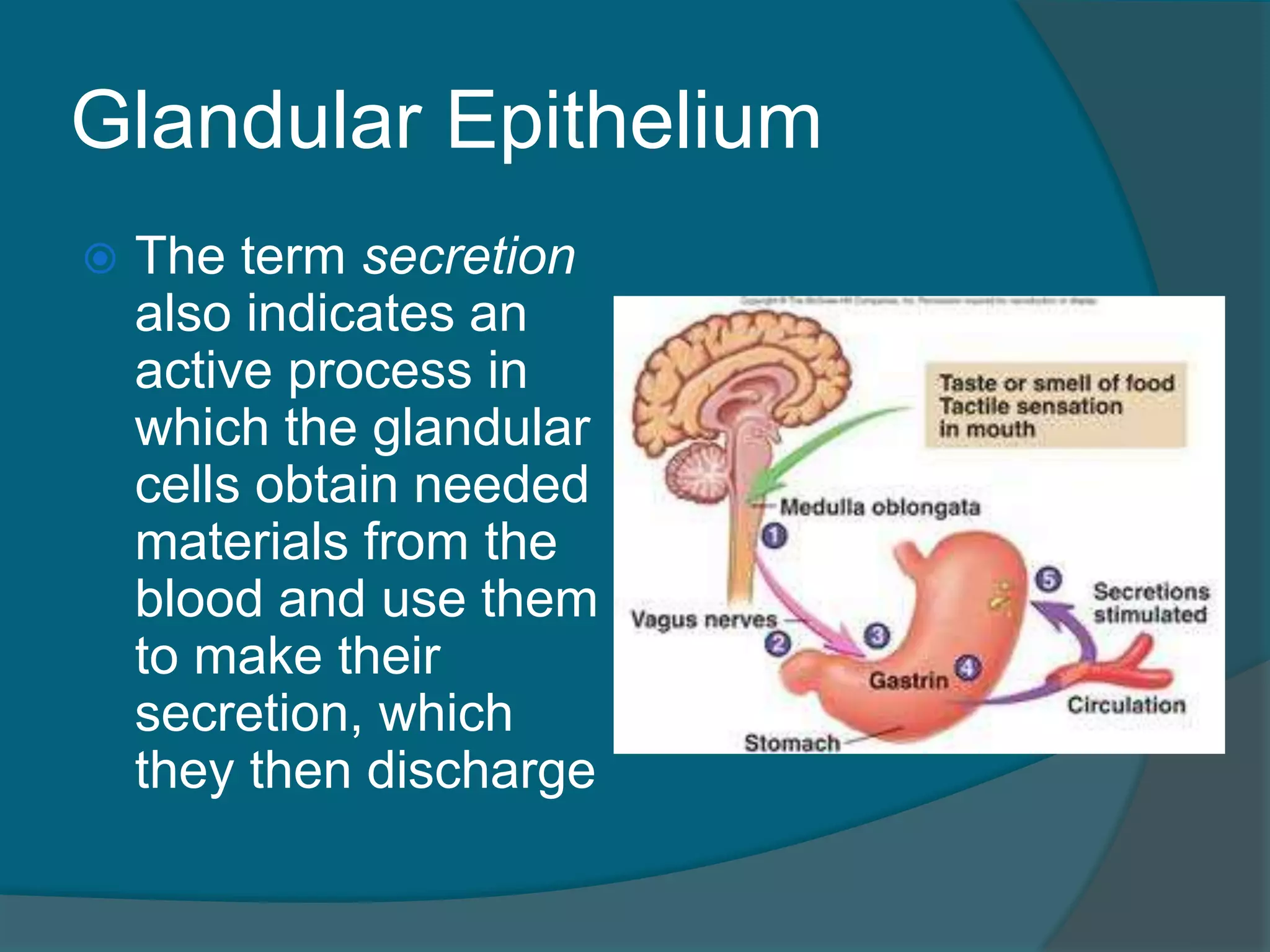
Glandular Epithelium
Theterm secretion
also indicates an
active process in
which the glandular
cells obtain needed
materials from the
blood and use them
to make their
secretion, which
they then discharge
134.
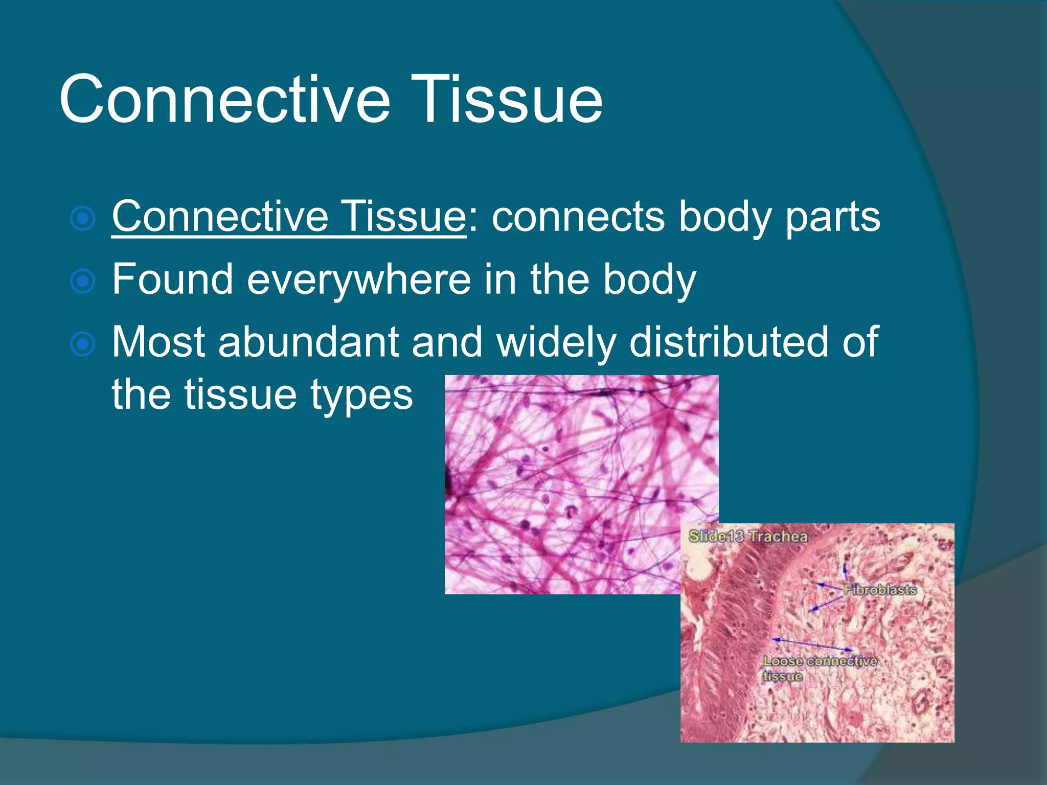
Connective Tissue
ConnectiveTissue: connects body parts
Found everywhere in the body
Most abundant and widely distributed of
the tissue types
135.
Connective Tissue
Thecharacteristics of connective tissue
include:
1. Variations in blood supply
Most connective tissue is well vascularized
Exceptions – Ligaments, Tendons,
Cartilages
○ As a result these heal very slowly
2. Extracellular Matrix
Varying amounts of a nonliving substance
outside the cells
136.
Connective Tissue
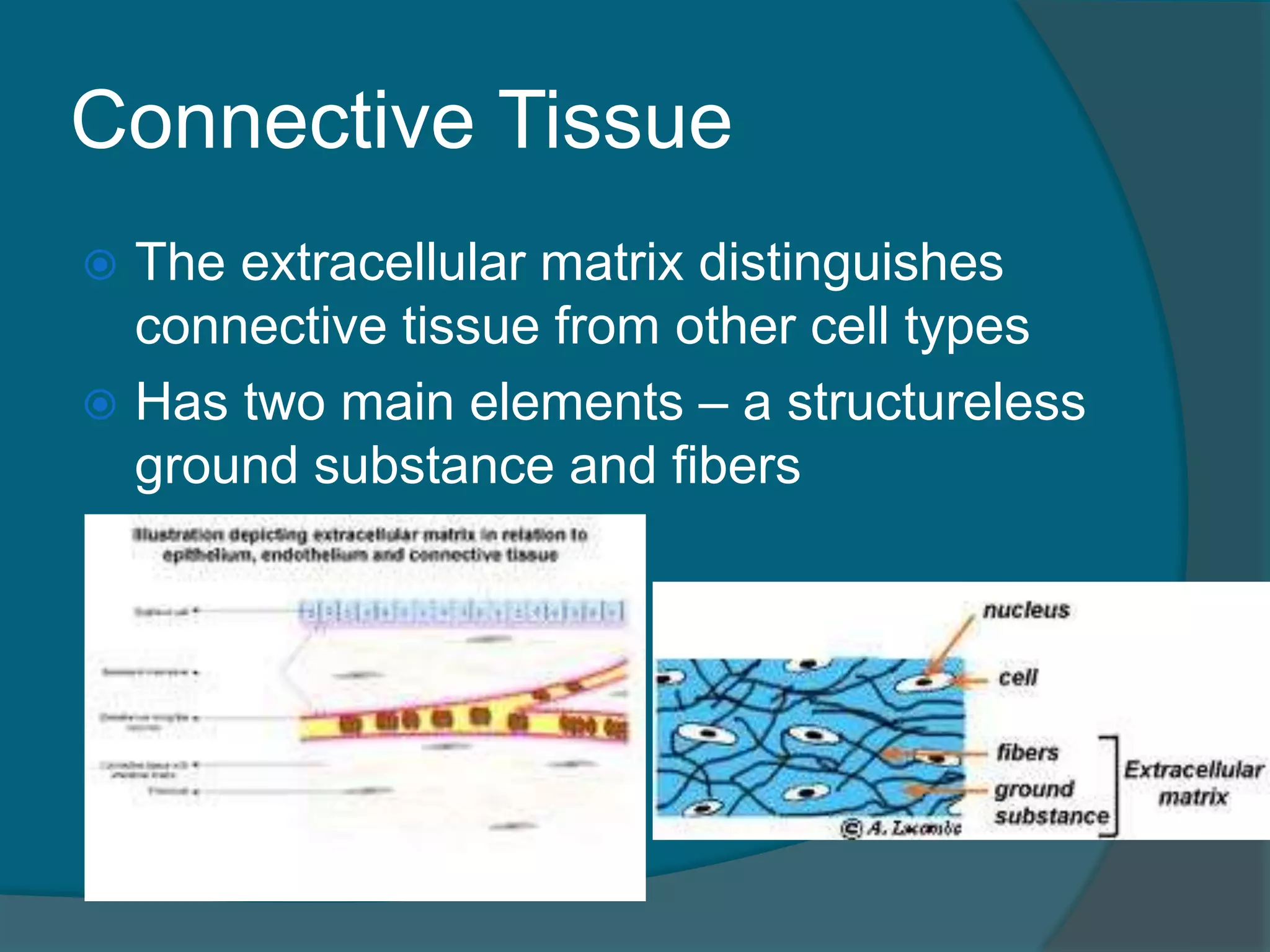
Theextracellular matrix distinguishes
connective tissue from other cell types
Has two main elements – a structureless
ground substance and fibers
137.
Connective Tissue
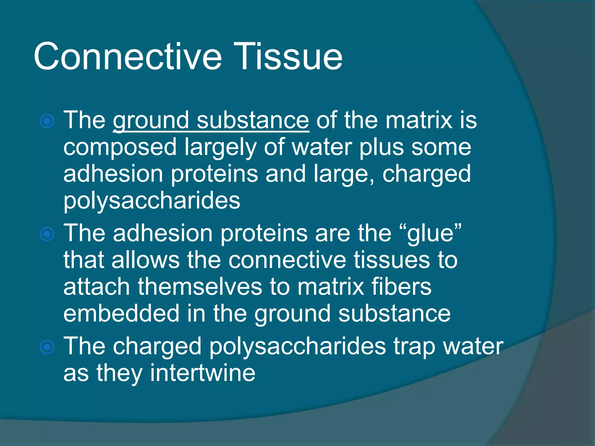
Theground substance of the matrix is
composed largely of water plus some
adhesion proteins and large, charged
polysaccharides
The adhesion proteins are the “glue”
that allows the connective tissues to
attach themselves to matrix fibers
embedded in the ground substance
The charged polysaccharides trap water
as they intertwine
138.
Connective Tissue
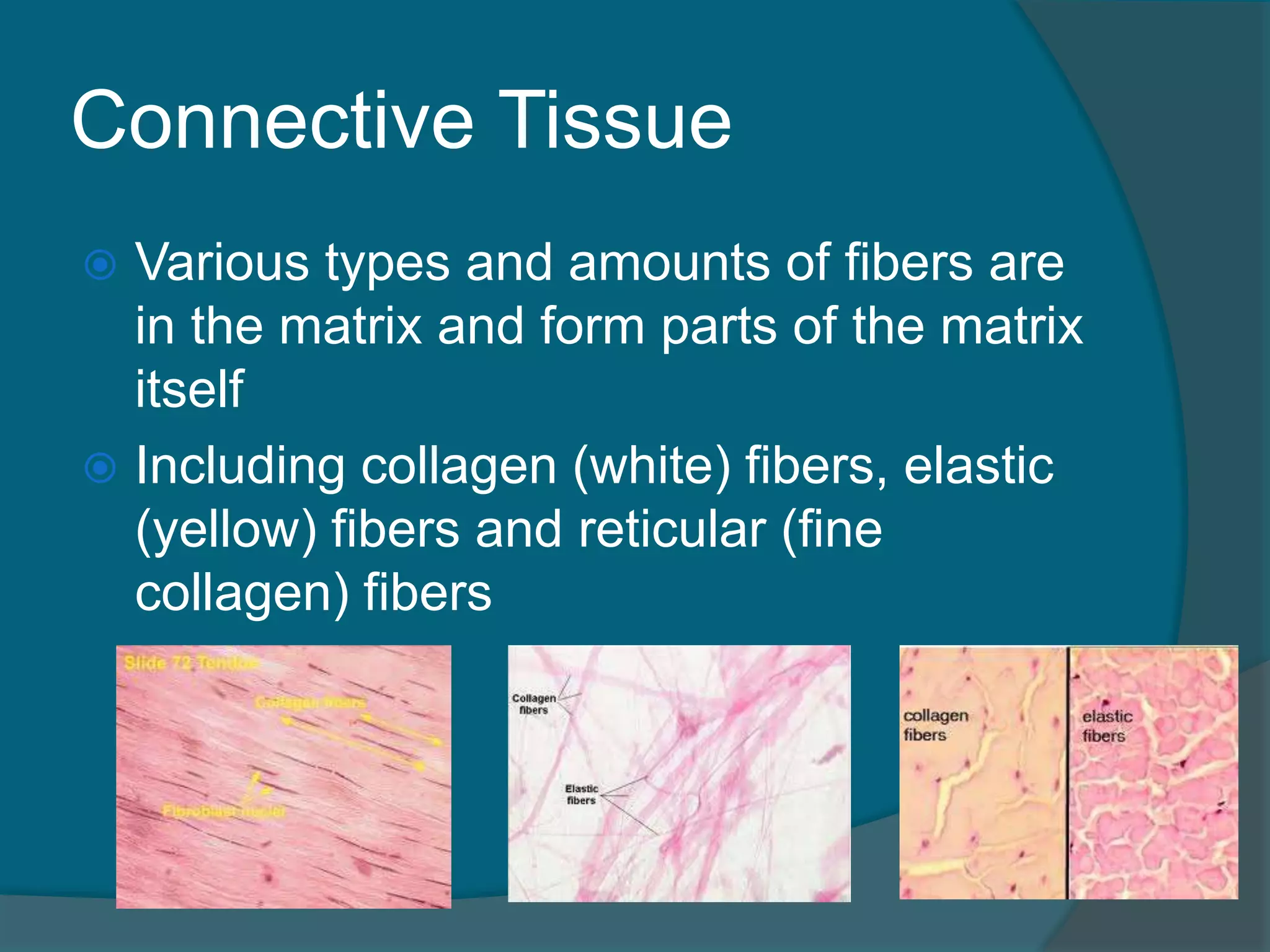
Varioustypes and amounts of fibers are
in the matrix and form parts of the matrix
itself
Including collagen (white) fibers, elastic
(yellow) fibers and reticular (fine
collagen) fibers
139.
Connective Tissues
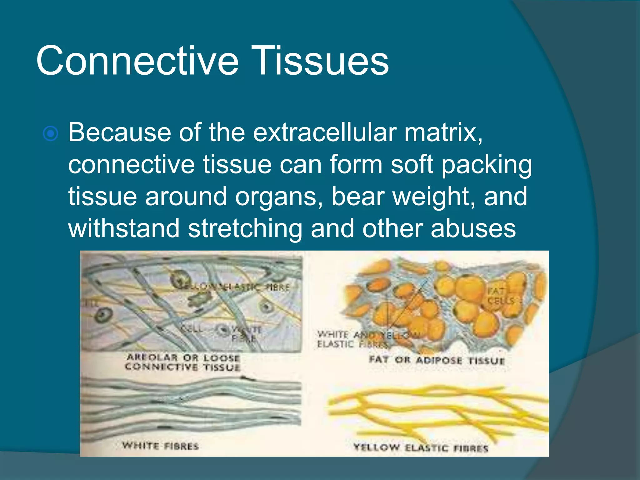
Becauseof the extracellular matrix,
connective tissue can form soft packing
tissue around organs, bear weight, and
withstand stretching and other abuses
140.
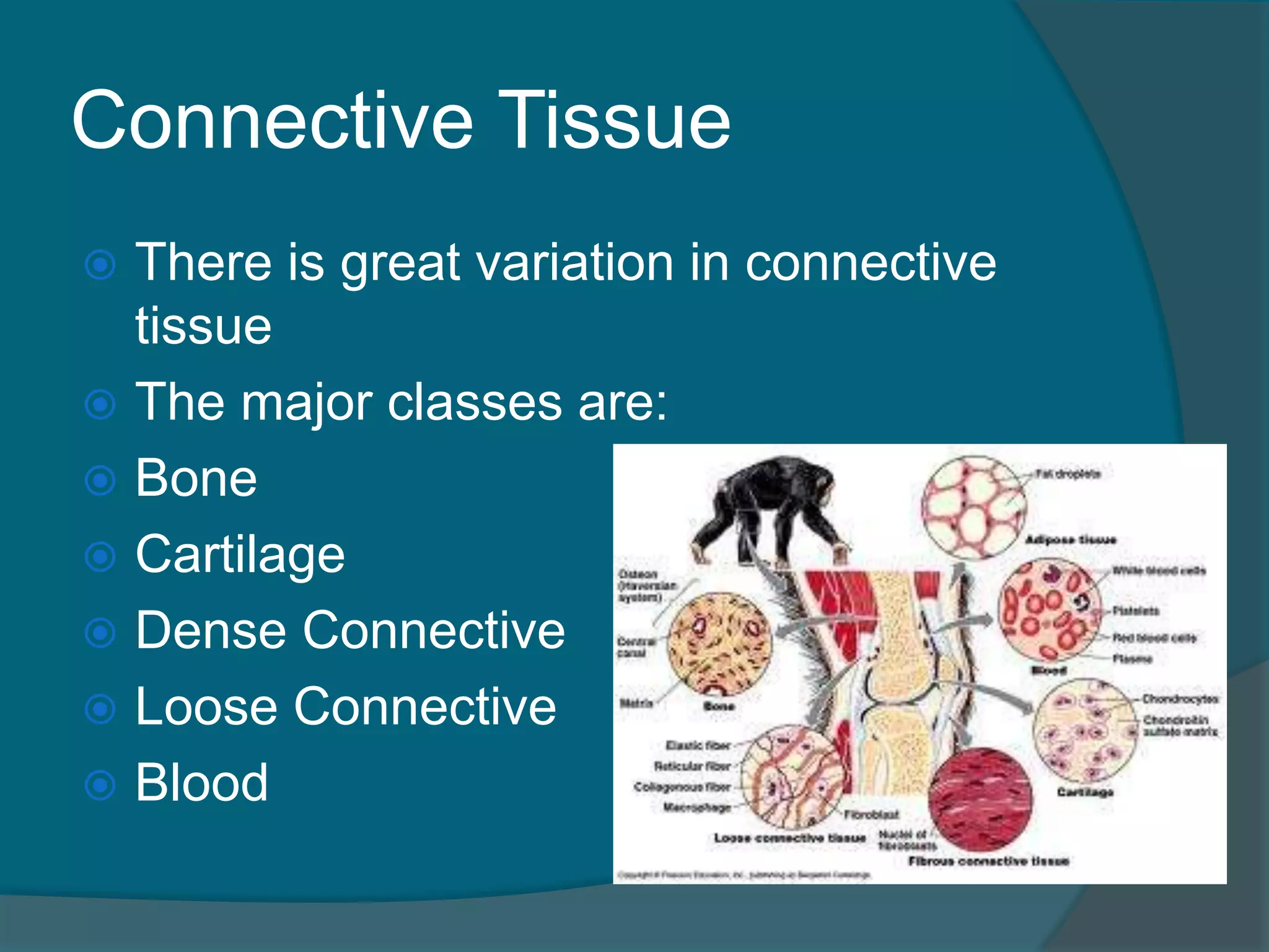
Connective Tissue
Thereis great variation in connective
tissue
The major classes are:
Bone
Cartilage
Dense Connective
Loose Connective
Blood
141.
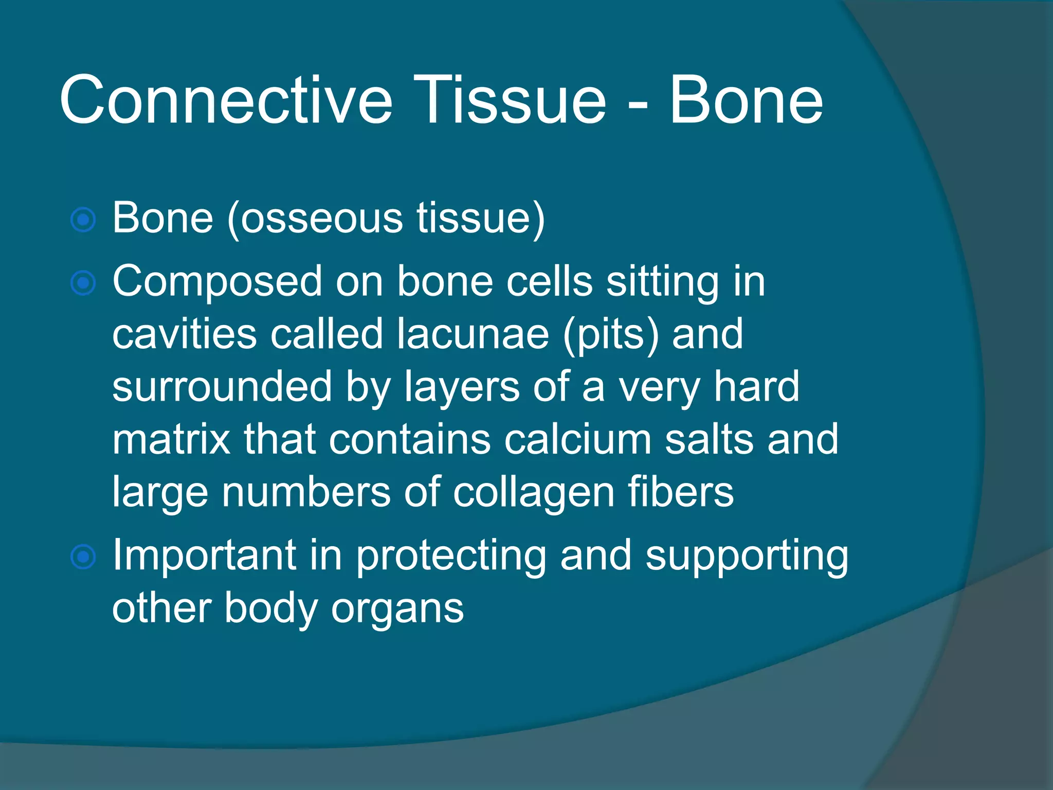
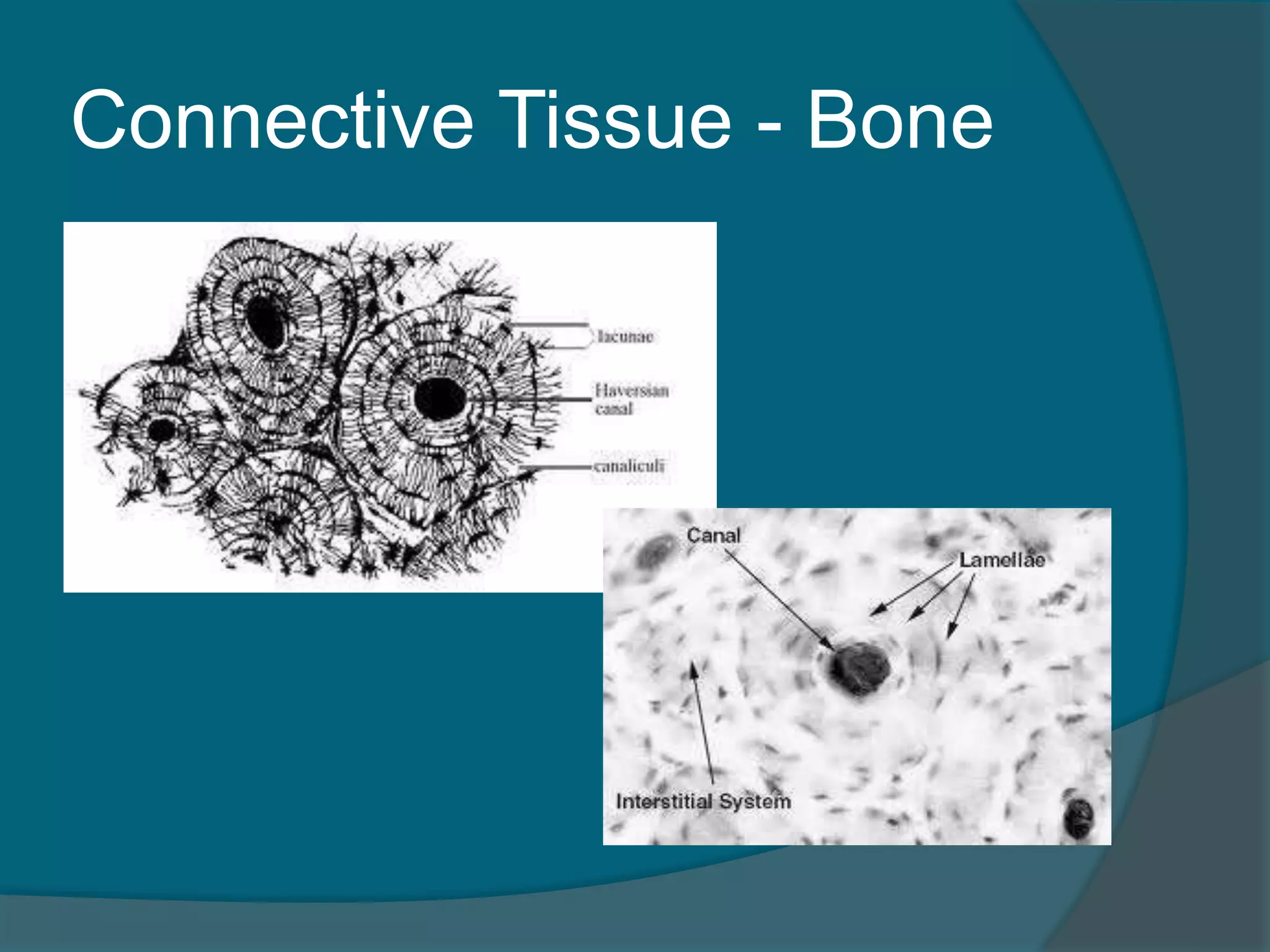
Connective Tissue -Bone
Bone (osseous tissue)
Composed on bone cells sitting in
cavities called lacunae (pits) and
surrounded by layers of a very hard
matrix that contains calcium salts and
large numbers of collagen fibers
Important in protecting and supporting
other body organs
Connective Tissue -
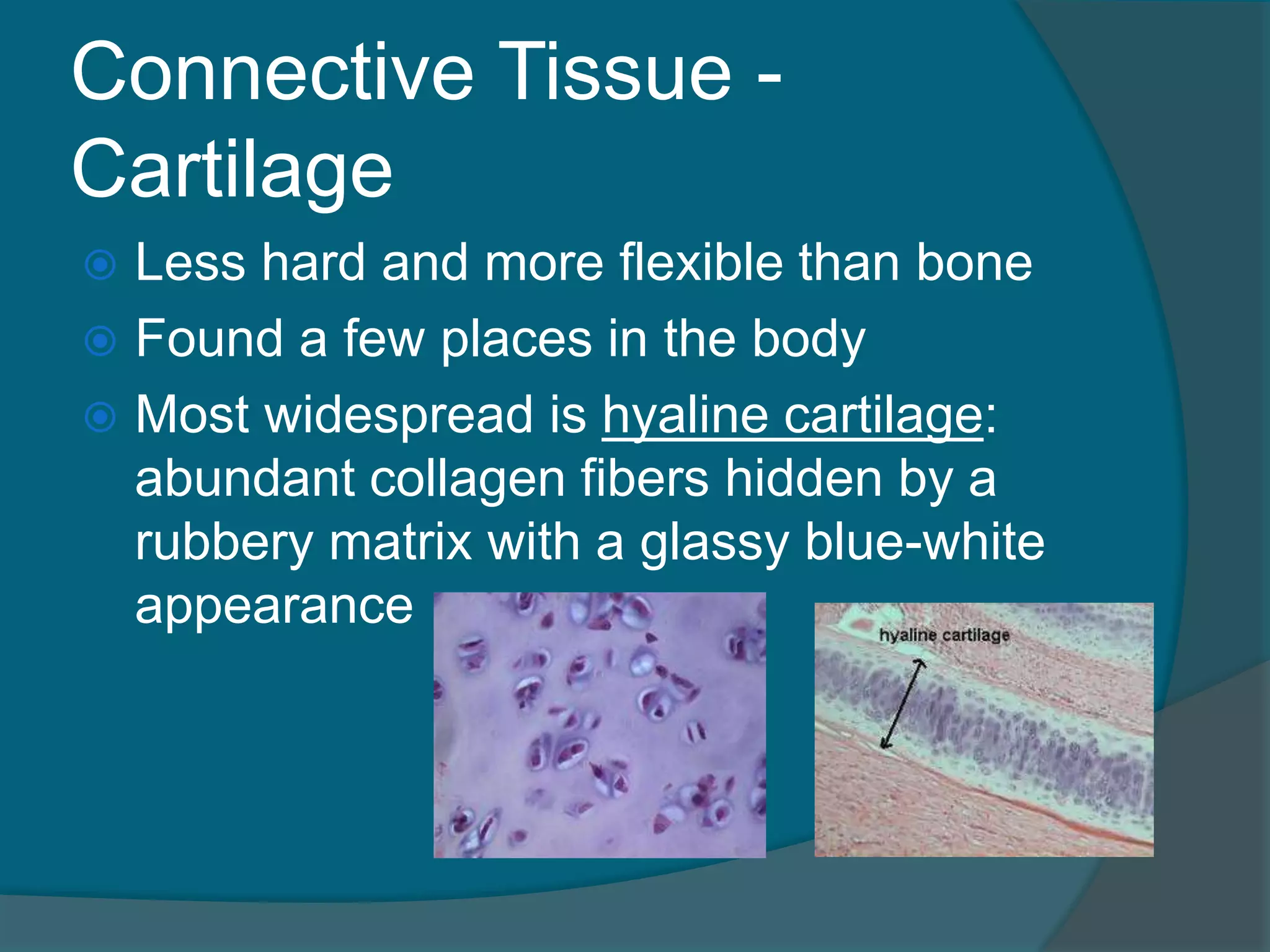
Cartilage
Less hard and more flexible than bone
Found a few places in the body
Most widespread is hyaline cartilage:
abundant collagen fibers hidden by a
rubbery matrix with a glassy blue-white
appearance
144.
Connective Tissue -
Cartilage
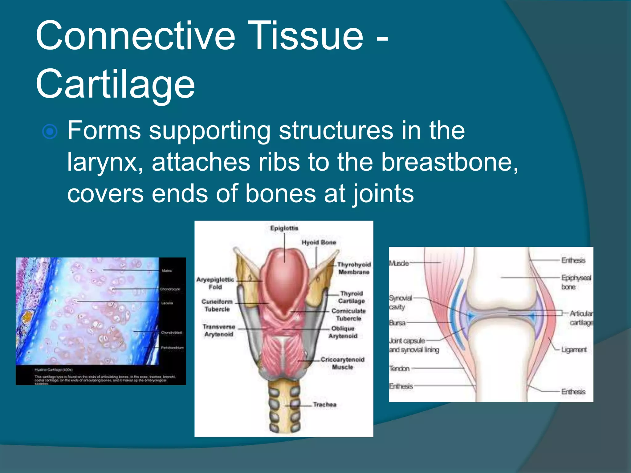
Forms supporting structures in the
larynx, attaches ribs to the breastbone,
covers ends of bones at joints
145.
Connective Tissue
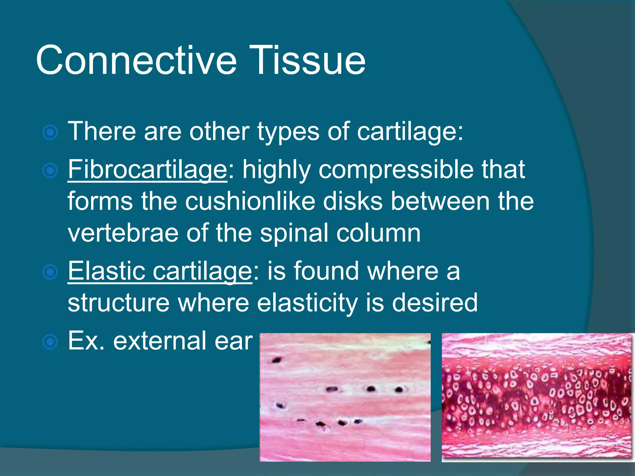
Thereare other types of cartilage:
Fibrocartilage: highly compressible that
forms the cushionlike disks between the
vertebrae of the spinal column
Elastic cartilage: is found where a
structure where elasticity is desired
Ex. external ear
146.
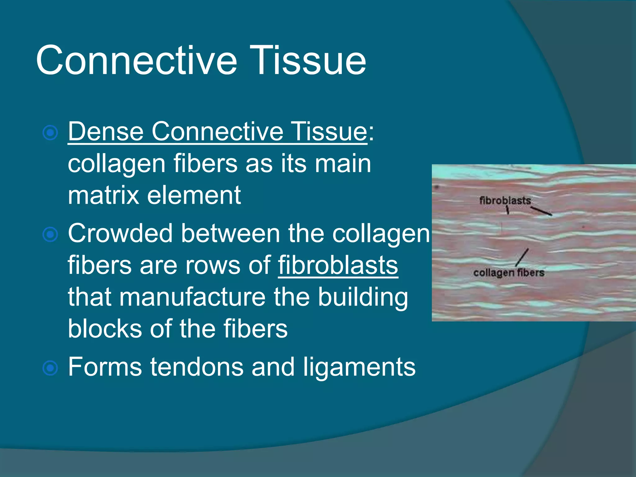
Connective Tissue
DenseConnective Tissue:
collagen fibers as its main
matrix element
Crowded between the collagen
fibers are rows of fibroblasts
that manufacture the building
blocks of the fibers
Forms tendons and ligaments
147.
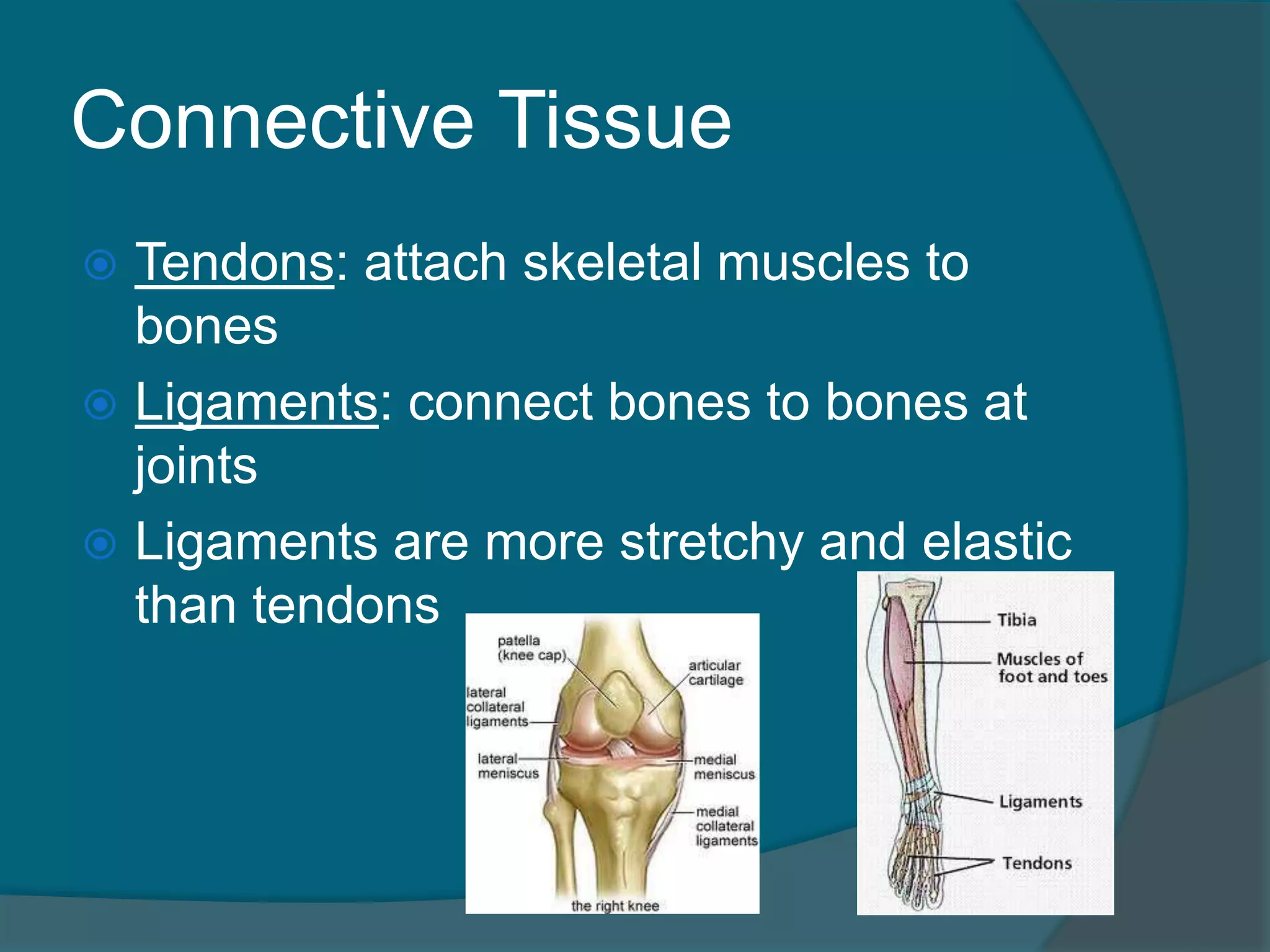
Connective Tissue
Tendons:attach skeletal muscles to
bones
Ligaments: connect bones to bones at
joints
Ligaments are more stretchy and elastic
than tendons
148.
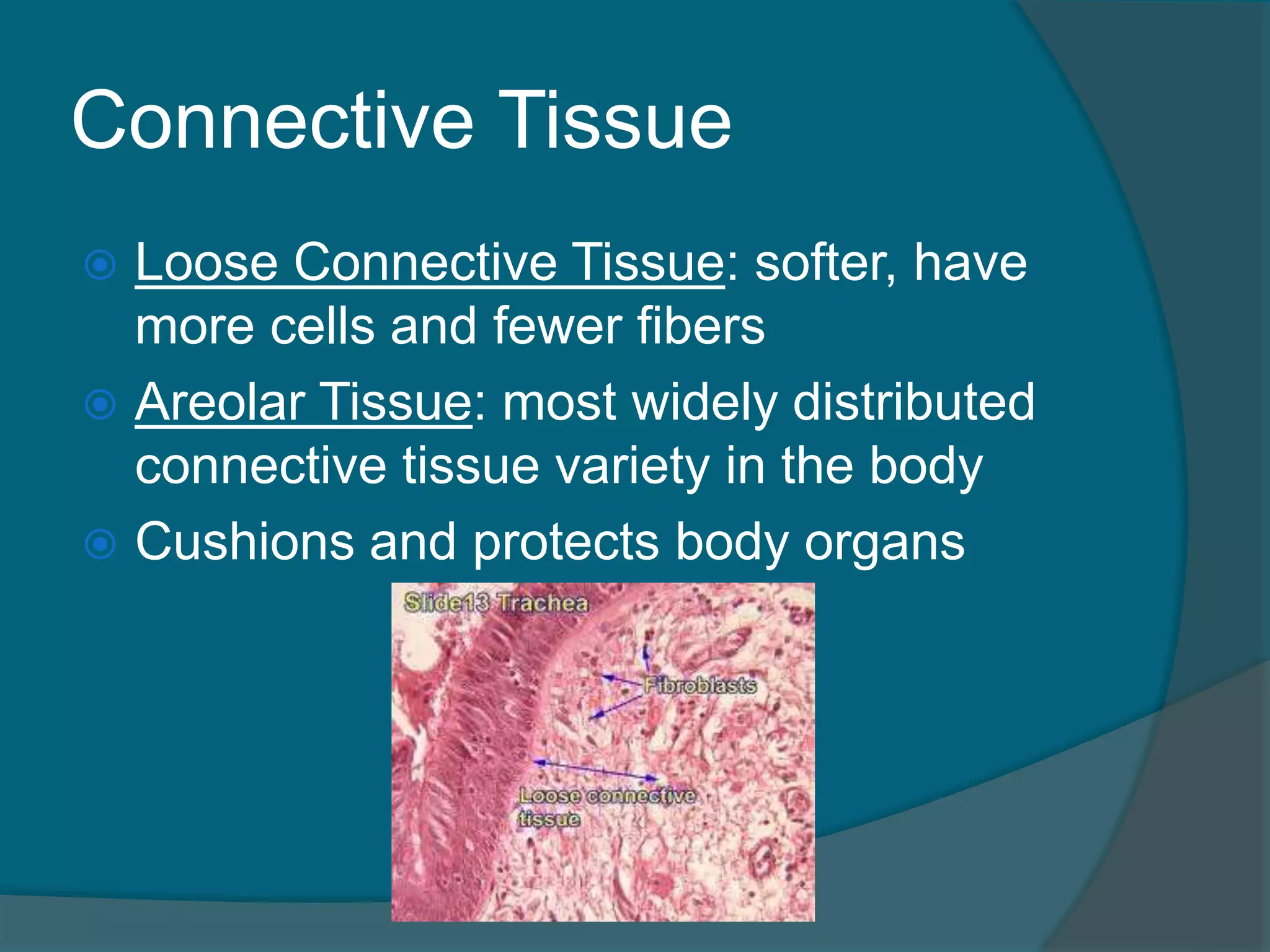
Connective Tissue
LooseConnective Tissue: softer, have
more cells and fewer fibers
Areolar Tissue: most widely distributed
connective tissue variety in the body
Cushions and protects body organs
149.
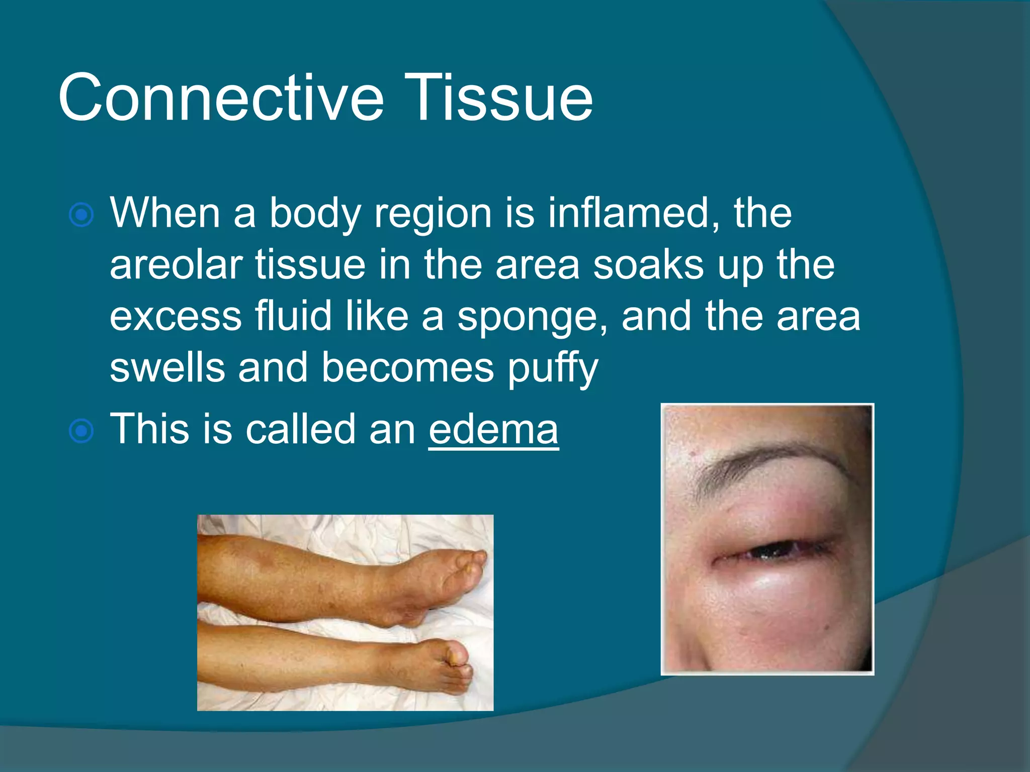
Connective Tissue
Whena body region is inflamed, the
areolar tissue in the area soaks up the
excess fluid like a sponge, and the area
swells and becomes puffy
This is called an edema
150.
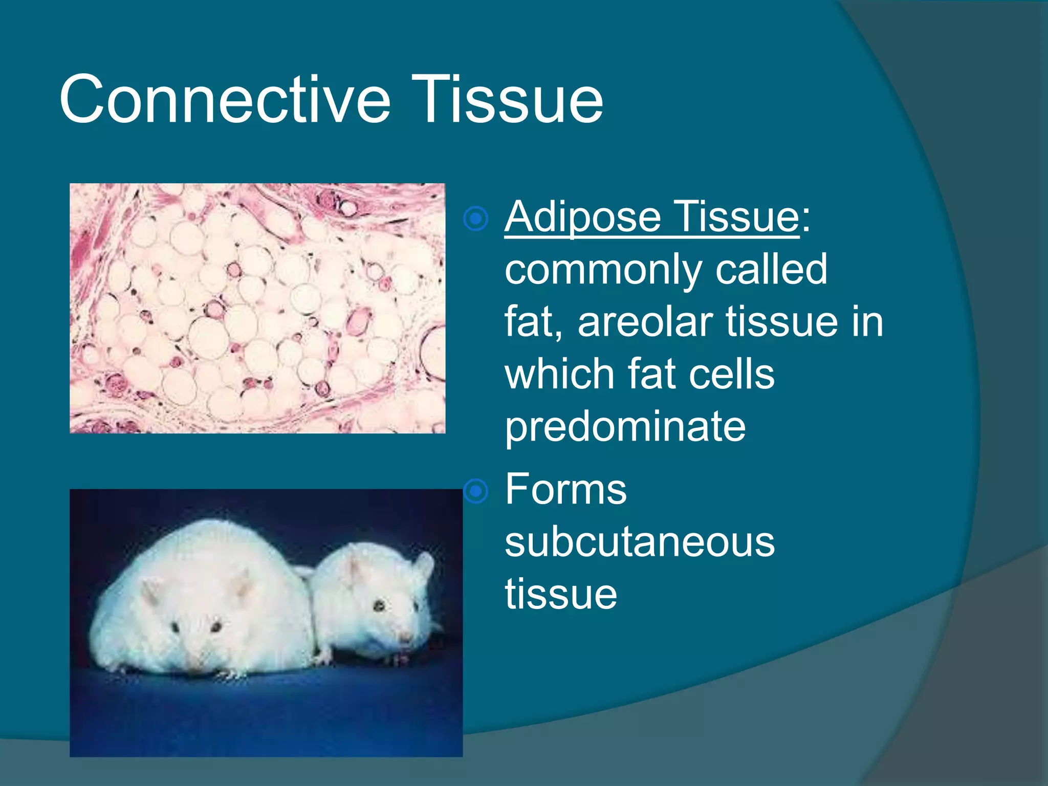
Connective Tissue
AdiposeTissue:
commonly called
fat, areolar tissue in
which fat cells
predominate
Forms
subcutaneous
tissue
151.
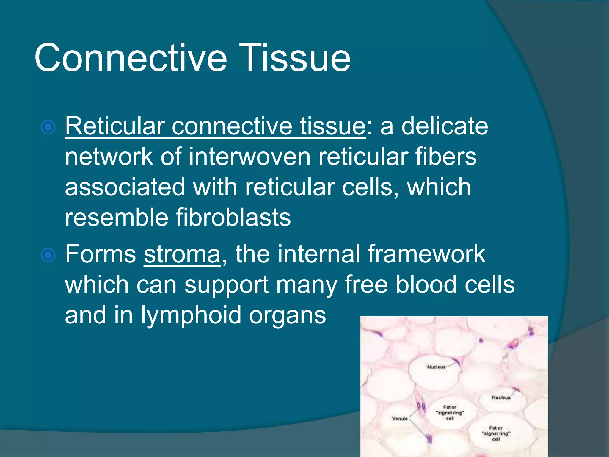
Connective Tissue
Reticularconnective tissue: a delicate
network of interwoven reticular fibers
associated with reticular cells, which
resemble fibroblasts
Forms stroma, the internal framework
which can support many free blood cells
and in lymphoid organs
152.
Connective Tissue
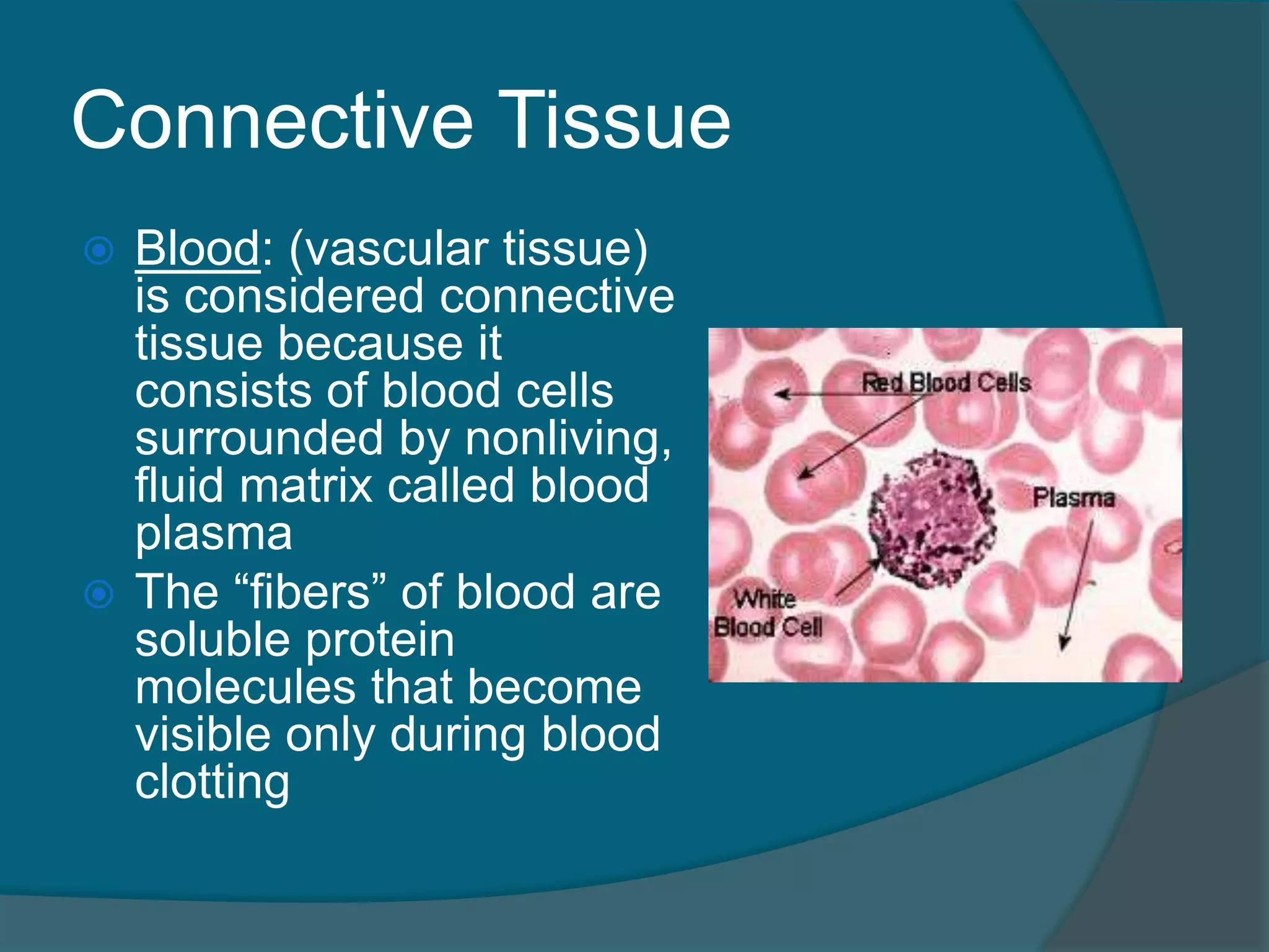
Blood:(vascular tissue)
is considered connective
tissue because it
consists of blood cells
surrounded by nonliving,
fluid matrix called blood
plasma
The “fibers” of blood are
soluble protein
molecules that become
visible only during blood
clotting
153.
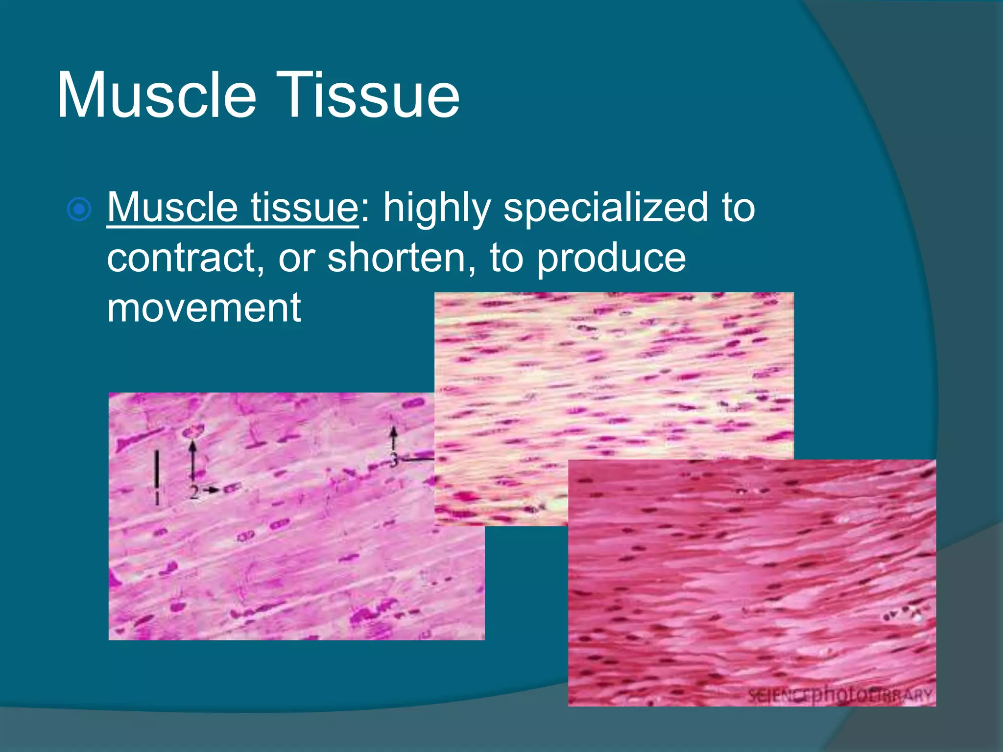
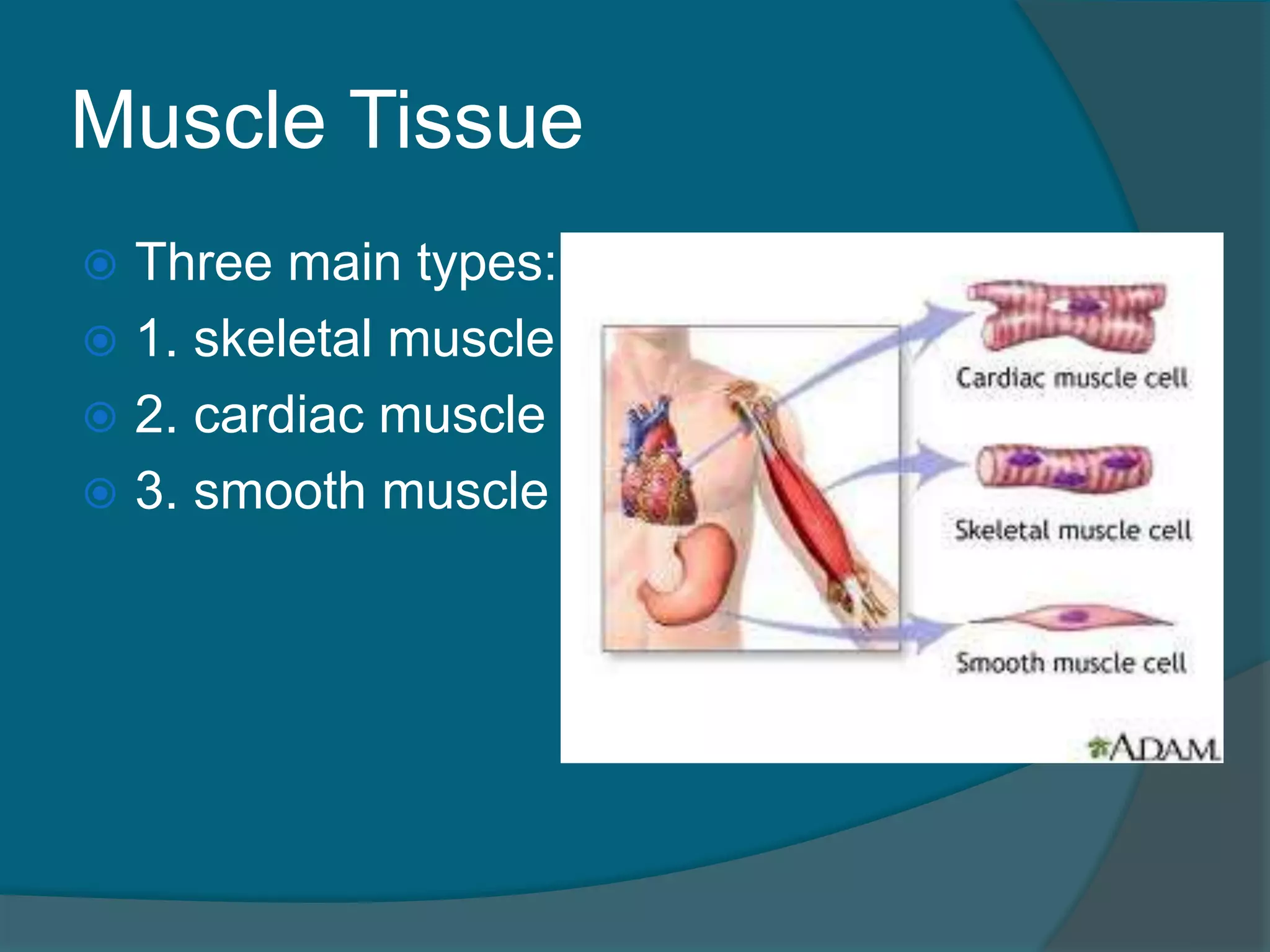
Muscle Tissue
Muscletissue: highly specialized to
contract, or shorten, to produce
movement
Muscle Tissue
SkeletalMuscle
attached to the skeleton
can be controlled voluntarily
when contracted they pull on
bones or skin
the cells of skeletal muscle
are long, cylindrical,
multinucleate and have
obvious striations
156.
Muscle Tissue
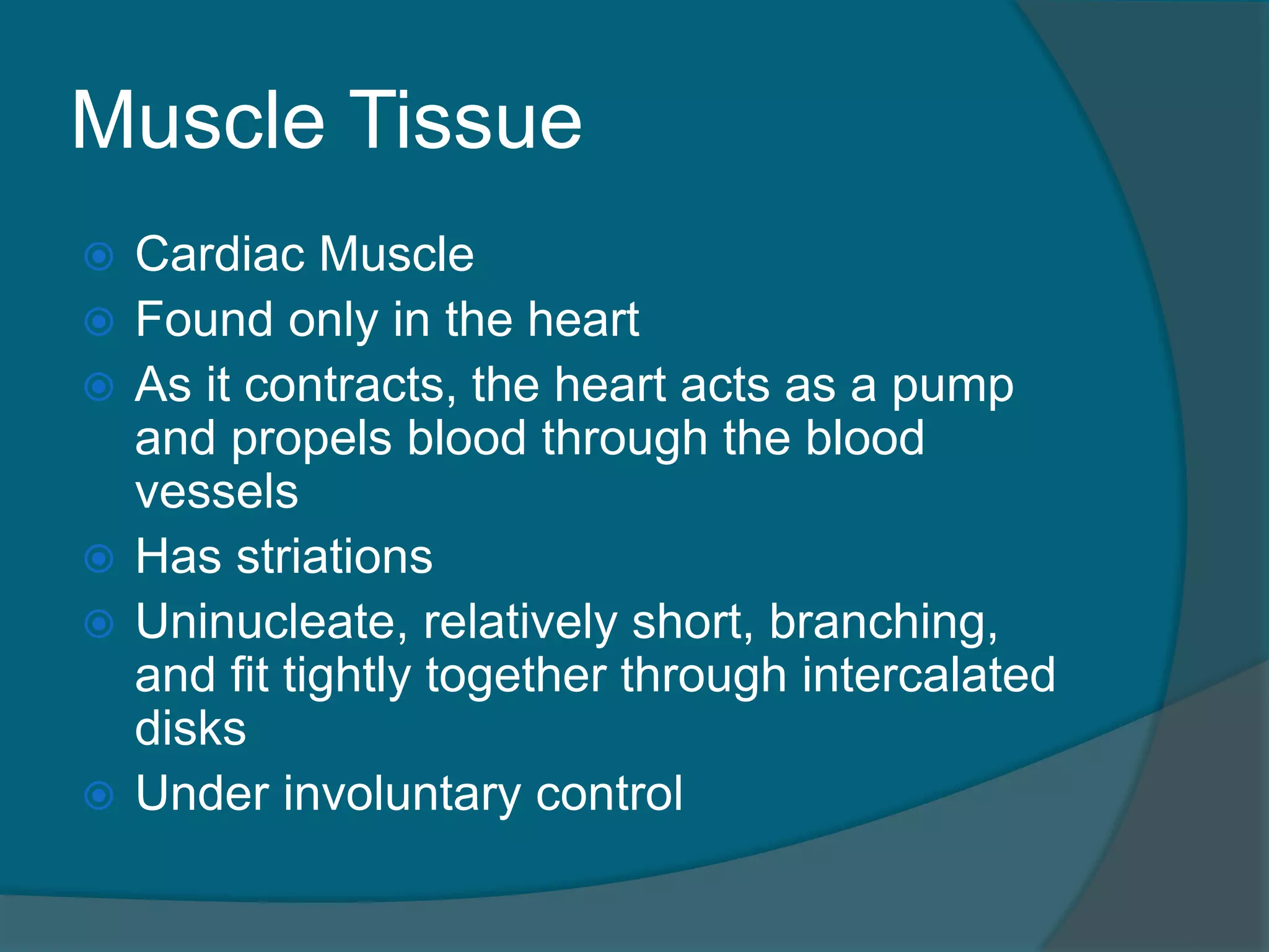
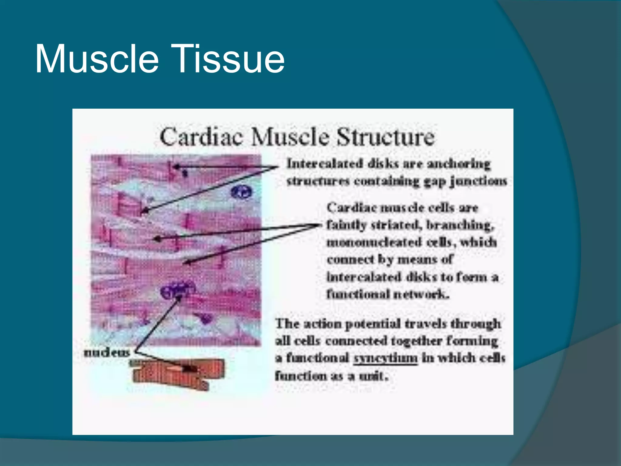
CardiacMuscle
Found only in the heart
As it contracts, the heart acts as a pump
and propels blood through the blood
vessels
Has striations
Uninucleate, relatively short, branching,
and fit tightly together through intercalated
disks
Under involuntary control
Muscle Tissue
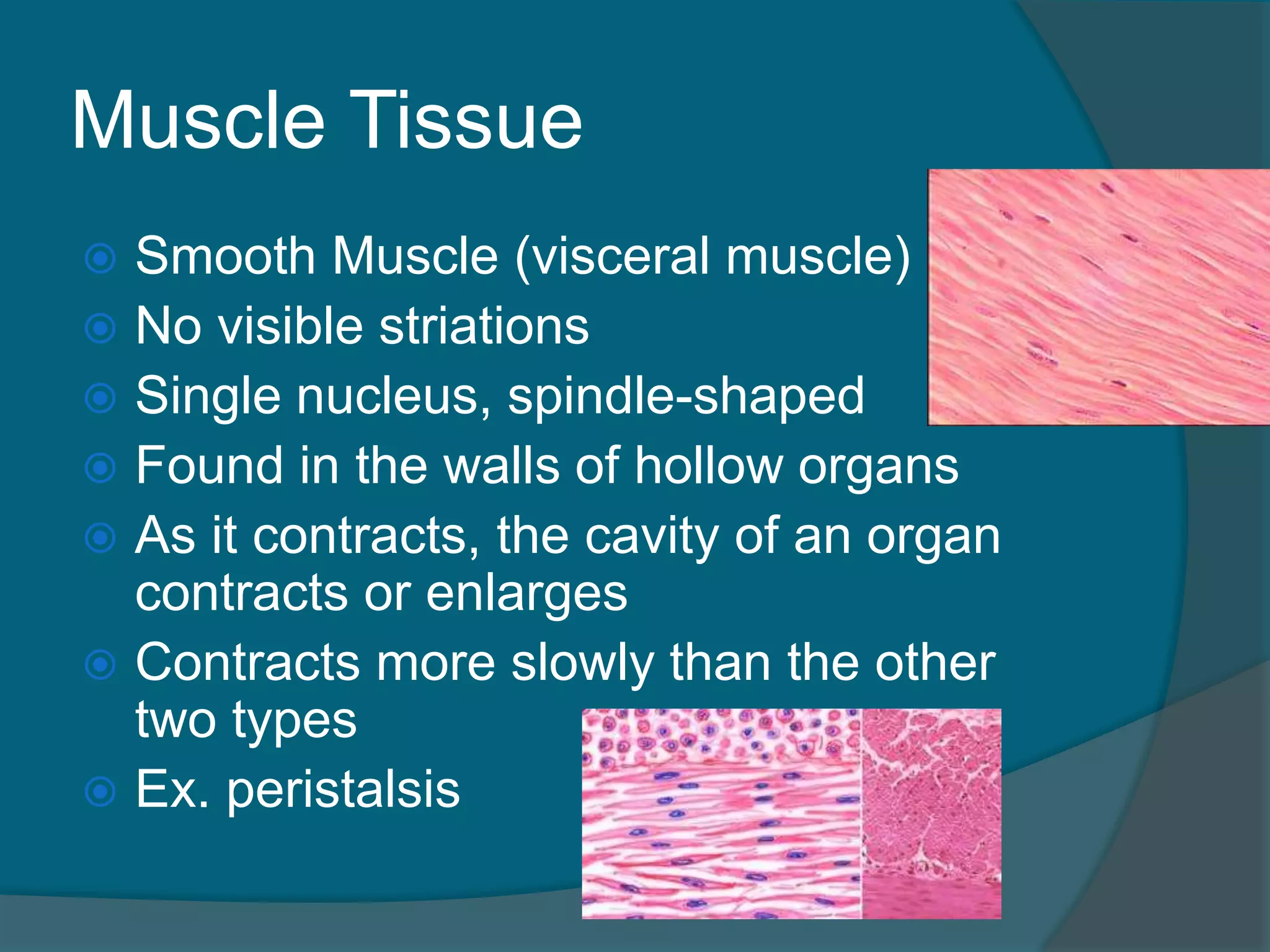
SmoothMuscle (visceral muscle)
No visible striations
Single nucleus, spindle-shaped
Found in the walls of hollow organs
As it contracts, the cavity of an organ
contracts or enlarges
Contracts more slowly than the other
two types
Ex. peristalsis
159.
Nervous Tissue
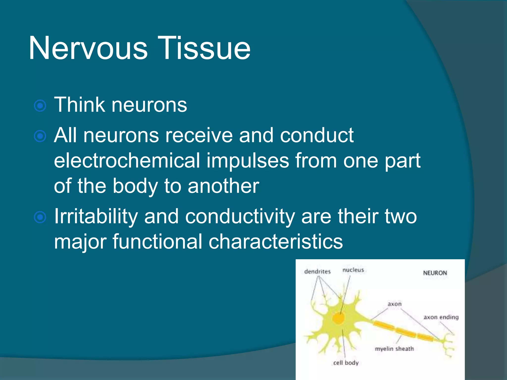
Thinkneurons
All neurons receive and conduct
electrochemical impulses from one part
of the body to another
Irritability and conductivity are their two
major functional characteristics
160.
Nervous Tissue
Drawnout cytoplasm, allow for long
signal transmission
With supporting cells, neurons make up
the structures of the nervous system
162.
Tissue Repair
Tissuerepair occurs in two major ways:
Regeneration – replacement of
destroyed tissue by the same kind of
cells
Fibrosis – involves repair by dense
connective tissue by the formation of
scar tissue
Depends on the type of tissue damaged and
the severity of the injury
163.
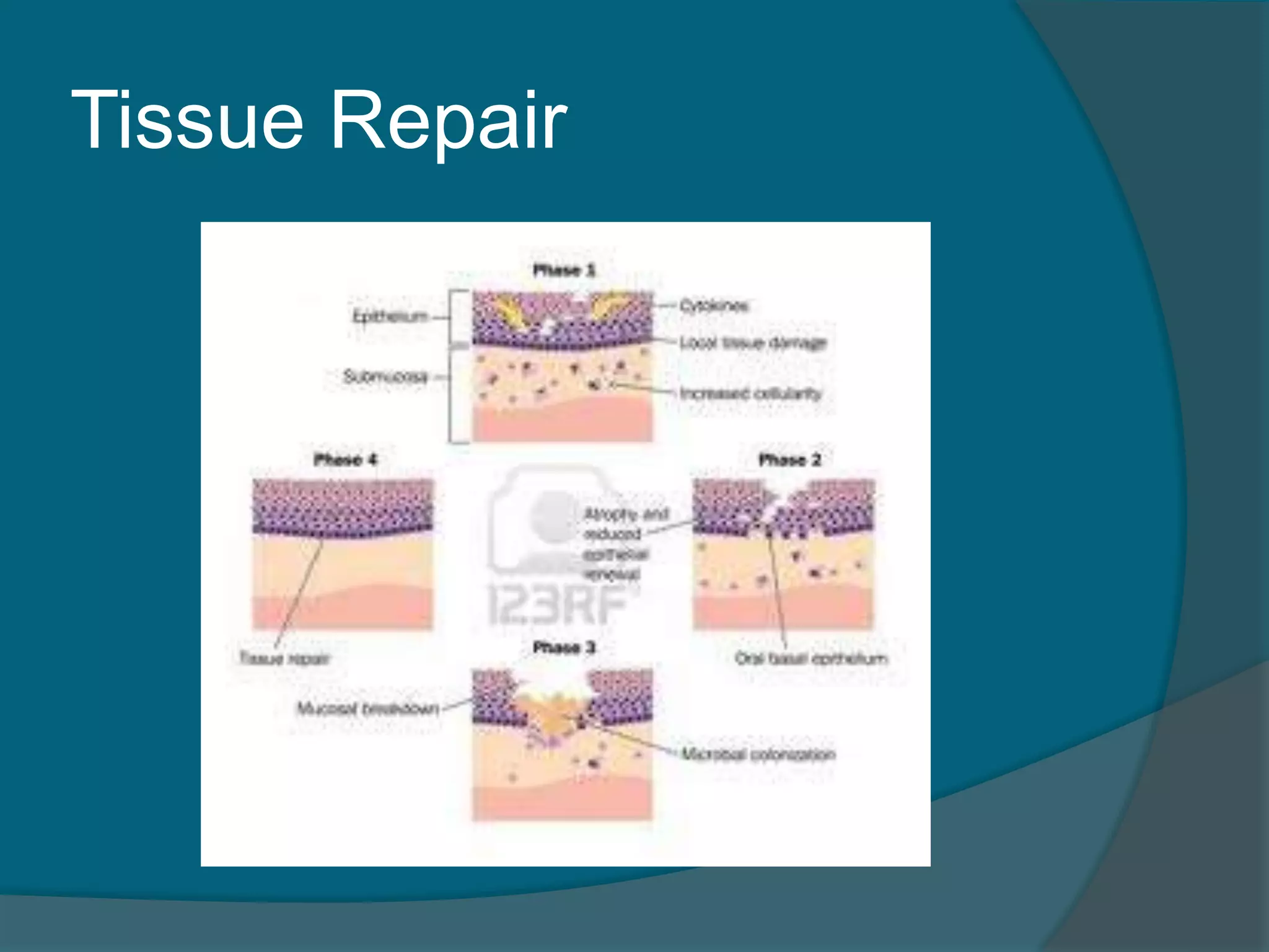
Tissue Repair
Tissueinjury sets the following steps in
motion:
1. capillaries become permeable
Fluid rich in clotting proteins seep into the
injured areas
2. granulation tissue forms
Delicate pink tissue composed largely of new
capillaries
3. surface epithelium regenerates
Makes its way across the granulation tissue
Three other important
terms:
Neoplasm: an abnormal mass of
proliferating cells
Benign or malignant
Hyperplasia: when certain body tissues
may enlarge because there is some
local irritant or condition that stimulates
the cells
Atrophy: a decrease in size in an organ
or body area that loses its normal
stimulation