Downloaded 317 times
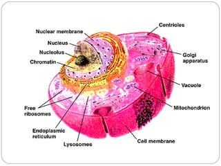
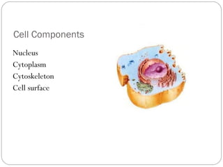
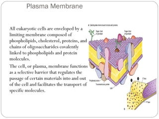
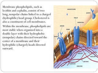
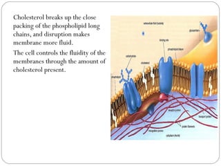
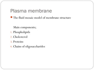
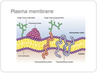
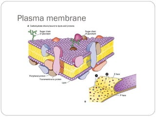
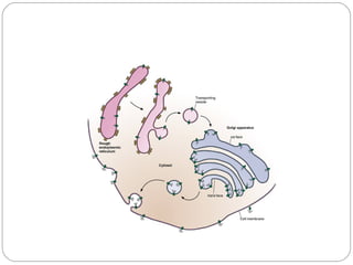
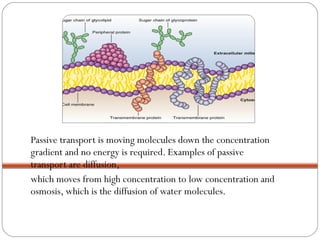
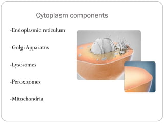
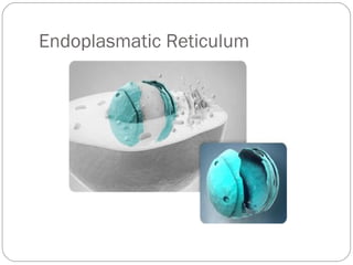
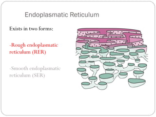
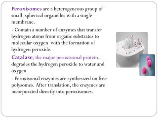
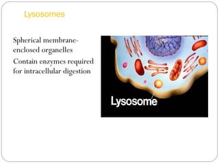

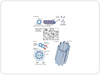
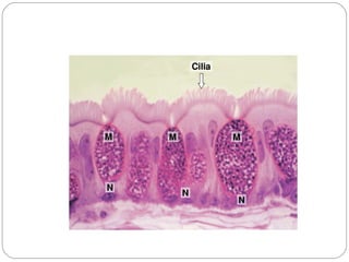

The plasma membrane surrounds eukaryotic cells and is composed of phospholipids, cholesterol, proteins, and carbohydrates. Phospholipids form a double layer with hydrophobic tails facing the center and hydrophilic heads facing out. Cholesterol makes the membrane more fluid. The plasma membrane regulates what passes in and out of the cell. Inside the cell are organelles including the nucleus that contains DNA, endoplasmic reticulum that synthesizes proteins, Golgi apparatus that modifies proteins, mitochondria that generate energy, lysosomes that digest waste, and peroxisomes that break down fatty acids.


