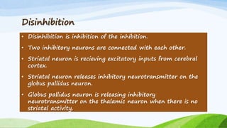
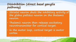
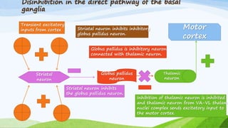
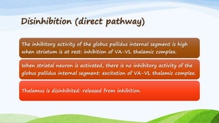
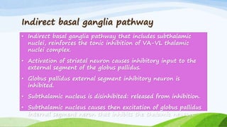
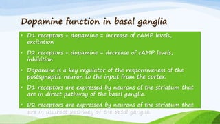
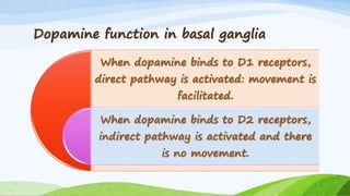
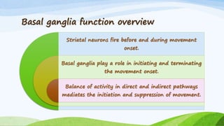
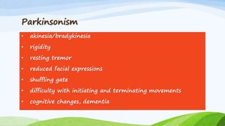
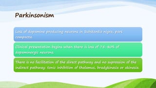
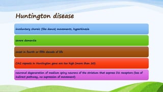
Basal ganglia
function
Domina Petric, MD
Disinhibition in the body
movement loop: direct
and indirect basal ganglia
pathways
I.
Disinhibition
• Disinhibition is inhibition of the inhibition.
• Two inhibitory neurons are connected with each other.
• Striatal neuron is recieving excitatory inputs from cerebral
cortex.
• Striatal neuron releases inhibitory neurotransmitter on the
globus pallidus neuron.
• Globus pallidus neuron is releasing inhibitory
neurotransmitter on the thalamic neuron when there is no
striatal activity.
Disinhibition (direct basal ganglia
pathway)
• Striatal neuron shuts the inhibitory activity of
the globus pallidus neuron on the thalamic
neuron.
• Thalamic neuron then releases excitatory
neurotransmitter for cortical target.
• In the motor loop, cortical target is motor
cortex.
• Motor cortex creates action potential that is
generated down the corticospinal tract.
Disinhibition in the direct pathway of the basal
ganglia
Transient excitatory
inputs from cortex
Striatal
neuron
Striatal neuron inhibits
the globus pallidus neuron.
Globus pallidus
neuron
Globus pallidus is inhibitory neuron
connected with thalamic neuron.
Striatal neuron inhibits inhibitory
globus pallidus neuron.
Thalamic
neuron
Inhibition of thalamic neuron is inhibited
and thalamic neuron from VA-VL thalami
nuclei complex sends excitatory input to
the motor cortex.
Motor
cortex
Disinhibition (direct pathway)
The inhibitory activity of the globus pallidus internal segment is high
when striatum is at rest: inhibition of VA-VL thalamic complex.
When striatal neuron is activated, there is no inhibitory activity of the
globus pallidus internal segment: excitation of VA-VL thalamic complex.
Thalamus is disinhibited: released from inhibition.
Indirect basal ganglia pathway
• Indirect basal ganglia pathway that includes subthalamic
nuclei, reinforces the tonic inhibition of VA-VL thalamic
nuclei complex.
• Activation of striatal neuron causes inhibitory input to the
external segment of the globus pallidus.
• Globus pallidus external segment inhibitory neuron is
inhibited.
• Subthalamic nucleus is disinhibited: released from inhibition.
• Subthalamic nucleus causes then excitation of globus pallidus
internal segment neron that inhibits the thalamic neuron.
Dopamine function in basal ganglia
• D1 receptors + dopamine = increase of cAMP levels,
excitation
• D2 receptors + dopamine = decrease of cAMP levels,
inhibition
• Dopamine is a key regulator of the responsiveness of the
postsynaptic neuron to the input from the cortex.
• D1 receptors are expressed by neurons of the striatum that
are in direct pathway of the basal ganglia.
• D2 receptors are expressed by neurons of the striatum that
are in indirect pathway of the basal ganglia.
Dopamine function in basal ganglia
When dopamine binds to D1 receptors,
direct pathway is activated: movement is
facilitated.
When dopamine binds to D2 receptors,
indirect pathway is activated and there
is no movement.
Basal ganglia function overview
Striatal neurons fire before and during movement
onset.
Basal ganglia play a role in initiating and terminating
the movement onset.
Balance of activity in direct and indirect pathways
mediates the initiation and suppression of movement.
Movement disorders
II.
Parkinsonism
• akinesia/bradykinesia
• rigidity
• resting tremor
• reduced facial expressions
• shuffling gate
• difficulty with initiating and terminating movements
• cognitive changes, dementia
Parkinsonism
Loss of dopamine producing neurons in Substantia nigra, pars
compacta.
Clinical presentation begins when there is loss of 75-80% of
dopaminergic neurons.
There is no facilitation of the direct pathway and no supression of the
indirect pathway: tonic inhibition of thalamus, bradykinesia or akinesia.
Huntington disease
involuntary choreic (like dance) movements, hyperkinesia
severe dementia
onset in fourth or fifth decade of life
CAG repeats in Huntington gene are too high (more than 20)
neuronal degeneration of medium spiny neurons of the striatum that express D2 receptors (loss of
indirect pathway, no supression of movement)
Literature
https://www.coursera.org/learn/medical-
neuroscience/lecture: Leonard E. White,
PhD, Duke University

Basal ganglia function

  • 1.
  • 2.
    Disinhibition in thebody movement loop: direct and indirect basal ganglia pathways I.
  • 3.
    Disinhibition • Disinhibition isinhibition of the inhibition. • Two inhibitory neurons are connected with each other. • Striatal neuron is recieving excitatory inputs from cerebral cortex. • Striatal neuron releases inhibitory neurotransmitter on the globus pallidus neuron. • Globus pallidus neuron is releasing inhibitory neurotransmitter on the thalamic neuron when there is no striatal activity.
  • 4.
    Disinhibition (direct basalganglia pathway) • Striatal neuron shuts the inhibitory activity of the globus pallidus neuron on the thalamic neuron. • Thalamic neuron then releases excitatory neurotransmitter for cortical target. • In the motor loop, cortical target is motor cortex. • Motor cortex creates action potential that is generated down the corticospinal tract.
  • 5.
    Disinhibition in thedirect pathway of the basal ganglia Transient excitatory inputs from cortex Striatal neuron Striatal neuron inhibits the globus pallidus neuron. Globus pallidus neuron Globus pallidus is inhibitory neuron connected with thalamic neuron. Striatal neuron inhibits inhibitory globus pallidus neuron. Thalamic neuron Inhibition of thalamic neuron is inhibited and thalamic neuron from VA-VL thalami nuclei complex sends excitatory input to the motor cortex. Motor cortex
  • 6.
    Disinhibition (direct pathway) Theinhibitory activity of the globus pallidus internal segment is high when striatum is at rest: inhibition of VA-VL thalamic complex. When striatal neuron is activated, there is no inhibitory activity of the globus pallidus internal segment: excitation of VA-VL thalamic complex. Thalamus is disinhibited: released from inhibition.
  • 7.
    Indirect basal gangliapathway • Indirect basal ganglia pathway that includes subthalamic nuclei, reinforces the tonic inhibition of VA-VL thalamic nuclei complex. • Activation of striatal neuron causes inhibitory input to the external segment of the globus pallidus. • Globus pallidus external segment inhibitory neuron is inhibited. • Subthalamic nucleus is disinhibited: released from inhibition. • Subthalamic nucleus causes then excitation of globus pallidus internal segment neron that inhibits the thalamic neuron.
  • 8.
    Dopamine function inbasal ganglia • D1 receptors + dopamine = increase of cAMP levels, excitation • D2 receptors + dopamine = decrease of cAMP levels, inhibition • Dopamine is a key regulator of the responsiveness of the postsynaptic neuron to the input from the cortex. • D1 receptors are expressed by neurons of the striatum that are in direct pathway of the basal ganglia. • D2 receptors are expressed by neurons of the striatum that are in indirect pathway of the basal ganglia.
  • 9.
    Dopamine function inbasal ganglia When dopamine binds to D1 receptors, direct pathway is activated: movement is facilitated. When dopamine binds to D2 receptors, indirect pathway is activated and there is no movement.
  • 10.
    Basal ganglia functionoverview Striatal neurons fire before and during movement onset. Basal ganglia play a role in initiating and terminating the movement onset. Balance of activity in direct and indirect pathways mediates the initiation and suppression of movement.
  • 11.
  • 12.
    Parkinsonism • akinesia/bradykinesia • rigidity •resting tremor • reduced facial expressions • shuffling gate • difficulty with initiating and terminating movements • cognitive changes, dementia
  • 13.
    Parkinsonism Loss of dopamineproducing neurons in Substantia nigra, pars compacta. Clinical presentation begins when there is loss of 75-80% of dopaminergic neurons. There is no facilitation of the direct pathway and no supression of the indirect pathway: tonic inhibition of thalamus, bradykinesia or akinesia.
  • 14.
    Huntington disease involuntary choreic(like dance) movements, hyperkinesia severe dementia onset in fourth or fifth decade of life CAG repeats in Huntington gene are too high (more than 20) neuronal degeneration of medium spiny neurons of the striatum that express D2 receptors (loss of indirect pathway, no supression of movement)
  • 15.