Sporulation is the process of spores formation by bacteria during unfavorable condition
Germination is the process of spores return to give vegetative cells under favorable condition.
Sporulation is the process of spores formation by bacteria during unfavorable condition
Germination is the process of spores return to give vegetative cells under favorable condition.
Growth of bacteria is affected by many factors such as nutrition concentration and other environmental factors.
Some of the important factors affecting bacterial growth are:
Nutrition concentration
Temperature
Gaseous concentration
pH
Ions and salt concentration
Available water
Acid fast staining is differential staining technique which differentiate bacteria into two group- acid fast bacteria and non acid bacteria. It used to identify acid-fast organisms such as members of the genus Mycobacterium .
The physical factors affects the growth of microorganism.
1) Temperature
Temperature is the most important factor that influences the rate of enzyme catalysed reactions and rate of growth.
For every organisms there is an optimum temperature for growth and minimum temperature for inhibiting the growth.
Most extreme the microbes need liquid water to grow.(330C).
some algae and fungi grow at 55-60 degreeC.
Prokaryotes are grow at 100 degreeC.
Based on temperature the microorganisms are classified into two 4.
Immunity
Definitions
Components of Immune system
Types
Innate immunity and Mechanism
Adaptive immunity and Mechanism
2. Antigen
Origin of Antigen
Immunogen
3. Antibody- Immunoglobulin
- Structure
- Classification
- Function of each antibody
Bacteria are a type of biological cell. They constitute a large domain of prokaryotic microorganisms. Typically a few micrometres in length, bacteria have a number of shapes, ranging from spheres to rods and spirals. Bacteria were among the first life forms to appear on Earth, and are present in most of its habitats
Bacteria are microbes with a cell structure simpler than that of many other organisms. Their control centre, containing the genetic information, is contained in a single loop of DNA. Some bacteria have an extra circle of genetic material called a plasmid rather than a nucleus. The plasmid often contains genes that give the bacterium some advantage over other bacteria. For example it may contain a gene that makes the bacterium resistant to a certain antibiotic.
Bacteria are classified into five groups according to their basic shapes: spherical (cocci), rod (bacilli), spiral (spirilla), comma (vibrios) or corkscrew (spirochaetes). They can exist as single cells, in pairs, chains or clusters.
Ultrastructure and characterstic features of bacteria.Archana Shaw
Ultrastructure and characterstic features of bacteria: BACTERIA AS A MODEL ORGANISM
THIS WAS MY PRESENTATION TOPIC IN CLASS. THOUGHT OF SHARING IT AND HOPE IT HELPS.
PULP
• FUNCTIONS OF THE PULP
• Formative: Elaboration of dentin to form the tooth
• Protective: Protection against and repairing of the effectsof noxious stimuli
• Nutritive: Preserving the vitality of all the cellular elements
• Sensory: Perception of stimuli
• ZONES OF PULP
• Starting at the periphery, the pulp is divided into four zones:
• Odontoblastic zone, which surrounds the periphery of
• the pulp
• Cell-free zone
• Cell-rich zone
• Central zone
ODONTOBLASTIC ZONE
• The primary function of the odontoblasts throughout the life of the pulp is the production and deposition of dentin.
• The crowded arrangement of the coronal odontoblasts is due to the rapid reduction of the pulp chamber by the deposition of dentin, which compresses the existing cells to a stratified layer.
• This crowding of odontoblasts produces more cells per unit area and, therefore, more dentinal tubules (45,000/mm2) in the pulpal side than in the enamel side (20,000/mm2).
• The unmyelinated nerves for sensory perception are also found in the pulpal end of the periodontoblastic space of the dentinal tubules
• The incremental lines represent rest periods in dentinogenesis, whereas the interglobular dentin and the granular layer of Tomes probably represent a defect in matrix formation.
• The accentuated incremental line that occurs at birth is called the neonatal line.
• In some areas in the mature dentin, the matrix has not calcified or is hypocalcified. These areas are called interglobular dentin.
• One also sees spaces in the root dentin near the cementodentinal junction called the granular layer of Tomes.
• The dentinal tubules extend from the predentin border to the dentinoenamel and the dentinocemental junctions.
• Dentinal Tubules are conical in shape, with a 2.5 μm mean diameter in the pulpal wall and a 0.9 μm mean diameter in the dentinoenamel or dentinocemental junctions because of the deposition of the peritubular dentin.
• The continuous deposition of peritubular dentinformation of the sclerotic dentin, which has a glassy appearance under transmitted light.
• Primary dentin is elaborated before the teeth erupt and is divided into mantle and circumpulpal dentin.
• Mantle dentin, the first calcified layer of the dentin deposited against the enamel, forms the dentinal side of the dentinoenamel junction.
• Circumpulpal dentin is the dentin formed after the layer of mantle dentin.
• Secondary dentin is elaborated after eruption of the teeth. It can be differentiated from primary dentin by the sharpbending of the tubules producing a line of demarcation.
• secondary dentin is deposited in greater quantities in the floor and roof of the pulp chamber than on the walls.
• Tertiary Dentin
• Two types of tertiary dentin are recognized:
• Tertiary dentin formed by primary odontoblasts following a mild stimulus is called reactionary dentin.
• Tertiary dentin formed by newly differentiated or secondary odontoblasts is termed reparative dentin.
• Reparative dentin,
• Cementum is deposited in a thin layer at the cementoenamel junction to form one of the following three configurations:
A butt joint (30%)
An overlap joint (60%)
A gap between cementum and enamel (10%) (this gap may produce cervical sensitivity or may predispose the tooth to cervical caries)
• The continued incremental deposition of cementum in the apical third maintains the length of the tooth, constricts the apical foramen, and deviates the apical foramen from the center of the apex.
• The surface of the bony crypt becomes known as the lamina dura radiographically.
• The sensory nerve fibers traverse the dental papilla and, on reaching the coronal pulp, they branch toward the periphery to form a plexus of nerves called the plexus of Raschkow.
• This plexus of Raschkow is located in the subodontoblastic zone of the coronal pulp.
• These sensory nerve fibers are myelinated; therefore, they are enclosed in a sheath made of Schwann’s cells.
• Human premolars receive almost 2300 axons at the root apex, of which about 13% are myelinated and 87% are nonmyelinated fibers.
• Most apical myelinated axons are fast-conducting Aδ fibers.
• These fibers are probably activated by a hydrodynamic mechanism and conduct impulses that are perceived as a short, well-localized, sharp pain.
• Most C fibers are slow conducting and fine sensory afferents
• TOOTH DEVELOPMENT (EMBRYOLOGY)
• . The initiation of tooth development-6th week of IUL
• . The oral stratified squamous epithelium covers the primordia of the future maxillary and mandibular processes in a horseshoe-shaped pattern.
• . Tooth development starts when stratified squamous epithelium begins to thicken and forms the dental lamina.
• . The stratified squamous oral epithelium covers an embryonic connective tissue that is called the ectomesenchyme because of its derivation from the neural crest cells. condensed area of ectomesenchyme forms the future dental papilla and subsequently the pulp.
• . BUD STAGE (FORMATION OF ENAMEL ORGAN)
• . CAP STAGE (OUTER AND INNER ENAMEL EPITHELIUM)
• The convex surface consists of the cuboidal epithelial cells and is called the outer enamel epithelium. The concave surface, called the inner enamel epithelium, consists of elongated epithelial cells with polarized nuclei that later differentiate into ameloblasts.
• . The inner and the outer enamel epithelium, the cells begin to separate due to the deposition of intercellular mucoid fluid rich in glycogen that forms a branch reticular arrangement called the stellate reticulum.
• . The ectomesenchyme surrounding the dental papilla and the enamel organ condenses and becomes more fibrous, it is called the dental follicle or the dental sac—the precursor of the cementum, the periodontal ligament (PDL), and the alveolar bone.
• . BELL STAGE (CERVICAL LOOP)
• . The junction of the outer and the inner enamel epithelium at the rim of the enamel organ becomes a distinct zone called the cervical loop.
• . The buds of the permanent molars, which have no primary predecessors.
• . The squamous cells between the stellate reticulum and the inner enamel epithelium form the stratum intermedium.
• . Stratum intermedium → Enamel
• Ectomesenchyme → Dentin
• Dental papilla → Pulp
• Dental follicle or dental sac → Cementum, the periodontal ligament(PDL), and the alveolar bone.
• Primary dentin is formed in increments of 4–8 μm per day and is continually deposited until the end of tooth development.
• . large-diameter collagen fibers known as von Korff fibers are deposited at right angles to the basement membrane in the extracellular matrix of the acellular zone. This process creates the organic matrixof the first-formed dentin or mantle dentin.
• The organic matrix or predentin is deposited around the odontoblastic processes. The predentin later calcifies and thereby forms the dentinal tubules.
• Primary dentin differs from the mantle dentin in which the matrix originates solely(only) in the odontoblasts.
• The mineralization of primary dentin originates from the previous mineralized dentin.
PERITUBULAR DENTIN
As the incremental deposition of dentin continues toward the center of the pulp, the diameter of the odontoblastic processes is reduced peripherally.
more mineralized and it is harder than primary dentin, is called peritubular dentin.70% inorganic material
• Dentin consist
tooth development, formation of enamel root formation, hertwing epithelial root sheath
development of the enamel ,dentin, pulp ,root .
preparation of NEET MDS
• AMELOGENESIS
• The preameloblasts differentiate into tall columnar epithelial cells with their nuclei polarized toward the stratum intermedium –Ameloblast
• The inner enamelepithelium is being resorbed and dentin is being deposited to follow the contour established by the basement membrane. This process forms the future dentinoenamel junction
• The deposition of enamel matrix causes the ameloblasts to migrate peripherally and form conic projections – Tomes process
• This maturation process begins in the dentinoenamel junction and progresses peripherally to the enamel surface.
• During the final phase of the maturation process, the ameloblasts join the stratified epithelium to form the reduced enamel epithelium and to cover and protect the enamel until eruption of the tooth.
• DEVELOPMENT OF THE ROOT
• Outer and inner enamel epithelia proliferate to form the HERS(Hertwig’s Epithelial Root Sheath)
• Hertwig’s epithelial root sheath- which determines the size and shape of the root of the tooth.
• the Hertwig’s epithelial root sheath proliferateshorizontally between the dentinal papilla and the dentalfollicle; this process partially encloses the dental papilla anddelineates the apical foramen or foramina. This proliferationis called the epithelial diaphragm.
• In multirooted teeth, the epithelial diaphragm guides the formation of the furca, roots, root canals, and apical foramina.
• CEMENTOBLAST
• These mesenchymal cells differentiate into cementoblasts, which are round, plump cells that have basophilic cytoplasm.
• The cell rests of Malassez remain dormant in the mature PDL and have the potential of proliferating into periradicular cysts if stimulated by chronic inflammation.
• Accessory canals are more prevalent in the apical third of the root.
details about the dental caries
Introduction
Definition
Etiology of dental caries
Clinical features of dental caries
Classification of caries
Theory of dental caries
Management of dental caries
#dentalcaries
THE HUMORAL THEORY
Vital Theory
Chemical theory
Parasitic theory
Miller Chemico Parasitic Theory
Proteolysis Chelation Theory
Sucrose Chelation Theory
types of dental caries
senile caries
backward caries
reversible caries
pit and fissure caries
root caries
1.Introduction
2.Definition
3.Classification
a) based on source or origin
b) based on nature of waste
#Non hazardous or solid waste
#Hazardous waste
4. Health hazards of accumulated solid waste
Dept of Oral Medicine & Radiology
Ameloblastoma A case report
Dhananjay Singh
CHIEF COMPLAINT
HISTORY OF PRESENT ILLNESS
dental history
medical history
diagnosis
investigation
final diagnosis
treatment
clinical features
oral medicine
radiology
xray
oral diagnosis
All details about the dental cements
Introduction
Definitions
Ideal properties
Classification
Based on Ingredients & Application(craig)
Based on Bonding mechanism(william O’Brien)
Based on setting reaction (Anusavice)
Silicate cement
Zinc phosphate cement
all details about the chemistry of polymerization
Addition polymerization
Chemical Stages of polymerization
induction
propagation
chain transfer
termination
Inhibition of polymerization
Ring-opening polymerization,
Step growth/Condensation polymerization
Copolymerization
Acrylic resins
Methylmethacrylate
Polymethylmethacrylate{PMMA}
Multifunctional Methacrylate and Acrylate resins
All details about the dental polymer
Components and Composition
Molecular Weight
Polydispersity
Structure Of Polymer
Mechanical And Physical Properties Of Polymer,
Rheometric Properties
Solvation and dissolution Properties
Thermal Properties
Requirement Of Dental Resins
Dental Use Of Resins
cavity liners details it applied and action and how to use in cavity for the restorations of filling materials,
manipulation of cavity liners
others types of cavity liners
details about the metabolism of drugs, non synthetic reaction, synthetic reaction, inhibition of drug metabolism, pre systemic metabolism, microsomal enzyme induction,
The Indian economy is classified into different sectors to simplify the analysis and understanding of economic activities. For Class 10, it's essential to grasp the sectors of the Indian economy, understand their characteristics, and recognize their importance. This guide will provide detailed notes on the Sectors of the Indian Economy Class 10, using specific long-tail keywords to enhance comprehension.
For more information, visit-www.vavaclasses.com
The French Revolution, which began in 1789, was a period of radical social and political upheaval in France. It marked the decline of absolute monarchies, the rise of secular and democratic republics, and the eventual rise of Napoleon Bonaparte. This revolutionary period is crucial in understanding the transition from feudalism to modernity in Europe.
For more information, visit-www.vavaclasses.com
Ethnobotany and Ethnopharmacology:
Ethnobotany in herbal drug evaluation,
Impact of Ethnobotany in traditional medicine,
New development in herbals,
Bio-prospecting tools for drug discovery,
Role of Ethnopharmacology in drug evaluation,
Reverse Pharmacology.
Operation “Blue Star” is the only event in the history of Independent India where the state went into war with its own people. Even after about 40 years it is not clear if it was culmination of states anger over people of the region, a political game of power or start of dictatorial chapter in the democratic setup.
The people of Punjab felt alienated from main stream due to denial of their just demands during a long democratic struggle since independence. As it happen all over the word, it led to militant struggle with great loss of lives of military, police and civilian personnel. Killing of Indira Gandhi and massacre of innocent Sikhs in Delhi and other India cities was also associated with this movement.
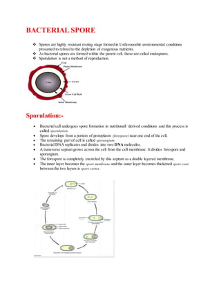
1. BACTERIAL SPORE
Spores are highly resistant resting stage formed in Unfavourable environmental conditions
presumed to related to the depletion of exogenous nutrients.
As bacterial spores are formed within the parent cell, these are called endospores.
Sporulation is not a method of reproduction.
Sporulation:-
Bacterial cell undergoes spore formation in nutritionall derived conditions and this process is
called sporulation.
Spore develops from a portion of protoplasm (forespore) near one end of the cell.
The remaining part of cell is called sporangium.
Bacterial DNA replicates and divides into two DNA molecules.
A transverse septum grows across the cell from the cell membrane. It divides forespore and
sporangium.
The forespore is completely encircled by this septum as a double layered membrane.
The inner layer becomes the spore membrane and the outer layer becomes thickened spore coat.
between the two layers is spore cortex.
2. Morphologyof Spore:-
The cell membrane grows inward and forms spore wall around the core (forespore).
The inner-most layer of the spore wall forms the spore membrane from which the cell wall of
future vegetative bacterium develops.
Outside this membrane is thick layer, the cortex and a multilayered tough spore coat.
Some spores have an additional apparently rather loose, outer covering called exosporium.
Shape and Positionof Spores:-
o The precise position, shape and relative size of spore are constant within a particular species.
o Spores may be central, subterminal or terminal.
o They may be oval or spherical in shape.
Resistance:-
Bacterial spores are extremely resistant to ordinary boiling, disinfectants and heating.
The high resistance of spore due to hi content o calcium and dipicolinic acid;
low water content; he thick impervious cortex and spore coats;
their low metabolic and enzymatic activity.
However,spores of all medically important bacteria are destroyed by autoclaving at 121 °C for 15
minute.
Germination:-
The process of conversion of a spore into vegetative cell under suitable conditions is known as germination.
3. Uses ofspores:-
Spore of certain species of bacteria are employed indicator for proper sterilisation.
e.g. Bacillus stearothermophilus,which is destroyed a temperature of 121 °C for 10-20 minutes.
Spore Forming Bacteria:-
Obligate aerobes- genus Bacillus e.g. B. anthracis and B. subtilis.
Obligate anaerobes- genus Clostridia e. g. Cl. tetani,Cl. welchii,Cl. Botulinum.
Both Bacillus and Clostridia are gram positive bacteria.
DR. DHANANJAY SINGH