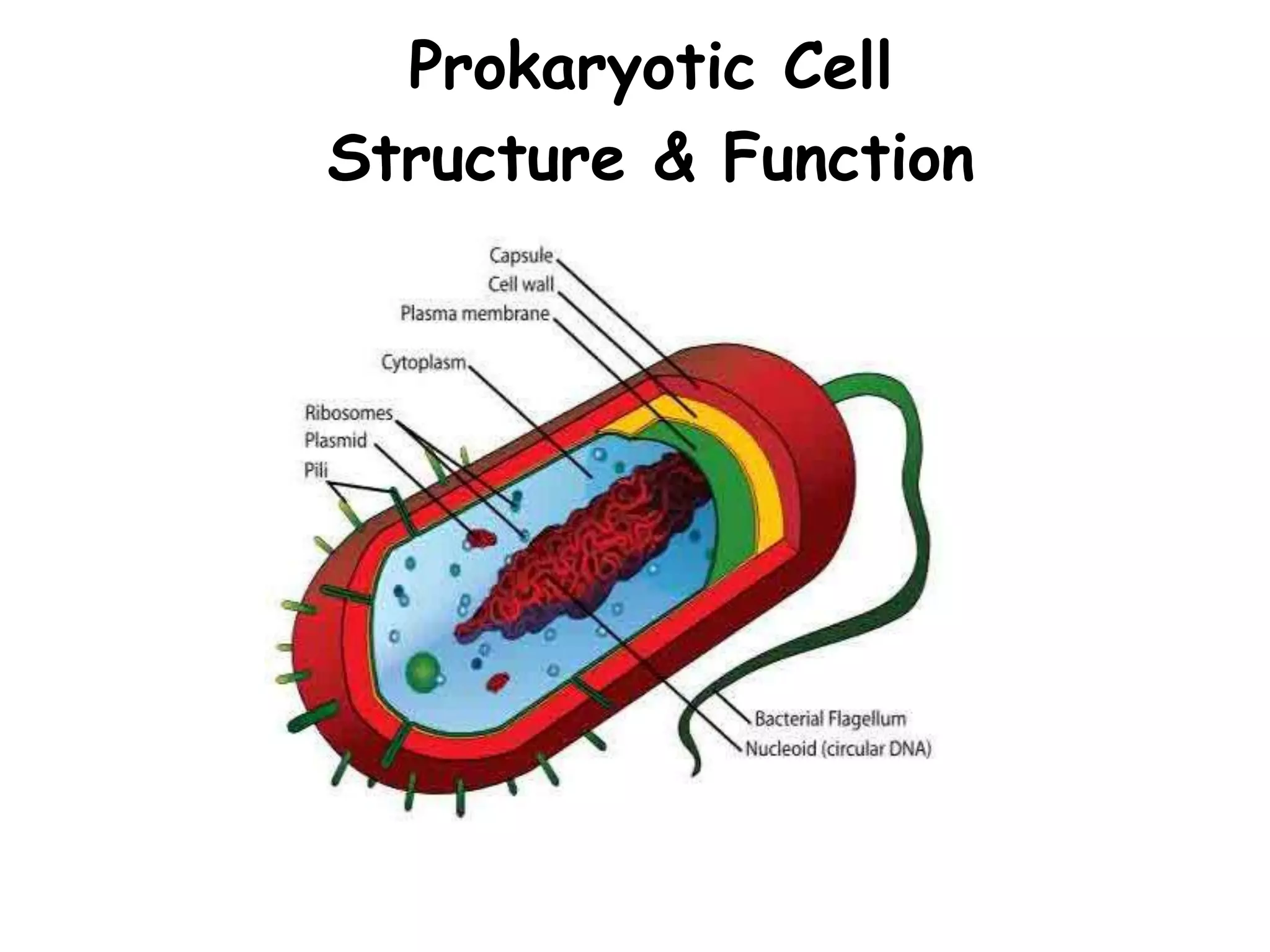
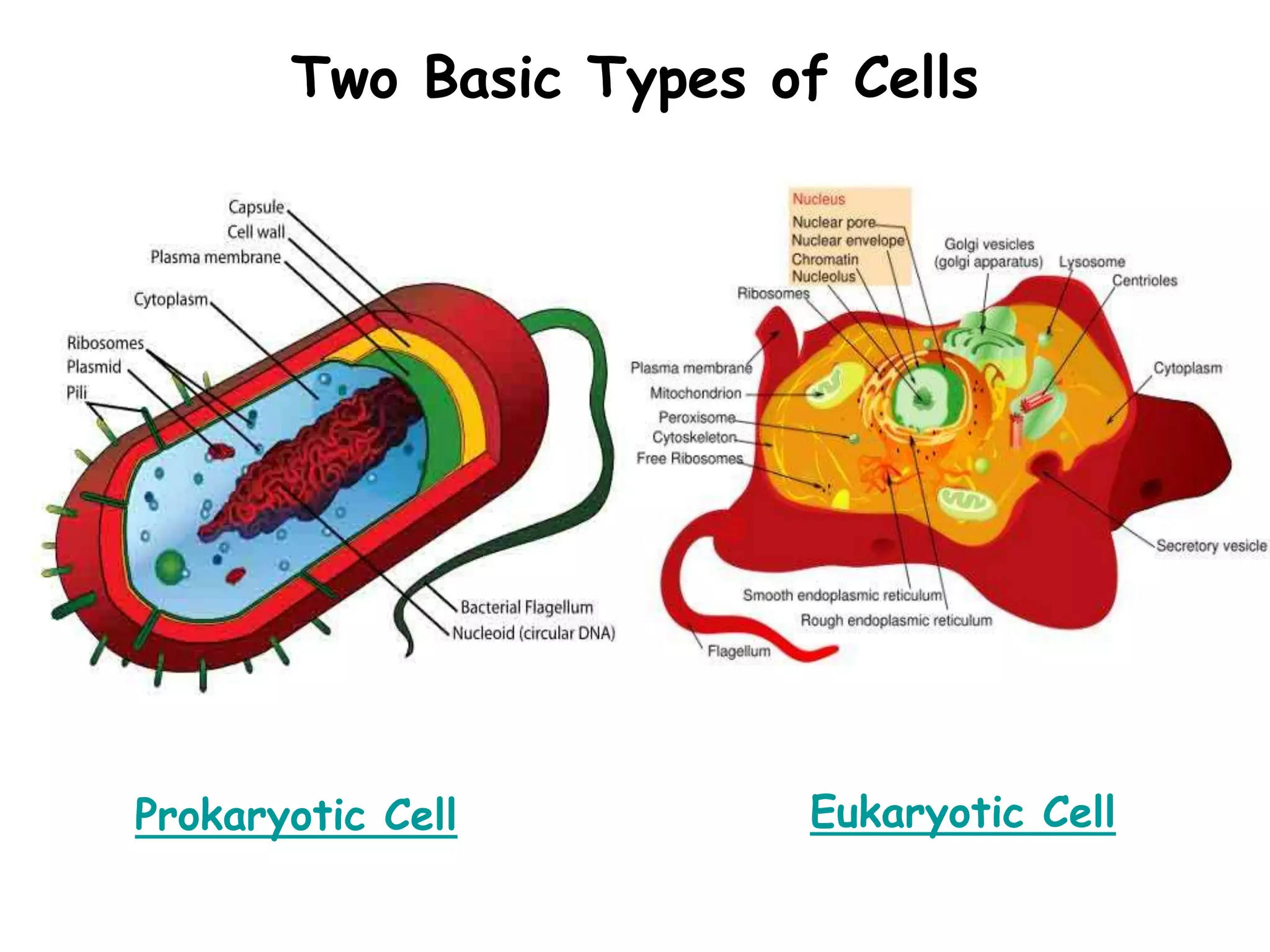
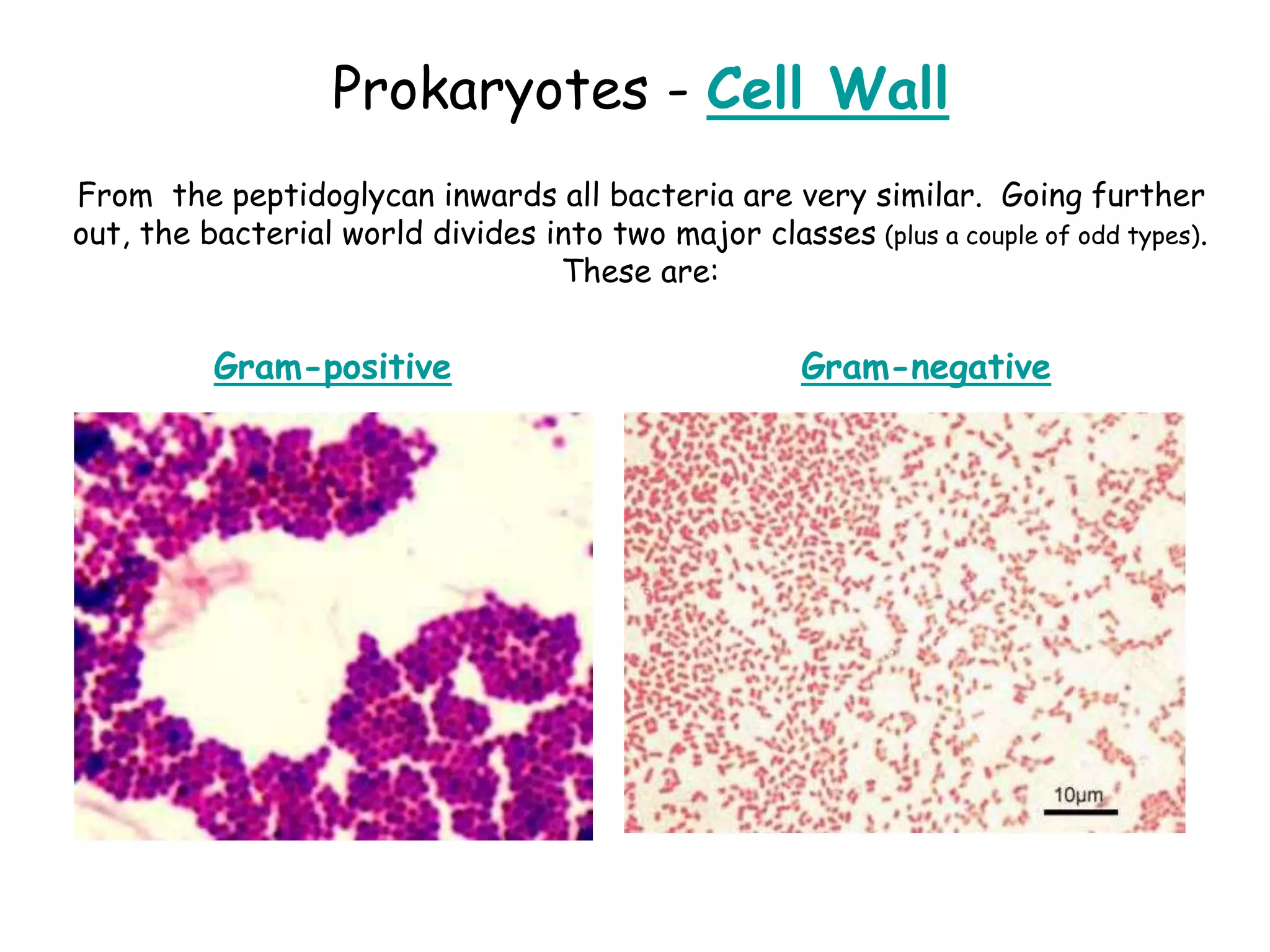
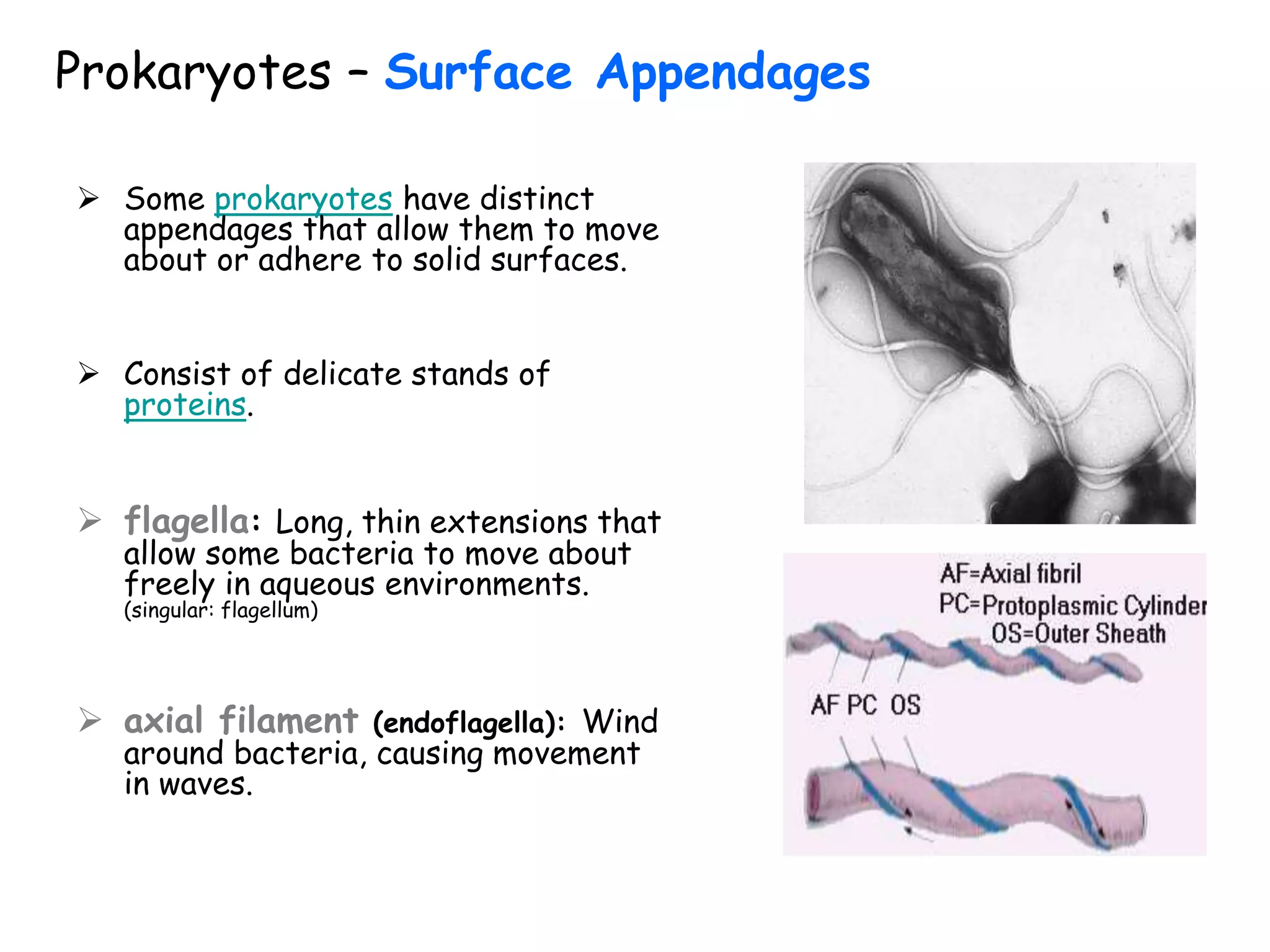
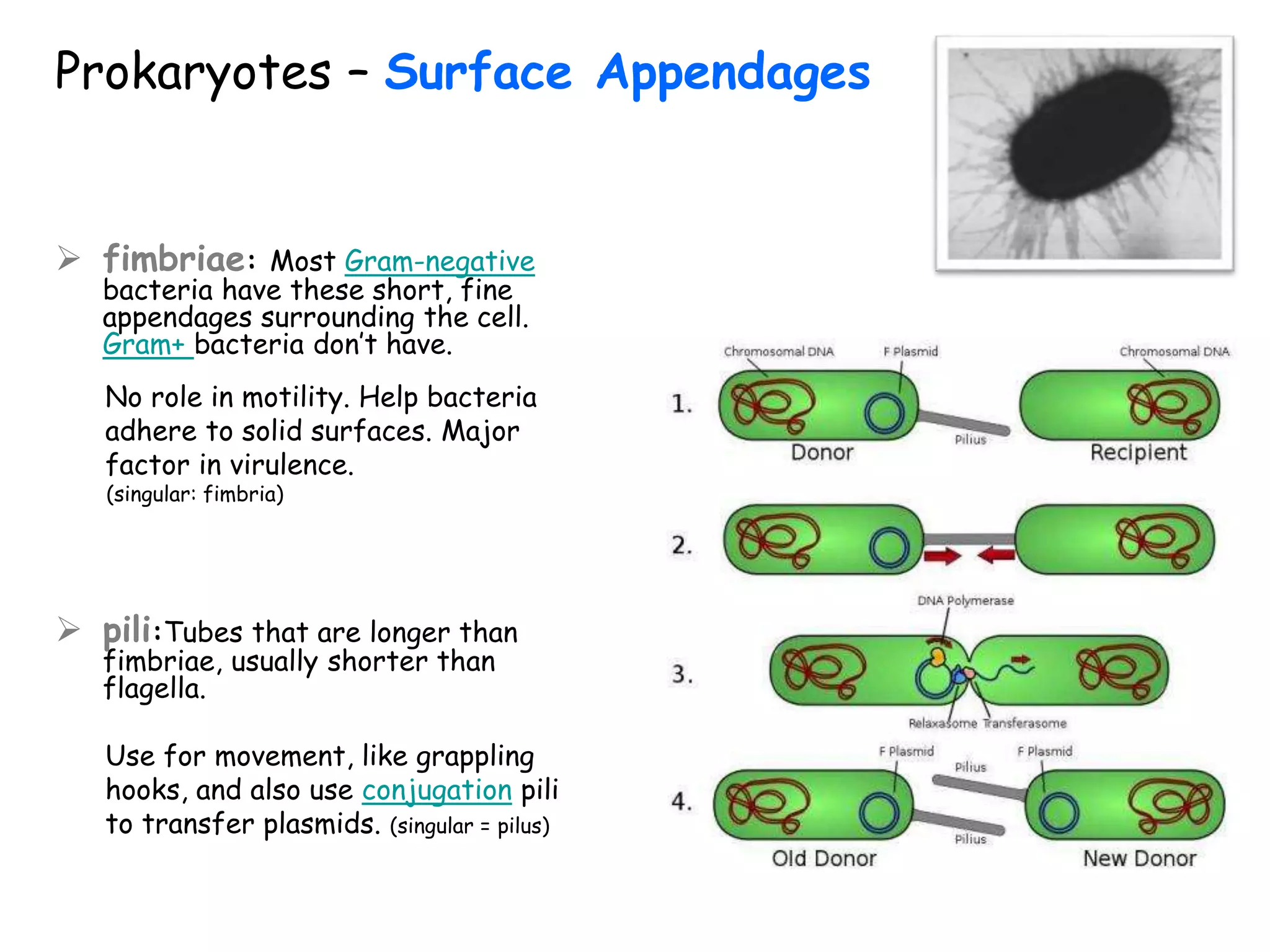
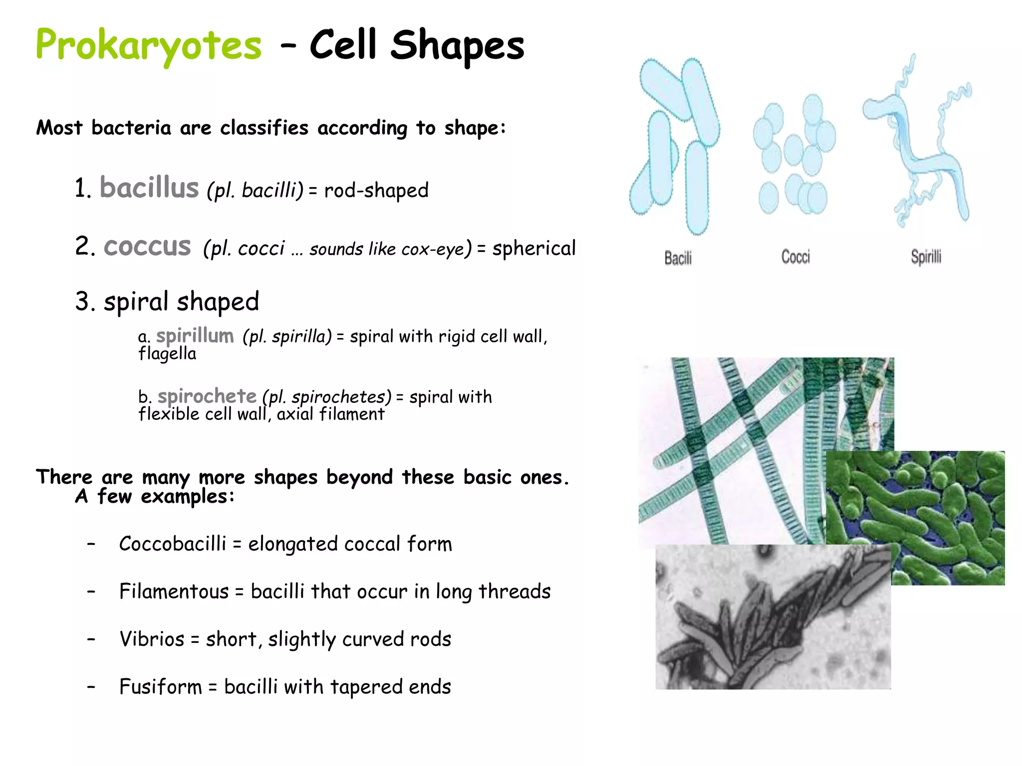
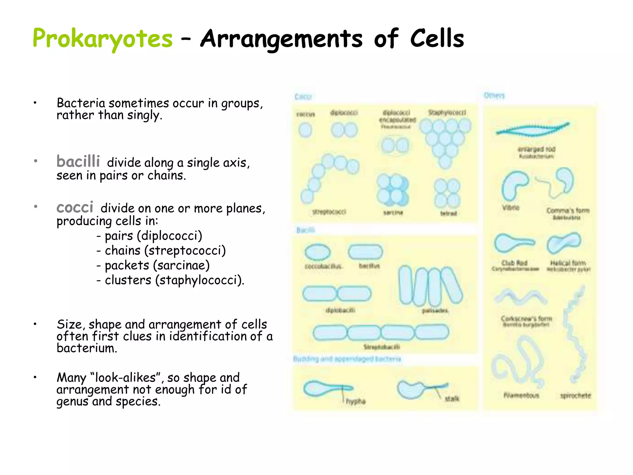
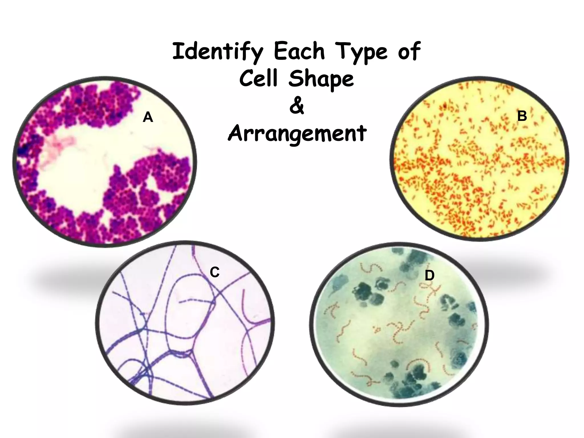
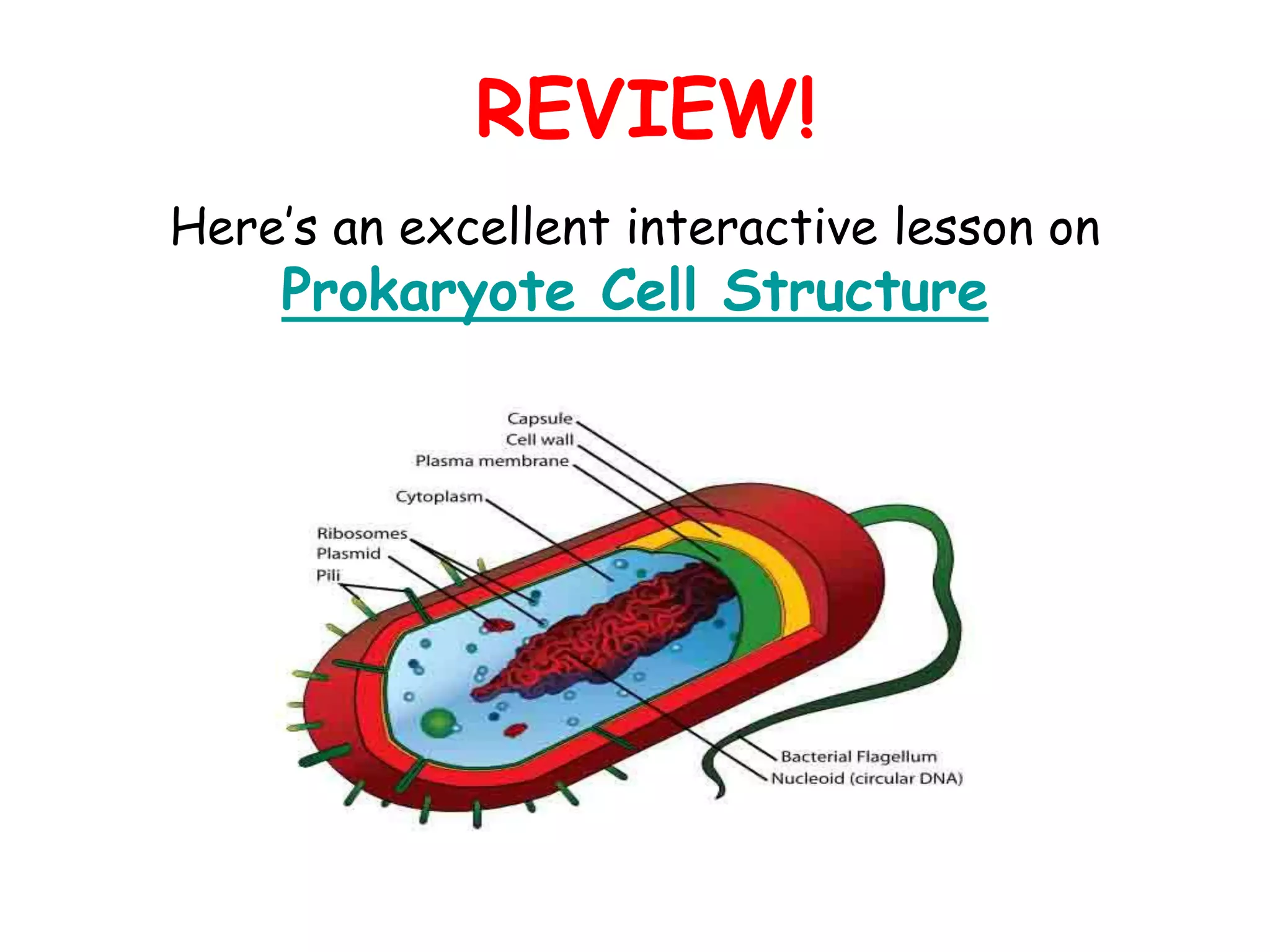
Introduction to Biotechnology
Subject Code- BT-201
Department of Biotechnology
Delhi Technological University
UNIT-2
Engineering perspective to
Biology
Topic-
Cellular architecture of prokaryotic cell
Prokaryotic Cell
Structure & Function
Two Basic Types of Cells
Eukaryotic Cell
Prokaryotic Cell
Size of Living Things
1 m = 100 cm = 1,000mm = 1,000,000 µm = 1,000,000,000nm
1mm = 1000 µm = 1000000nm
1 µm = 1000nm
Prokaryotes
Tell me about Prokaryotes…
Binary Fission
Check out these quick
animated lessons on
binary fission:
(Please watch both, as each provides
different and useful information.)
Binary Fission Animation
from ClassZone
Binary Fission Animation
From McGraw-Hill
Prokaryote Genetics
Nucleoid
• Region of cytoplasm where prokaryote’s genome
(DNA) is located.
• Usually a singular, circular chromosome.
Plasmid
• Small extra piece of chromosome/genetic material.
• 5 - 100 genes
• Not critical to everyday functions.
• Can provide genetic information to promote:
- Antibiotic resistance
- Virulence factors
(molecules produced by pathogen that specifically influence
host's function to allow the pathogen to thrive)
- Promote conjugation
(transfer of genetic material between bacteria through cell-
to-cell contact)
Prokaryotes
Cytoplasm
• Also known as proto-plasm.
• Gel-like matrix of water, enzymes,
nutrients, wastes, and gases and
contains cell structures.
• Location of growth, metabolism, and
replication.
Granules
• Bacteria’s way of storing nutrients.
• Staining of some granules aids in
identification.
Prokaryotes
Cytoskeleton
 Cellular "scaffolding" or
"skeleton" within the
cytoplasm.
 Major advance in
prokaryotic cell biology in
the last decade has been
discovery of the
prokaryotic cytoskeleton.
 Up until recently, thought
to be a feature only of
eukaryotic cells.
Prokaryotes
Ribosomes
 Found within cytoplasm or
attached to plasma membrane.
 Made of protein & rRNA.
 Composed of two subunits.
 Cell may contain thousands .
 Q: What do ribosomes do?
 Q: What’s the relationship
between the job that the
ribosomes do and the genetic
instructions (nucleic acids) of the
cell?
Click here for
animation of
ribosome building a
protein.
 Separates the cell from its
environment.
 Phospholipid molecules
oriented so that hydrophilic
water-loving heads directed
outward and hydrophobic
water-hating tails directed
inward.
 Proteins embedded in two
layers of lipids (lipid bilayer).
 Membrane is semi-permeable.
Q: What does that mean?
Prokaryotes
Plasma Membrane
Prokaryotes – Plasma Membrane as a Barrier
Osmosis
 Is the diffusion of water
across a semi-permeable
membrane.
 Environment surrounding
cells may contain amounts of
dissolved substances
(solutes) that are…
- equal to
- less than
- greater than
…those found within the cell.
CELL
Plasma
membrane
Liquid
environment
outside the cell.
Liquid
environment
inside the cell.
Prokaryotes – Plasma Membrane as a Barrier
Tonicity and Osmosis
 isotonic: equal concentration of a solute
inside and outside of cell.
 hypertonic: a higher concentration of
solute.
 hypotonic: a lower concentration of
solute.
Water will always move
toward a hypertonic
environment!!
REVIEW!
• How Osmosis Works animation
• Diffusion, Osmosis & Active
Transport Lecture Main Page of the
Virtual Cell Biology Classroom on the
Science Prof Online website
• H2O important reactant in many metabolic
reactions.
• Most cells die in absence of water.
• Cell walls of bacteria and plants prevent
them from exploding in a hypotonic
environment, but most bacteria are
vulnerable in hypertonic environments.
• Many bacteria can be plasmolyzed by
high concentrations of solutes.
• You salty perspiration protects you from
bacteria that cannot handle the high
sodium chloride concentration.
• The water moves out of the bacterium
and it dies of ‘hyperosmostic shock’
(desiccation).
Cells & Water:
Osmotic Pressure
Plasma Membrane as a Barrier
ACTIVE TRANSPORT
• How most molecules move across the plasma membrane.
• Analogous to a pump moving water uphill.
• Types of active transport are classified by type of energy used to drive molecules
across membranes.
• ATP Driven Active Transport
Energy from adenosine triphosphate (ATP) drives substances across the plasma membrane with
the aid of carrier molecules.
From the peptidoglycan inwards all bacteria are very similar. Going further
out, the bacterial world divides into two major classes (plus a couple of odd types).
These are:
Gram-positive Gram-negative
Prokaryotes - Cell Wall
 Peptidoglycan is a huge polymer of interlocking chains of
alternating monomers.
 Provides rigid support while freely permeable to solutes.
 Backbone of peptidoglycan molecule composed of two amino
sugar derivatives of glucose. The “glycan” part of peptidoglycan:
- N-acetylglucosamine (NAG)
- N-acetlymuramic acid (NAM)
 NAG / NAM strands are
connected by interlocking
peptide bridges.
The “peptid” part
of peptidoglycan.
Bacterial Cell Wall
Prokaryotes - Cell Wall
Gram-Positive & Gram-Negative
Prokaryotes - Cell Wall
Gram-Positive & Gram-Negative
FYI: The bacterial plasma membrane and cell wall together are called the
cell envelope.
Q: Why are these differences in bacterial cell
wall structure so important?
Prokaryotes - Glycocalyx
Some bacteria have an additional layer
outside of the cell wall called the
glycocalyx.
This additional layer can come in
one of two forms:
1. Slime Layer
- Glycoproteins loosely associated with the
cell wall.
- Slime layer causes bacteria to adhere to
solid surfaces and helps prevent the cell from
drying out.
- Streptococcus
The slime layer of Gram+ Streptococcus mutans
allows it to accumulate on tooth enamel (yuck mouth
and one of the causes of cavities).
Other bacteria in the mouth become trapped in the
slime and form a biofilm & eventually a buildup of
plaque.
Mannitol
Salt
Prokaryotes - Glycocalyx
2. Capsule
• Polysaccharides firmly attached to
the cell wall.
• Capsules adhere to solid surfaces and
to nutrients in the environment.
• Adhesive power of capsules is a
major factor in the initiation of some
bacterial diseases.
• Capsule also protect bacteria from
being phagocytized by cells of the
hosts immune system.
Prokaryotes - Endospores
 Dormant, tough, non-reproductive structure
produced by small number of bacteria.
 Resistant to radiation, desiccation, lysozyme,
temperature, starvation, and chemical
disinfectants.
 Endospores are commonly found in soil and
water, where they may survive for very long
periods of time.
 Q: How and why do endospores form?
Watch the animated lesson “Bacterial Spore
Formation” to find out. Link also provides quiz
questions to test your understanding of the
material.
 Q: What are the two endospore producing
bacterial genera that were introduced in
our History of Microbiology lecture?
An endospore stained bacterial smear of
Bacillus subtilis showing endospores as
green and vegetative cells as red.
GRAM-POSITIVE
Obligate or facultative anaerobes,
endospore producers
bacillus-shaped
Q: Which two groups of bacteria produce
endospores?
Common in soil. Only a few species cause
disease in humans.
Extremely diverse group of bacteria,
includes:
- causative agent of anthrax (Bacillus
anthracis)
- species that synthesize important
antibiotics, and enzymes for detergents.
Due to extreme tolerance to both heat and
disinfectants, used to test heat
sterilization techniques and chemical
disinfectants.
Bacterial Genus: __________
This is our lab
friend Bacillus
subtilis.
Gram Stain
Endospore
Stain
Prokaryotes – Surface Appendages
 Some prokaryotes have distinct
appendages that allow them to move
about or adhere to solid surfaces.
 Consist of delicate stands of
proteins.
 flagella: Long, thin extensions that
allow some bacteria to move about
freely in aqueous environments.
(singular: flagellum)
 axial filament (endoflagella): Wind
around bacteria, causing movement
in waves.
Prokaryotes – Surface Appendages
 fimbriae: Most Gram-negative
bacteria have these short, fine
appendages surrounding the cell.
Gram+ bacteria don’t have.
No role in motility. Help bacteria
adhere to solid surfaces. Major
factor in virulence.
(singular: fimbria)
 pili:Tubes that are longer than
fimbriae, usually shorter than
flagella.
Use for movement, like grappling
hooks, and also use conjugation pili
to transfer plasmids. (singular = pilus)
Meet the Microbe! Neisseria and its Fimbiriae
• Gram- diplococci, resemble coffee beans when viewed
microscopically.
• Neisseria gonorrhoeae causes sexually transmitted disease
gonorrhoeae.
• Antibiotics applied to the eyes of neonates as a preventive
measure against gonorrhoea.
• One of the most communicable disease in the U.S.
• 125 cases per 100,000. Teens 15-19 yo 634 cases per 100,000.
Young adults 20-25 460 per 100,000.
• N. meningitidis most common causes of bacterial meningitis in
young adults.
Q: What makes Neisseria so tough?
• Lipopolysaccharide (LPS) of the cell wall of Neisseria acts as
an endotoxin.
• Polysaccharide capsule prevents host phagocytosis and aids in
evasion of the host immune response.
• Use fimbriae to attach onto host cells; avirulent without.
Fimbriae have adhesion proteins (adhesins) on their tips that
match, lock and key, with proteins on host epithelial cell surface.
Prokaryotes – Cell Shapes
Most bacteria are classifies according to shape:
1. bacillus (pl. bacilli) = rod-shaped
2. coccus (pl. cocci … sounds like cox-eye) = spherical
3. spiral shaped
a. spirillum (pl. spirilla) = spiral with rigid cell wall,
flagella
b. spirochete (pl. spirochetes) = spiral with
flexible cell wall, axial filament
There are many more shapes beyond these basic ones.
A few examples:
– Coccobacilli = elongated coccal form
– Filamentous = bacilli that occur in long threads
– Vibrios = short, slightly curved rods
– Fusiform = bacilli with tapered ends
Prokaryotes – Arrangements of Cells
• Bacteria sometimes occur in groups,
rather than singly.
• bacilli divide along a single axis,
seen in pairs or chains.
• cocci divide on one or more planes,
producing cells in:
- pairs (diplococci)
- chains (streptococci)
- packets (sarcinae)
- clusters (staphylococci).
• Size, shape and arrangement of cells
often first clues in identification of a
bacterium.
• Many “look-alikes”, so shape and
arrangement not enough for id of
genus and species.
Identify Each Type of
Cell Shape
&
Arrangement
A B
C D
REVIEW!
Here’s an excellent interactive lesson on
Prokaryote Cell Structure
Confused?
Here are links to fun resources that further explain aerobic
respiration:
• Cell Structure: Prokaryotes Main Page on the Virtual
Micrboiology Classroom of Science Prof Online.
• Prokaryotic Cell: Structures, Functions & Diagrams, an article
from SPO.
• Prokaryotic & Eukaryotic: Two Types of Biological Cells, an article
from SPO.
• “Got the Time” music video by Anthrax.
• Prokaryotic Cell interactive diagram from Cells Alive website.
• “How big is a…” interactive diagram from Cells Alive website.
• Cell Structure tutorials and quizzes from Interactive Concepts in
Biochemistry.
• How Osmosis Works, animation from McGraw-Hill.
• “Germs”. Music by Weird Al Yankovic. Video by RevLucio.
• Bacterial Pathogen Pronunciation Station, a webpage with links
to audio files containing the pronunciation of the bacterial names, created by
Neal R. Chamberlain, Ph.D.
• Biology4Kids – Cell Biology Main Page by Raders.
(You must be in PPT slideshow view to click on links.)
From the Virtual Microbiology Classroom on ScienceProfOnline.com

Unit-2(Lecture- 5th) Cellular architecture of Prokaryotic Cell.pptx

  • 1.
    Introduction to Biotechnology SubjectCode- BT-201 Department of Biotechnology Delhi Technological University
  • 2.
  • 3.
  • 4.
  • 5.
    Two Basic Typesof Cells Eukaryotic Cell Prokaryotic Cell
  • 6.
    Size of LivingThings 1 m = 100 cm = 1,000mm = 1,000,000 µm = 1,000,000,000nm 1mm = 1000 µm = 1000000nm 1 µm = 1000nm
  • 7.
    Prokaryotes Tell me aboutProkaryotes… Binary Fission Check out these quick animated lessons on binary fission: (Please watch both, as each provides different and useful information.) Binary Fission Animation from ClassZone Binary Fission Animation From McGraw-Hill
  • 8.
    Prokaryote Genetics Nucleoid • Regionof cytoplasm where prokaryote’s genome (DNA) is located. • Usually a singular, circular chromosome. Plasmid • Small extra piece of chromosome/genetic material. • 5 - 100 genes • Not critical to everyday functions. • Can provide genetic information to promote: - Antibiotic resistance - Virulence factors (molecules produced by pathogen that specifically influence host's function to allow the pathogen to thrive) - Promote conjugation (transfer of genetic material between bacteria through cell- to-cell contact)
  • 9.
    Prokaryotes Cytoplasm • Also knownas proto-plasm. • Gel-like matrix of water, enzymes, nutrients, wastes, and gases and contains cell structures. • Location of growth, metabolism, and replication. Granules • Bacteria’s way of storing nutrients. • Staining of some granules aids in identification.
  • 10.
    Prokaryotes Cytoskeleton  Cellular "scaffolding"or "skeleton" within the cytoplasm.  Major advance in prokaryotic cell biology in the last decade has been discovery of the prokaryotic cytoskeleton.  Up until recently, thought to be a feature only of eukaryotic cells.
  • 11.
    Prokaryotes Ribosomes  Found withincytoplasm or attached to plasma membrane.  Made of protein & rRNA.  Composed of two subunits.  Cell may contain thousands .  Q: What do ribosomes do?  Q: What’s the relationship between the job that the ribosomes do and the genetic instructions (nucleic acids) of the cell? Click here for animation of ribosome building a protein.
  • 12.
     Separates thecell from its environment.  Phospholipid molecules oriented so that hydrophilic water-loving heads directed outward and hydrophobic water-hating tails directed inward.  Proteins embedded in two layers of lipids (lipid bilayer).  Membrane is semi-permeable. Q: What does that mean? Prokaryotes Plasma Membrane
  • 13.
    Prokaryotes – PlasmaMembrane as a Barrier Osmosis  Is the diffusion of water across a semi-permeable membrane.  Environment surrounding cells may contain amounts of dissolved substances (solutes) that are… - equal to - less than - greater than …those found within the cell. CELL Plasma membrane Liquid environment outside the cell. Liquid environment inside the cell.
  • 14.
    Prokaryotes – PlasmaMembrane as a Barrier Tonicity and Osmosis  isotonic: equal concentration of a solute inside and outside of cell.  hypertonic: a higher concentration of solute.  hypotonic: a lower concentration of solute. Water will always move toward a hypertonic environment!! REVIEW! • How Osmosis Works animation • Diffusion, Osmosis & Active Transport Lecture Main Page of the Virtual Cell Biology Classroom on the Science Prof Online website
  • 15.
    • H2O importantreactant in many metabolic reactions. • Most cells die in absence of water. • Cell walls of bacteria and plants prevent them from exploding in a hypotonic environment, but most bacteria are vulnerable in hypertonic environments. • Many bacteria can be plasmolyzed by high concentrations of solutes. • You salty perspiration protects you from bacteria that cannot handle the high sodium chloride concentration. • The water moves out of the bacterium and it dies of ‘hyperosmostic shock’ (desiccation). Cells & Water: Osmotic Pressure
  • 16.
    Plasma Membrane asa Barrier ACTIVE TRANSPORT • How most molecules move across the plasma membrane. • Analogous to a pump moving water uphill. • Types of active transport are classified by type of energy used to drive molecules across membranes. • ATP Driven Active Transport Energy from adenosine triphosphate (ATP) drives substances across the plasma membrane with the aid of carrier molecules.
  • 17.
    From the peptidoglycaninwards all bacteria are very similar. Going further out, the bacterial world divides into two major classes (plus a couple of odd types). These are: Gram-positive Gram-negative Prokaryotes - Cell Wall
  • 18.
     Peptidoglycan isa huge polymer of interlocking chains of alternating monomers.  Provides rigid support while freely permeable to solutes.  Backbone of peptidoglycan molecule composed of two amino sugar derivatives of glucose. The “glycan” part of peptidoglycan: - N-acetylglucosamine (NAG) - N-acetlymuramic acid (NAM)  NAG / NAM strands are connected by interlocking peptide bridges. The “peptid” part of peptidoglycan. Bacterial Cell Wall
  • 19.
    Prokaryotes - CellWall Gram-Positive & Gram-Negative
  • 20.
    Prokaryotes - CellWall Gram-Positive & Gram-Negative FYI: The bacterial plasma membrane and cell wall together are called the cell envelope.
  • 21.
    Q: Why arethese differences in bacterial cell wall structure so important?
  • 22.
    Prokaryotes - Glycocalyx Somebacteria have an additional layer outside of the cell wall called the glycocalyx. This additional layer can come in one of two forms: 1. Slime Layer - Glycoproteins loosely associated with the cell wall. - Slime layer causes bacteria to adhere to solid surfaces and helps prevent the cell from drying out. - Streptococcus The slime layer of Gram+ Streptococcus mutans allows it to accumulate on tooth enamel (yuck mouth and one of the causes of cavities). Other bacteria in the mouth become trapped in the slime and form a biofilm & eventually a buildup of plaque. Mannitol Salt
  • 23.
    Prokaryotes - Glycocalyx 2.Capsule • Polysaccharides firmly attached to the cell wall. • Capsules adhere to solid surfaces and to nutrients in the environment. • Adhesive power of capsules is a major factor in the initiation of some bacterial diseases. • Capsule also protect bacteria from being phagocytized by cells of the hosts immune system.
  • 24.
    Prokaryotes - Endospores Dormant, tough, non-reproductive structure produced by small number of bacteria.  Resistant to radiation, desiccation, lysozyme, temperature, starvation, and chemical disinfectants.  Endospores are commonly found in soil and water, where they may survive for very long periods of time.  Q: How and why do endospores form? Watch the animated lesson “Bacterial Spore Formation” to find out. Link also provides quiz questions to test your understanding of the material.  Q: What are the two endospore producing bacterial genera that were introduced in our History of Microbiology lecture? An endospore stained bacterial smear of Bacillus subtilis showing endospores as green and vegetative cells as red.
  • 25.
    GRAM-POSITIVE Obligate or facultativeanaerobes, endospore producers bacillus-shaped Q: Which two groups of bacteria produce endospores? Common in soil. Only a few species cause disease in humans. Extremely diverse group of bacteria, includes: - causative agent of anthrax (Bacillus anthracis) - species that synthesize important antibiotics, and enzymes for detergents. Due to extreme tolerance to both heat and disinfectants, used to test heat sterilization techniques and chemical disinfectants. Bacterial Genus: __________ This is our lab friend Bacillus subtilis. Gram Stain Endospore Stain
  • 26.
    Prokaryotes – SurfaceAppendages  Some prokaryotes have distinct appendages that allow them to move about or adhere to solid surfaces.  Consist of delicate stands of proteins.  flagella: Long, thin extensions that allow some bacteria to move about freely in aqueous environments. (singular: flagellum)  axial filament (endoflagella): Wind around bacteria, causing movement in waves.
  • 27.
    Prokaryotes – SurfaceAppendages  fimbriae: Most Gram-negative bacteria have these short, fine appendages surrounding the cell. Gram+ bacteria don’t have. No role in motility. Help bacteria adhere to solid surfaces. Major factor in virulence. (singular: fimbria)  pili:Tubes that are longer than fimbriae, usually shorter than flagella. Use for movement, like grappling hooks, and also use conjugation pili to transfer plasmids. (singular = pilus)
  • 28.
    Meet the Microbe!Neisseria and its Fimbiriae • Gram- diplococci, resemble coffee beans when viewed microscopically. • Neisseria gonorrhoeae causes sexually transmitted disease gonorrhoeae. • Antibiotics applied to the eyes of neonates as a preventive measure against gonorrhoea. • One of the most communicable disease in the U.S. • 125 cases per 100,000. Teens 15-19 yo 634 cases per 100,000. Young adults 20-25 460 per 100,000. • N. meningitidis most common causes of bacterial meningitis in young adults. Q: What makes Neisseria so tough? • Lipopolysaccharide (LPS) of the cell wall of Neisseria acts as an endotoxin. • Polysaccharide capsule prevents host phagocytosis and aids in evasion of the host immune response. • Use fimbriae to attach onto host cells; avirulent without. Fimbriae have adhesion proteins (adhesins) on their tips that match, lock and key, with proteins on host epithelial cell surface.
  • 29.
    Prokaryotes – CellShapes Most bacteria are classifies according to shape: 1. bacillus (pl. bacilli) = rod-shaped 2. coccus (pl. cocci … sounds like cox-eye) = spherical 3. spiral shaped a. spirillum (pl. spirilla) = spiral with rigid cell wall, flagella b. spirochete (pl. spirochetes) = spiral with flexible cell wall, axial filament There are many more shapes beyond these basic ones. A few examples: – Coccobacilli = elongated coccal form – Filamentous = bacilli that occur in long threads – Vibrios = short, slightly curved rods – Fusiform = bacilli with tapered ends
  • 30.
    Prokaryotes – Arrangementsof Cells • Bacteria sometimes occur in groups, rather than singly. • bacilli divide along a single axis, seen in pairs or chains. • cocci divide on one or more planes, producing cells in: - pairs (diplococci) - chains (streptococci) - packets (sarcinae) - clusters (staphylococci). • Size, shape and arrangement of cells often first clues in identification of a bacterium. • Many “look-alikes”, so shape and arrangement not enough for id of genus and species.
  • 31.
    Identify Each Typeof Cell Shape & Arrangement A B C D
  • 32.
    REVIEW! Here’s an excellentinteractive lesson on Prokaryote Cell Structure
  • 33.
    Confused? Here are linksto fun resources that further explain aerobic respiration: • Cell Structure: Prokaryotes Main Page on the Virtual Micrboiology Classroom of Science Prof Online. • Prokaryotic Cell: Structures, Functions & Diagrams, an article from SPO. • Prokaryotic & Eukaryotic: Two Types of Biological Cells, an article from SPO. • “Got the Time” music video by Anthrax. • Prokaryotic Cell interactive diagram from Cells Alive website. • “How big is a…” interactive diagram from Cells Alive website. • Cell Structure tutorials and quizzes from Interactive Concepts in Biochemistry. • How Osmosis Works, animation from McGraw-Hill. • “Germs”. Music by Weird Al Yankovic. Video by RevLucio. • Bacterial Pathogen Pronunciation Station, a webpage with links to audio files containing the pronunciation of the bacterial names, created by Neal R. Chamberlain, Ph.D. • Biology4Kids – Cell Biology Main Page by Raders. (You must be in PPT slideshow view to click on links.) From the Virtual Microbiology Classroom on ScienceProfOnline.com