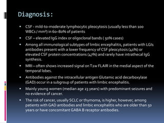
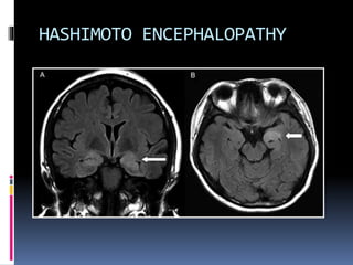
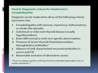
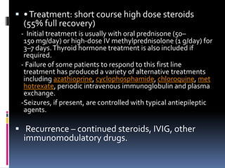
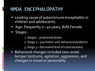
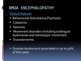
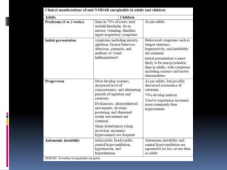
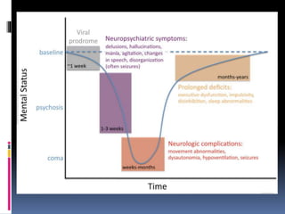
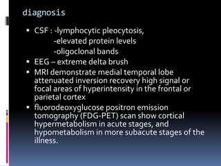
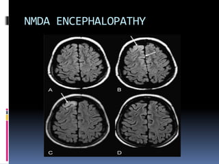
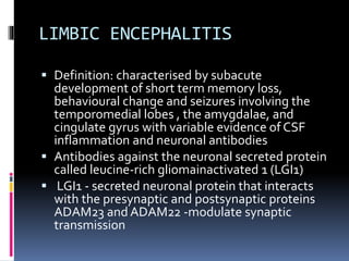
AUTOIMMUNE ENCEPHALITIS
Rusudan Genebashvili
Definition
 The term autoimmune encephalitis is used to
describe a group of disorders:
- Characterized by symptoms of the CNS
(limbic, extra-limbic, basal ganglia,
autonomic structures or more wide-spread)
due to autoantibodies against neuronal
surface or synaptic proteins, which are likely
to mediate the disease.
HASHIMOTO ENCEPHALOPATHY
 Defined by the detection of thyroid
peroxidase (TPO) antibodies in patients with
acute or subacute encephalitis that responds
to steroids.
 “Encephalopathy associated with
autoimmune thyroid disease” is considered
more accurate- due to normal thyroid
function.
 Very vague symptoms- unclear course
 Clinical features : non specific
- Stroke-like symptoms
-Tremor, myoclonus
-Transient aphasia
- Sleep and behaviour abnormalities
- Hallucinations, seizures and ataxia.
Diagnosis:
 Clinical triad of neuropsychiatric symptoms,
detection of antimicrosomal or
antithyroglobulin antibodies, and exclusion of
other causes.
 Antithyroid peroxidase, antithyroglobulin,
 lesser extent thyroid-stimulating hormone
receptor
 blocking antibodies.
 α-enolase
Diagnosis:
 Autoimmune cerebral vasculitis perhaps
related to immune complex deposition.
 CSF show moderately elevated protein, may
be positive for anti thyroid Ab, OCB seen
 EEG: slowing, triphasic waves, epileptiform
discharges.
 MRI usually normal, occasionally non-specific
sub cortical white matterT2 signal changes.
 Thyroid status may be normal.
HASHIMOTO ENCEPHALOPATHY
 •Treatment: short course high dose steroids
(55% full recovery)
- Initial treatment is usually with oral prednisone (50–
150 mg/day) or high-dose IV methylprednisolone (1 g/day) for
3–7 days.Thyroid hormone treatment is also included if
required.
- Failure of some patients to respond to this first line
treatment has produced a variety of alternative treatments
including azathioprine, cyclophosphamide, chloroquine, met
hotrexate, periodic intravenous immunoglobulin and plasma
exchange.
-Seizures, if present, are controlled with typical antiepileptic
agents.
 Recurrence – continued steroids, IVIG, other
immunomodulatory drugs.
NMDA ENCEPHALOPATHY
 Leading cause of autoimmune encephalitis in
children and adolescents.
 Age- frequently 2– 40 years, 80% Female
 Stages :
1. Stage1 – prodromal phase
2. Stage 2 – psychiatric and behavioral problems
3. Stage 3 – Decreased level of consciousness
 Behavioral changes included new-onset
temper tantrums, agitation, aggression, and
changes in mood or personality
NMDA ENCEPHALOPATHY
Clinical features
 Behavioural disturbance,Psychosis
 Catatonia
 Seizures
 Movement disorders including orolingual
dyskinesias and stereotypic movement.
 Dysautonomia.
 Ovarian teratoma is associated in up to 50%
of the cases
diagnosis
 CSF : -lymphocytic pleocytosis,
-elevated protein levels
-oligoclonal bands
 EEG – extreme delta brush
 MRI demonstrate medial temporal lobe
attenuated inversion recovery high signal or
focal areas of hyperintensity in the frontal or
parietal cortex
 fluorodeoxyglucose positron emission
tomography (FDG-PET) scan show cortical
hypermetabolism in acute stages, and
hypometabolism in more subacute stages of the
illness.
NMDA ENCEPHALOPATHY
19 year old with NMDA encephalitis
Delta Brush
treatment
 first line of immunotherapies including
corticosteroids, intravenous immunoglobulin,
or plasma exchange
 Rituximab and cyclophosphamide, alone or
combined, are often effective in adults
 Approximately 80% of patients have
substantial or full recovery.
LIMBIC ENCEPHALITIS
 Definition: characterised by subacute
development of short term memory loss,
behavioural change and seizures involving the
temporomedial lobes , the amygdalae, and
cingulate gyrus with variable evidence of CSF
inflammation and neuronal antibodies
 Antibodies against the neuronal secreted protein
called leucine-rich gliomainactivated 1 (LGI1)
 LGI1 - secreted neuronal protein that interacts
with the presynaptic and postsynaptic proteins
ADAM23 and ADAM22 -modulate synaptic
transmission
 In 1968 , as Para neoplastic syndrome
 median age of patients is 60 years
 Severe short term memory loss
 hyponatremia and myoclonic-like
movements, described as faciobrachial
dystonic seizures
 Other antibodies associated- antibodies
against intracellular antigens (eg, Hu, CRMP5,
Ma2) or against cell surface or synaptic
proteins (eg (AMPA) [GABA(B)] [mGluR5])
 Ophelia syndrome (- association with
Hodgkin’s lymphoma)
Diagnosis:
 CSF - mild-to-moderate lymphocytic pleocytosis (usually less than 100
WBCs / mm³) in 60–80% of patients
 CSF – elevated IgG index or oligoclonal bands ( 50% cases)
 Among all immunological subtypes of limbic encephalitis, patients with LGI1
antibodies present with a lower frequency of CSF pleocytosis (41%) or
elevated CSF protein concentrations (47%) and rarely have intrathecal IgG
synthesis.
 MRI – often shows increased signal onT2w FLAIR in the medial aspect of the
temporal lobes.
 Antibodies against the intracellular antigen Glutamic acid decarboxylase
(GAD) occur in a subgroup of patients with limbic encephalitis.
 Mainly young women (median age 23 years) with predominant seizures and
no evidence of cancer.
 The risk of cancer, usually SCLC or thymoma, is higher, however, among
patients with GAD antibodies and limbic encephalitis who are older than 50
years or have concomitant GABA B receptor antibodies.
Treatment:
 Treatments that have been tried
include intravenous immunoglobulin,
plasmapheresis, corticosteroids, cyclophosph
amide and rituximab
 If an associated tumour is found, then
recovery is not possible until the tumour is
removed
Thank you 
Rusudan Genebashvili

Autoimmune encephalitis

  • 1.
  • 2.
    Definition  The termautoimmune encephalitis is used to describe a group of disorders: - Characterized by symptoms of the CNS (limbic, extra-limbic, basal ganglia, autonomic structures or more wide-spread) due to autoantibodies against neuronal surface or synaptic proteins, which are likely to mediate the disease.
  • 3.
    HASHIMOTO ENCEPHALOPATHY  Definedby the detection of thyroid peroxidase (TPO) antibodies in patients with acute or subacute encephalitis that responds to steroids.  “Encephalopathy associated with autoimmune thyroid disease” is considered more accurate- due to normal thyroid function.  Very vague symptoms- unclear course
  • 4.
     Clinical features: non specific - Stroke-like symptoms -Tremor, myoclonus -Transient aphasia - Sleep and behaviour abnormalities - Hallucinations, seizures and ataxia.
  • 5.
    Diagnosis:  Clinical triadof neuropsychiatric symptoms, detection of antimicrosomal or antithyroglobulin antibodies, and exclusion of other causes.  Antithyroid peroxidase, antithyroglobulin,  lesser extent thyroid-stimulating hormone receptor  blocking antibodies.  α-enolase
  • 6.
    Diagnosis:  Autoimmune cerebralvasculitis perhaps related to immune complex deposition.  CSF show moderately elevated protein, may be positive for anti thyroid Ab, OCB seen  EEG: slowing, triphasic waves, epileptiform discharges.  MRI usually normal, occasionally non-specific sub cortical white matterT2 signal changes.  Thyroid status may be normal.
  • 7.
  • 10.
     •Treatment: shortcourse high dose steroids (55% full recovery) - Initial treatment is usually with oral prednisone (50– 150 mg/day) or high-dose IV methylprednisolone (1 g/day) for 3–7 days.Thyroid hormone treatment is also included if required. - Failure of some patients to respond to this first line treatment has produced a variety of alternative treatments including azathioprine, cyclophosphamide, chloroquine, met hotrexate, periodic intravenous immunoglobulin and plasma exchange. -Seizures, if present, are controlled with typical antiepileptic agents.  Recurrence – continued steroids, IVIG, other immunomodulatory drugs.
  • 11.
    NMDA ENCEPHALOPATHY  Leadingcause of autoimmune encephalitis in children and adolescents.  Age- frequently 2– 40 years, 80% Female  Stages : 1. Stage1 – prodromal phase 2. Stage 2 – psychiatric and behavioral problems 3. Stage 3 – Decreased level of consciousness  Behavioral changes included new-onset temper tantrums, agitation, aggression, and changes in mood or personality
  • 12.
    NMDA ENCEPHALOPATHY Clinical features Behavioural disturbance,Psychosis  Catatonia  Seizures  Movement disorders including orolingual dyskinesias and stereotypic movement.  Dysautonomia.  Ovarian teratoma is associated in up to 50% of the cases
  • 15.
    diagnosis  CSF :-lymphocytic pleocytosis, -elevated protein levels -oligoclonal bands  EEG – extreme delta brush  MRI demonstrate medial temporal lobe attenuated inversion recovery high signal or focal areas of hyperintensity in the frontal or parietal cortex  fluorodeoxyglucose positron emission tomography (FDG-PET) scan show cortical hypermetabolism in acute stages, and hypometabolism in more subacute stages of the illness.
  • 16.
  • 17.
    19 year oldwith NMDA encephalitis Delta Brush
  • 19.
    treatment  first lineof immunotherapies including corticosteroids, intravenous immunoglobulin, or plasma exchange  Rituximab and cyclophosphamide, alone or combined, are often effective in adults  Approximately 80% of patients have substantial or full recovery.
  • 20.
    LIMBIC ENCEPHALITIS  Definition:characterised by subacute development of short term memory loss, behavioural change and seizures involving the temporomedial lobes , the amygdalae, and cingulate gyrus with variable evidence of CSF inflammation and neuronal antibodies  Antibodies against the neuronal secreted protein called leucine-rich gliomainactivated 1 (LGI1)  LGI1 - secreted neuronal protein that interacts with the presynaptic and postsynaptic proteins ADAM23 and ADAM22 -modulate synaptic transmission
  • 21.
     In 1968, as Para neoplastic syndrome  median age of patients is 60 years  Severe short term memory loss  hyponatremia and myoclonic-like movements, described as faciobrachial dystonic seizures  Other antibodies associated- antibodies against intracellular antigens (eg, Hu, CRMP5, Ma2) or against cell surface or synaptic proteins (eg (AMPA) [GABA(B)] [mGluR5])  Ophelia syndrome (- association with Hodgkin’s lymphoma)
  • 22.
    Diagnosis:  CSF -mild-to-moderate lymphocytic pleocytosis (usually less than 100 WBCs / mm³) in 60–80% of patients  CSF – elevated IgG index or oligoclonal bands ( 50% cases)  Among all immunological subtypes of limbic encephalitis, patients with LGI1 antibodies present with a lower frequency of CSF pleocytosis (41%) or elevated CSF protein concentrations (47%) and rarely have intrathecal IgG synthesis.  MRI – often shows increased signal onT2w FLAIR in the medial aspect of the temporal lobes.  Antibodies against the intracellular antigen Glutamic acid decarboxylase (GAD) occur in a subgroup of patients with limbic encephalitis.  Mainly young women (median age 23 years) with predominant seizures and no evidence of cancer.  The risk of cancer, usually SCLC or thymoma, is higher, however, among patients with GAD antibodies and limbic encephalitis who are older than 50 years or have concomitant GABA B receptor antibodies.
  • 25.
    Treatment:  Treatments thathave been tried include intravenous immunoglobulin, plasmapheresis, corticosteroids, cyclophosph amide and rituximab  If an associated tumour is found, then recovery is not possible until the tumour is removed
  • 26.