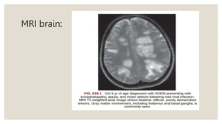
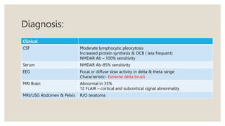
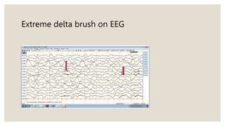
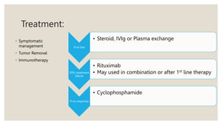
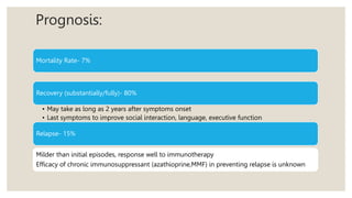
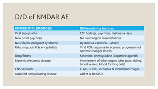
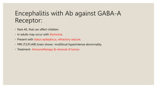
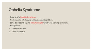
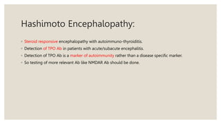
AUTOIMMUNE
ENCEPHALITIS
SPEAKER – DR CHAYANIKA MISHRA
INTRODUCTION:
◦ Autoimmune encephalitis a term used in children presenting with neurological syndrome associated
with serum and/ or cerebrospinal fluid antibodies directed against ion channels, receptors and
associated proteins.
◦ It comprises group of clinical syndrome that can occur at all ages but preferentially affect younger
adult and children.
◦ Auto antibodies against:
1. Neuronal cell surface protein
2. Synaptic receptors involved in transmission ,plasticity ,excitability.
◦ Triggers:
1. Tumors
2. Post viral infections
3. Post vaccination
AUTOIMMUNE ENCEPHALITIS IN CHILDREN
◦ ADEM
◦ Anti NMDAR encephalitis
◦ Encephalitis a/w GABA R antibodies
◦ NMOSD
◦ Opsoclonus-myoclonus & cerebellar - brainstem encephalitis
◦ Bickerstaff encephalitis
◦ Hashimoto encephalitis
◦ Rasmussen encephalitis
◦ Basal ganglia encephalitis
◦ CLIPPERS
◦ ROHHAD
ADEM:
◦ MC cause of autoimmune encephalitis in children and adolescents.
◦ Acute onset of polyfocal neurological deficit accompanied by encephalopathy and
changes compatible with demyelination on MRI brain.
EPIDEMIOLOGY:
◦ It can occur at any age most cases reported 5-8 yrs, slight male predominance.
◦ Usually it is monophasic, if recurrence occur 3 month or longer after the first episode termed as
MDEM (Multiphasic Disseminated Encephalo-Myelitis).
◦ 50% ADEM MOG (Myelin Oligodendrocyte Glycoprotein) Ab +, almost all case of MDEM MOG Ab +.
◦ ADEM can be followed by demyelination in a new location & if MOG-Ab negative - MS
◦ ADEM f/b relapse in specific location like
Optic nerve -ADEM-ON
◦ Optic nerve and spinal cord- NMOSD . Both frequently a/w MOG-Ab positive.
PATHOGENESIS:
Molecular
mimicry
• Post infectious
• Influenzae
• EBV
• CMV
• Varicella
• Enterovirus
• Measles, mumps, rubella
• HSV
• Mycoplasma pneumoniae
• Post vaccination
• Rabies
• Smallpox
• Measles
• Mumps
• Rubella
• JE-B
• DPT
• Influenzae
CLINICAL MANIFESTATION:
◦ Initial symptoms include- lethargy ,fever ,headache ,vomiting ,meningeal sign and seizure.
◦ Encephalopathy:
1. Is hallmark
2. Ranging from change in behaviour , persistent irritability to coma.
◦ Focal neurological deficit:
1. Difficult to ascertain in obtunded or very young child ,
2. Common neurological signs- visual loss, ataxia, motor and sensory deficit, bowel bladder dysfunction
if spinal cord demyelination occurs.
◦ It is usually rapidly progressive over days may need ICU admission particularly when
1. Brain stem dysfunction
2. Raised ICT.
DIAGNOSIS:
Clinical
CSF • Often normal, pleocytosis with lymphocytic or
monocytic predominance.
• Proteins can be elevated but true OCB rare.
• MOG AB +ve
Serum • MOG Ab +ve
EEG • Generalised slowing, but focal slowing or epileptiform
discharge may present.
MRI (Imaging study of choice) • B/L large multifocal sometimes confluent edematous
mass like lesions with variable enhancement within
white & gray matter.
• Typically appears to be of similar age.
• Repeat MRI after 3- 12 months – improvement or
complete resolution.
CT Scan • Normal/Hypodense lesions
MRI brain:
Treatment:
Symptomatic treatment
• Empirical antibiotics and antivirals should be considered when infective evaluations are pending
High dose steroids
• IV methylprednisolone- 20-30 mg/kg for 5 days (max. 1000mg/day)
• Followed by oral prednisolone 1-2 mg/kg/day over 4-6 weeks (max. 40-60 mg/day)
For refractory or severe cases
Others options
• IVIG – 2 gm/kg over 2-5 days
• Plasmapheresis- 5-7 exchange every other days
Prognosis:
◦ Most children experiences full motor recovery.
◦ But residual defect may be seen- cognitive deficit & behavioural changes.
Differential diagnosis:
◦ Multiple sclerosis
◦ Leukodystrophy
◦ Vasculitis
◦ Tumor
◦ Ab associated disorder
Anti NMDAR Encephalitis:
(N-Methyl D-Asparatate Receptor)
◦ Second MCC of AE.
◦ Female > Male ( < 12 years Male > Female)
◦ Underlying teratoma (40% of female >12 yrs ovarian teratoma)
◦ Pathogens:
1. Mycoplasma pneumoniae
2. HSV-1
3. HHV-6
4. Enterovirus
5. Influenzae
HSV Encephalitis
(Ab against GluN
1 subunit of
NMDAR)
2-12 weeks after
treatment
completion
Relapse of
symptoms-
Choreoathetosis
post HSV
Encephalitis
Pathogenesis
• IgG Ab target the Glu N1 subunit of
NMDA receptor
Clinical features:
Usually evolve in stages
Prodormal symptoms
• Viral illness like
fever, headache.
Psychiatric
manifestation
• Rapidly progressive
anxiety
• Mood disturbances
• Catatonia
• Bizzare behaviour
• Memory deficit
• Language
disintegration
• Insomnia
After few days to weeks
• Decreased level of
consciousness
• Seizure
• Limb or oral
dyskinesia
• Choreoathetoid
movements
• Autonomic
instability (tachy-
brady, fluctuation of
BP, hypoventilation,
hyperthermia,
sialorrhea)
In Teenagers & young adults:
In toddlers & infants:
Psychiatric manifestation- may
be missed
• Irritability
• New onset temper tantrum
• Agitation
• Aggression
• Reduced speech
• Mutism
• Autistic like regression
After few days to weeks
• Decreased level of
consciousness
• Seizure
• Limb or oral dyskinesia
• Choreoathetoid movements
• Autonomic instability- less
sever
• Some children may develop
cerebellar ataxia &
hemiparesis
Diagnosis:
Clinical
CSF Moderate lymphocytic pleocytosis
Increased protein synthesis & OCB ( less frequent)
NMDAR Ab – 100% sensitivity
Serum NMDAR Ab-85% sensitivity
EEG Focal or diffuse slow activity in delta & theta range
Characteristic- Extreme delta brush
MRI Brain Abnormal in 35%
T2 FLAIR – cortical and subcortical signal abnormality
MRI/USG Abdomen & Pelvis R/O teratoma
Extreme delta brush on EEG
Treatment:
◦ Symptomatic
management
◦ Tumor Removal
◦ Immunotherapy
First line
• Steroid, IVIg or Plasma exchange
50% treatment
failure
• Rituximab
• May used in combination or after 1st line therapy
If no response
• Cyclophosphamide
Prognosis:
Mortality Rate- 7%
Recovery (substantially/fully)- 80%
• May take as long as 2 years after symptoms onset
• Last symptoms to improve social interaction, language, executive function
Relapse- 15%
Milder than initial episodes, response well to immunotherapy
Efficacy of chronic immunosuppressant (azathioprine,MMF) in preventing relapse is unknown
D/D of NMDAR AE
DIFFERENTIAL DIAGNOSIS Differentiating features
Viral Encephalitis CSF findings, psychosis, dyskinesia -less
New onset psychosis No neurological manifestations
Neuroleptic malignant syndrome Dyskinesia, catatonia - absent
Relapsing post HSV encephalitis Viral PCR, response to acyclovir, progression of
necrotic changes on MRI
Drug/Toxins Ketamine, phencyclidine (dopamine agonist)
Systemic rheumatic disease Involvement of other organs (skin, joint, kidney,
blood vessels, blood forming cells)
CNS vasculitis FLAIR T2 MRI- Ischemia & microhemorrhages
Acquired demyelinating disease ADEM & NMOSD
Encephalitis with Ab against GABA-A
Receptor:
◦ Rare AE, that can affect children.
◦ In adults may occur with thymoma.
◦ Present with status epilepticus, refractory seizure.
◦ MRI (T2/FLAIR) brain shows- multifocal hyperintense abnormality.
◦ Treatment- Immunotherapy & removal of tumor.
Ophelia Syndrome
◦ Occur in a/w Hodgkin lymphoma.
◦ Predominantly affect young adults, teenager & children.
◦ Some develops Ab against mGluR5 receptor involved in learning & memory.
◦ Management:
1. Removal of tumor
2. Immunotherapy
Hashimoto Encephalopathy:
◦ Steroid responsive encephalopathy with autoimmuno-thyroiditis.
◦ Detection of TPO Ab in patients with acute/subacute encephalitis.
◦ Detection of TPO Ab is a marker of autoimmunity rather than a disease specific marker.
◦ So testing of more relevant Ab like NMDAR Ab should be done.
Bickerstaff Encephalitis:
◦ Rapid progression (<4wks) of
1. B/L external ophthalmoplegia
2. Ataxia
3. Decreased level of consciousness
◦ Mostly affect adults, but children of 3 yrs old have been identified.
◦ Serum GQ1b IgG Ab found in 66% cases.
◦ May develop hyporeflexia and overlap with MFS.
◦ MRI T2 signal abnormality (30%)- Brainstem, thalamus & cerebellum.
◦ Good response to immunotherapy.
Rasmussen encephalitis:
◦ Inflamatory encephalopathy c/b
1. Refracory focal seizures
2. Cognitive deterioration
3. Focal neurological deficit.
◦ Frequently affect children 6-8 yr old but
adolescents &adults can be affected.
Etiology:
◦ Unknown.
◦ Theories proposed are neuronal antibody &
T- cell mechanisms - triggered by viral
infection.
MRI Brain- Cerebral hemiatrophy
Management of Rasmussen encephalitis:
1. High dose steroid, IVIG or plasma exchange.
2. Rituximab & intraventricular Gamma interferon effective in isolated cases.
3. Tacrolimus- better outcome of neurological function & slower progression of cerebral atrophy
but no effect on seizure control.
4. Adalimumab( monoclonal antibody against TNF-alpha) shows seizure control &preservation of
cognitive function in 50% cases.
5. Most effective treatment for controlling seizure is functional hemispherectomy i.e. surgical
disconnection of affected hemisphere.
Autoimmune limbic encephalitis:
◦ Inflamatory process of the limbic system including medial temporal lobe, amygdala, cingulate gyrus.
◦ Most commonly found in adults.
◦ Some patients there is underlying tumor i.e. leukemia, ganglioneuroblastoma, neuroblastoma, small cell
carcinoma of ovary.
◦ Autoantibodies are-
1. VGKCs /LGI1(Leucine rich glioma inactivated 1)
2. Caspr2 antibodies
MRI of limbic encephalitis
Opsoclonus-myoclonus and other type of brainstem-
cerebellar encephalitis:
◦ In children- 50% have underlying NEUROBLASTOMA.
◦ In teenagers and young adults underlying TERATOMA usually in ovaries.
◦ Initially present with- irritability, ataxia, falling, myoclonus, tremor and drooling.
◦ Later- hypotonia, Opsoclonus c/b rapid, chaotic, multidirectional eye movement without saccadic interval.
◦ Immunotherapy improves abnormal eye movement but residual behavioral, language, cognitive problems
persists.
◦ Delay in treatment appears to be a/w poor outcome , therefore in case of neuroblastoma removal of the
tumor should not delay the start of immunotherapy.
CLIPPERS:
◦ Chronic Lymphocytic Inflammation with Pontine Perivascular Enhancement Responsive to Steroids
◦ Pontine predominant encephalomyelitis
◦ Usually present with episodic diplopia and facial parasthesia.
◦ MRI Brain – symmetric curvilinear gadolinium enhancement around the pons & extend variably into
medulla, cerebellum, midbrain and spinal cord.
◦ Responds to high dose of steroids , may worsen during steroid tapering, requiring chronic steroid.
Brainstem & spinal cord
dysfunction
ROHHAD:
◦ Rapid-onset Obesity with Hypothalamic dysfunction, Hypoventilation & Autonomic Dysregulation.
◦ An autoimmune or
paraneoplastic etiology.
◦ Response to immunotherapy
in few patients.
Children having
normal
development till
2-4 yrs
Rapid onset of
hyperphagia,
weight gain,
abnormal
behaviour
• Social disinhibition,
impulsivity, lethargy,
outburst of euphoria &
laughing
Autonomic
dysfunction &
central
hypoventilation
• Abnormal pupillary
responses, thermal
dysregulation, GI
dysmotility
NMOSD
◦ Typical involvement of optic nerve & spinal cord.
◦ Encephalopathy in the form of diencephalic or area postrema syndromes.
◦ Can have AQP4 or MOG Ab. Some are seronegative.
◦ Involvement of brain areas rich in AQP4 (periaqueductal gray matter, hypothalamus, optic nerve
and central involvement of the spinal cord).
◦ High risk of relapses and long term disability.
◦ Requires chronic immunotherapy.
◦ Patients with MOG Ab have better outcome than those with AQP4 Ab or seronegative cases.
AUTOIMMUNE ENCEPHALITIS.pptx

AUTOIMMUNE ENCEPHALITIS.pptx

  • 1.
  • 2.
    INTRODUCTION: ◦ Autoimmune encephalitisa term used in children presenting with neurological syndrome associated with serum and/ or cerebrospinal fluid antibodies directed against ion channels, receptors and associated proteins. ◦ It comprises group of clinical syndrome that can occur at all ages but preferentially affect younger adult and children. ◦ Auto antibodies against: 1. Neuronal cell surface protein 2. Synaptic receptors involved in transmission ,plasticity ,excitability. ◦ Triggers: 1. Tumors 2. Post viral infections 3. Post vaccination
  • 3.
    AUTOIMMUNE ENCEPHALITIS INCHILDREN ◦ ADEM ◦ Anti NMDAR encephalitis ◦ Encephalitis a/w GABA R antibodies ◦ NMOSD ◦ Opsoclonus-myoclonus & cerebellar - brainstem encephalitis ◦ Bickerstaff encephalitis ◦ Hashimoto encephalitis ◦ Rasmussen encephalitis ◦ Basal ganglia encephalitis ◦ CLIPPERS ◦ ROHHAD
  • 4.
    ADEM: ◦ MC causeof autoimmune encephalitis in children and adolescents. ◦ Acute onset of polyfocal neurological deficit accompanied by encephalopathy and changes compatible with demyelination on MRI brain.
  • 5.
    EPIDEMIOLOGY: ◦ It canoccur at any age most cases reported 5-8 yrs, slight male predominance. ◦ Usually it is monophasic, if recurrence occur 3 month or longer after the first episode termed as MDEM (Multiphasic Disseminated Encephalo-Myelitis). ◦ 50% ADEM MOG (Myelin Oligodendrocyte Glycoprotein) Ab +, almost all case of MDEM MOG Ab +. ◦ ADEM can be followed by demyelination in a new location & if MOG-Ab negative - MS ◦ ADEM f/b relapse in specific location like Optic nerve -ADEM-ON ◦ Optic nerve and spinal cord- NMOSD . Both frequently a/w MOG-Ab positive.
  • 6.
    PATHOGENESIS: Molecular mimicry • Post infectious •Influenzae • EBV • CMV • Varicella • Enterovirus • Measles, mumps, rubella • HSV • Mycoplasma pneumoniae • Post vaccination • Rabies • Smallpox • Measles • Mumps • Rubella • JE-B • DPT • Influenzae
  • 7.
    CLINICAL MANIFESTATION: ◦ Initialsymptoms include- lethargy ,fever ,headache ,vomiting ,meningeal sign and seizure. ◦ Encephalopathy: 1. Is hallmark 2. Ranging from change in behaviour , persistent irritability to coma. ◦ Focal neurological deficit: 1. Difficult to ascertain in obtunded or very young child , 2. Common neurological signs- visual loss, ataxia, motor and sensory deficit, bowel bladder dysfunction if spinal cord demyelination occurs. ◦ It is usually rapidly progressive over days may need ICU admission particularly when 1. Brain stem dysfunction 2. Raised ICT.
  • 8.
    DIAGNOSIS: Clinical CSF • Oftennormal, pleocytosis with lymphocytic or monocytic predominance. • Proteins can be elevated but true OCB rare. • MOG AB +ve Serum • MOG Ab +ve EEG • Generalised slowing, but focal slowing or epileptiform discharge may present. MRI (Imaging study of choice) • B/L large multifocal sometimes confluent edematous mass like lesions with variable enhancement within white & gray matter. • Typically appears to be of similar age. • Repeat MRI after 3- 12 months – improvement or complete resolution. CT Scan • Normal/Hypodense lesions
  • 9.
  • 10.
    Treatment: Symptomatic treatment • Empiricalantibiotics and antivirals should be considered when infective evaluations are pending High dose steroids • IV methylprednisolone- 20-30 mg/kg for 5 days (max. 1000mg/day) • Followed by oral prednisolone 1-2 mg/kg/day over 4-6 weeks (max. 40-60 mg/day) For refractory or severe cases Others options • IVIG – 2 gm/kg over 2-5 days • Plasmapheresis- 5-7 exchange every other days
  • 11.
    Prognosis: ◦ Most childrenexperiences full motor recovery. ◦ But residual defect may be seen- cognitive deficit & behavioural changes. Differential diagnosis: ◦ Multiple sclerosis ◦ Leukodystrophy ◦ Vasculitis ◦ Tumor ◦ Ab associated disorder
  • 13.
    Anti NMDAR Encephalitis: (N-MethylD-Asparatate Receptor) ◦ Second MCC of AE. ◦ Female > Male ( < 12 years Male > Female) ◦ Underlying teratoma (40% of female >12 yrs ovarian teratoma) ◦ Pathogens: 1. Mycoplasma pneumoniae 2. HSV-1 3. HHV-6 4. Enterovirus 5. Influenzae HSV Encephalitis (Ab against GluN 1 subunit of NMDAR) 2-12 weeks after treatment completion Relapse of symptoms- Choreoathetosis post HSV Encephalitis
  • 14.
    Pathogenesis • IgG Abtarget the Glu N1 subunit of NMDA receptor
  • 15.
    Clinical features: Usually evolvein stages Prodormal symptoms • Viral illness like fever, headache. Psychiatric manifestation • Rapidly progressive anxiety • Mood disturbances • Catatonia • Bizzare behaviour • Memory deficit • Language disintegration • Insomnia After few days to weeks • Decreased level of consciousness • Seizure • Limb or oral dyskinesia • Choreoathetoid movements • Autonomic instability (tachy- brady, fluctuation of BP, hypoventilation, hyperthermia, sialorrhea) In Teenagers & young adults:
  • 16.
    In toddlers &infants: Psychiatric manifestation- may be missed • Irritability • New onset temper tantrum • Agitation • Aggression • Reduced speech • Mutism • Autistic like regression After few days to weeks • Decreased level of consciousness • Seizure • Limb or oral dyskinesia • Choreoathetoid movements • Autonomic instability- less sever • Some children may develop cerebellar ataxia & hemiparesis
  • 17.
    Diagnosis: Clinical CSF Moderate lymphocyticpleocytosis Increased protein synthesis & OCB ( less frequent) NMDAR Ab – 100% sensitivity Serum NMDAR Ab-85% sensitivity EEG Focal or diffuse slow activity in delta & theta range Characteristic- Extreme delta brush MRI Brain Abnormal in 35% T2 FLAIR – cortical and subcortical signal abnormality MRI/USG Abdomen & Pelvis R/O teratoma
  • 18.
  • 19.
    Treatment: ◦ Symptomatic management ◦ TumorRemoval ◦ Immunotherapy First line • Steroid, IVIg or Plasma exchange 50% treatment failure • Rituximab • May used in combination or after 1st line therapy If no response • Cyclophosphamide
  • 20.
    Prognosis: Mortality Rate- 7% Recovery(substantially/fully)- 80% • May take as long as 2 years after symptoms onset • Last symptoms to improve social interaction, language, executive function Relapse- 15% Milder than initial episodes, response well to immunotherapy Efficacy of chronic immunosuppressant (azathioprine,MMF) in preventing relapse is unknown
  • 21.
    D/D of NMDARAE DIFFERENTIAL DIAGNOSIS Differentiating features Viral Encephalitis CSF findings, psychosis, dyskinesia -less New onset psychosis No neurological manifestations Neuroleptic malignant syndrome Dyskinesia, catatonia - absent Relapsing post HSV encephalitis Viral PCR, response to acyclovir, progression of necrotic changes on MRI Drug/Toxins Ketamine, phencyclidine (dopamine agonist) Systemic rheumatic disease Involvement of other organs (skin, joint, kidney, blood vessels, blood forming cells) CNS vasculitis FLAIR T2 MRI- Ischemia & microhemorrhages Acquired demyelinating disease ADEM & NMOSD
  • 22.
    Encephalitis with Abagainst GABA-A Receptor: ◦ Rare AE, that can affect children. ◦ In adults may occur with thymoma. ◦ Present with status epilepticus, refractory seizure. ◦ MRI (T2/FLAIR) brain shows- multifocal hyperintense abnormality. ◦ Treatment- Immunotherapy & removal of tumor.
  • 23.
    Ophelia Syndrome ◦ Occurin a/w Hodgkin lymphoma. ◦ Predominantly affect young adults, teenager & children. ◦ Some develops Ab against mGluR5 receptor involved in learning & memory. ◦ Management: 1. Removal of tumor 2. Immunotherapy
  • 24.
    Hashimoto Encephalopathy: ◦ Steroidresponsive encephalopathy with autoimmuno-thyroiditis. ◦ Detection of TPO Ab in patients with acute/subacute encephalitis. ◦ Detection of TPO Ab is a marker of autoimmunity rather than a disease specific marker. ◦ So testing of more relevant Ab like NMDAR Ab should be done.
  • 25.
    Bickerstaff Encephalitis: ◦ Rapidprogression (<4wks) of 1. B/L external ophthalmoplegia 2. Ataxia 3. Decreased level of consciousness ◦ Mostly affect adults, but children of 3 yrs old have been identified. ◦ Serum GQ1b IgG Ab found in 66% cases. ◦ May develop hyporeflexia and overlap with MFS. ◦ MRI T2 signal abnormality (30%)- Brainstem, thalamus & cerebellum. ◦ Good response to immunotherapy.
  • 26.
    Rasmussen encephalitis: ◦ Inflamatoryencephalopathy c/b 1. Refracory focal seizures 2. Cognitive deterioration 3. Focal neurological deficit. ◦ Frequently affect children 6-8 yr old but adolescents &adults can be affected. Etiology: ◦ Unknown. ◦ Theories proposed are neuronal antibody & T- cell mechanisms - triggered by viral infection. MRI Brain- Cerebral hemiatrophy
  • 27.
    Management of Rasmussenencephalitis: 1. High dose steroid, IVIG or plasma exchange. 2. Rituximab & intraventricular Gamma interferon effective in isolated cases. 3. Tacrolimus- better outcome of neurological function & slower progression of cerebral atrophy but no effect on seizure control. 4. Adalimumab( monoclonal antibody against TNF-alpha) shows seizure control &preservation of cognitive function in 50% cases. 5. Most effective treatment for controlling seizure is functional hemispherectomy i.e. surgical disconnection of affected hemisphere.
  • 28.
    Autoimmune limbic encephalitis: ◦Inflamatory process of the limbic system including medial temporal lobe, amygdala, cingulate gyrus. ◦ Most commonly found in adults. ◦ Some patients there is underlying tumor i.e. leukemia, ganglioneuroblastoma, neuroblastoma, small cell carcinoma of ovary. ◦ Autoantibodies are- 1. VGKCs /LGI1(Leucine rich glioma inactivated 1) 2. Caspr2 antibodies
  • 29.
    MRI of limbicencephalitis
  • 30.
    Opsoclonus-myoclonus and othertype of brainstem- cerebellar encephalitis: ◦ In children- 50% have underlying NEUROBLASTOMA. ◦ In teenagers and young adults underlying TERATOMA usually in ovaries. ◦ Initially present with- irritability, ataxia, falling, myoclonus, tremor and drooling. ◦ Later- hypotonia, Opsoclonus c/b rapid, chaotic, multidirectional eye movement without saccadic interval. ◦ Immunotherapy improves abnormal eye movement but residual behavioral, language, cognitive problems persists. ◦ Delay in treatment appears to be a/w poor outcome , therefore in case of neuroblastoma removal of the tumor should not delay the start of immunotherapy.
  • 31.
    CLIPPERS: ◦ Chronic LymphocyticInflammation with Pontine Perivascular Enhancement Responsive to Steroids ◦ Pontine predominant encephalomyelitis ◦ Usually present with episodic diplopia and facial parasthesia. ◦ MRI Brain – symmetric curvilinear gadolinium enhancement around the pons & extend variably into medulla, cerebellum, midbrain and spinal cord. ◦ Responds to high dose of steroids , may worsen during steroid tapering, requiring chronic steroid. Brainstem & spinal cord dysfunction
  • 32.
    ROHHAD: ◦ Rapid-onset Obesitywith Hypothalamic dysfunction, Hypoventilation & Autonomic Dysregulation. ◦ An autoimmune or paraneoplastic etiology. ◦ Response to immunotherapy in few patients. Children having normal development till 2-4 yrs Rapid onset of hyperphagia, weight gain, abnormal behaviour • Social disinhibition, impulsivity, lethargy, outburst of euphoria & laughing Autonomic dysfunction & central hypoventilation • Abnormal pupillary responses, thermal dysregulation, GI dysmotility
  • 33.
    NMOSD ◦ Typical involvementof optic nerve & spinal cord. ◦ Encephalopathy in the form of diencephalic or area postrema syndromes. ◦ Can have AQP4 or MOG Ab. Some are seronegative. ◦ Involvement of brain areas rich in AQP4 (periaqueductal gray matter, hypothalamus, optic nerve and central involvement of the spinal cord). ◦ High risk of relapses and long term disability. ◦ Requires chronic immunotherapy. ◦ Patients with MOG Ab have better outcome than those with AQP4 Ab or seronegative cases.