SlideShare a Scribd company logo
Autoimmune
Associated
Seizures
Mohamed Abd-ElHady
MSc Neurology
March 2023
DEFINITIONS
SEIZURE OR EPILEPSY?
The term autoimmune epilepsy has been used in recent years in reference to any
seizures occurring in the context of autoimmune disorders. However, seizures are a
common clinical feature in autoimmune encephalitis but are typically not the only
clinical feature.
Also, after the resolution of the active phase of encephalitis, seizures may resolve,
thus not meeting the definition of epilepsy, which implies an ongoing predisposition
to seizures.
A more appropriate term is “acute symptomatic seizures secondary to autoimmune
encephalitis”
On the other hand, some patients with autoimmune encephalitis continue to have
seizures after the resolution of acute encephalitis.
In addition, some patients with chronic epilepsy may be discovered to have findings
suggesting an immune etiology despite a long history of seizures and the absence of a
clear prior episode of acute encephalitis.
In these cases, the concept of “epilepsy” is appropriate, given the presence of an
ongoing potential for seizures.
Hence, “autoimmune-associated epilepsy” has been proposed as the most appropriate
term for those situations.
PATHOPHYSIOLOGY AND DIAGNOSIS
The concept of immune epilepsy is that it results directly from an immune disorder in
which seizures are a core symptom of the disorder.
An immune etiology can be conceptualized as where there is evidence of autoimmune-
mediated central nervous system inflammation (antibody-, cytokine-, and T-cell),
targeting either neural cell surface or intracellular antigens.
The generation of seizures is due to disruption of the normal homeostatic balance
between neural excitation and inhibition.
(1)ACUTE SYMPTOMATIC SEIZURES
SECONDARY TO AUTOIMMUNE
ENCEPHALITIS
Autoimmune encephalitis classically presents with a cluster of symptoms, with
seizures being often the presenting symptom in antibody-mediated autoimmune
encephalitis.
It is crucial that we recognize the types of seizures that can indicate the presence of
neuronal cell surface antibody–mediated autoimmune encephalitis.
The most common seizure types in autoimmune encephalitis at presentation are focal
seizures, which tend to occur without impaired awareness or postictal confusion.
They tend to be much shorter in duration lasting seconds.
Seizure frequency in autoimmune encephalitis is also unusually high compared with
seizures in most patients with focal epilepsy due to other causes, often occurring
daily, and in some many times per day.
a preceding febrile illness and a history of systemic autoimmunity or malignancy
should also raise suspicion of an immune etiology.
Autoimmune encephalitis often affects the limbic structures, especially the mesial
temporal, insula, and perisylvian networks, so it is not unexpected that this is
reflected in the seizure symptoms.
Seizure and Clinical Characteristics
Seizure Clinical Features Reported inAutoimmune Encephalitis
Patients are typically between the ages of 40 and 80 years (median, 65 years).
Anti-LGI1 encephalitis most commonly presents with focal seizures. Facio-brachial
dystonic seizures (FBDS)are the most characteristic seizure type in this disorder, although
are absent in more than half of cases.
Facio-brachial dystonic seizures are brief, lasting seconds, occur multiple times per day,
and typically present with simultaneous contraction of the upper limb muscles and
ipsilateral facial and neck muscles.
These seizures usually precede the onset of other features suggestive of encephalitis.
EEG may show no ictal findings even during focal seizures in anti-LGI1 encephalitis.
MRI may be negative but may show abnormalities in the mesial temporal structures,
basal ganglia, and insula and, less commonly, in the extratemporal regions
LGI1 antibodies are absent in spinal fluid in up to 50% of patients even in clear cases of
anti-LGI1 encephalitis.
Anti–Leucine-Rich Glioma Inactivated Protein 1Antibody Encephalitis (Anti-LGI1)
Seizures are common in anti–NMDA receptor encephalitis, occurring in 70% of cases.
Seizures occur more commonly in the early phase of the disease in children and young men.
Later in the course, status epilepticus may occur in approximately one-quarter of cases.
Neurobehavioral abnormalities are typically the most prominent presenting feature, including
psychosis, speech disorder, catatonia, personality changes, impaired cognition, and memory
disturbance.
The seizure types present in anti–NMDA receptor encephalitis include:
Focal aware
focal impaired awareness
focal to bilateral tonic-clonic seizures, which occur in almost 80% of cases
Motor manifestations occur in 50% of patients with seizures, and multiple seizure types can
occur in the same patient
Anti–N-methyl-D-aspartate Receptor Encephalitis (Anti-NMDA)
The first EEG after presentation may show a normal background rhythm or only mild
slowing, which correlates with a better prognosis.
As the condition progress, the EEG usually becomes abnormal, typically showing prominent
slowing initially, then a generalized rhythmic delta activity is commonly present.
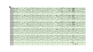
A characteristic EEG finding in anti–NMDA receptor encephalitis is the “extreme delta
brush” pattern, present in up to 30% of cases.
Anti–N-methyl-D-aspartate Receptor Encephalitis (Anti-NMDA)
Structural MRI is normal in up to 70% of anti–NMDA receptor encephalitis cases.
Abnormal findings most commonly involve the temporal lobe (in particular, the
hippocampus) however, other regions may be affected, including the frontal lobe, insula,
cingulate gyrus, and subcortical structures such as the thalamus and basal ganglia.
CSF is abnormal in the majority of anti–NMDA receptor encephalitis cases, showing
mononuclear pleocytosis and mild to moderate protein elevation, especially in the acute phase
of the illness.
CSF anti–NMDA receptor antibody testing is important because in some cases NMDA
receptor antibodies are found only in CSF and not in serum.
Anti–N-methyl-D-aspartate Receptor Encephalitis (Anti-NMDA)
antibodies targeting γ-aminobutyric acid B (GABAB) receptor, γ-aminobutyric acid A
(GABAA) receptor, anti–α-amino-3-hydroxy-5-methylisoxazole-4-propionic acid (AMPA)
receptor, dipeptidyl-peptidase–like protein 6 (DPPX), and metabotropic glutamate receptor 5
(mGluR5).
Anti-GABA B: most commonly presents with seizures and, in many cases, status epilepticus.
EEG and MRI may be normal and don’t provide information specific enough to allow clinical
diagnosis. The presence of anti-GABAB receptor antibodies in serum should be confirmed in
the CSF.
Anti-GABA A: commonly presents with drug-resistant seizures. may present with epilepsia
partialis continua or status epilepticus. Clear MRI abnormalities are present and are most
apparent on T2-weighted sequences, affecting both gray and white matter. These lesions are
often multifocal, non–diffusion-restricting, non-enhancing, and of a medium to large size.
Therefore, should also be considered in the differential diagnosis of fulminant MS and
ADEM.
OtherAntibody-Mediated Encephalitides
Anti-GAD65 antibody acute limbic encephalitis:
Relatively uncommon
more commonly associated with chronic focal epilepsy of temporal lobe origin.
Importantly, a diagnosis of anti-GAD65 encephalitis should not be made unless a concomitant
behavioral change or cognitive or memory impairment is present, and these features should help
clinicians differentiate the phenotypes of encephalitis and epilepsy.
OtherAntibody-Mediated Encephalitides
Symptoms of limbic encephalitis can also occur in the absence of CNS autoantibodies.
In such cases, provided other secondary causes of encephalitis are reasonably excluded, the
diagnosis of seronegative autoimmune limbic encephalitis can be made.
can occur as a paraneoplastic condition even in the absence of paraneoplastic antibodies.
Typically affects older men (median age, 62 years; age range, 40 to 79 years). The predominant
presenting symptom is short-term memory difficulties; in one-third of cases, this may be the
only symptom.
MRI changes with T2/FLAIR hyperintensities in both hippocampi are required to make the
diagnosis, and involvement of the insula, orbitofrontal cortex, or basal ganglia is also common.
SeronegativeAutoimmune Limbic Encephalitis
(2) SEIZURE AND CLINICAL
CHARACTERISTICS IN
AUTOIMMUNE ASSOCIATED
EPILEPSY
The term autoimmune-associated epilepsy denotes a condition characterized by an
enduring predisposition to unprovoked seizures in which evidence of an immune
etiology is present.
This can occur in several contexts:
A) Persistence of seizures after resolution of the active phase of encephalitis
B) In the setting of chronic unresolving encephalitis
C) Patients with epilepsy with compelling evidence of a CNS autoimmune condition and
where no alternative etiology for the epilepsy is identified.
Although seizures eventually resolve in most patients after timely treatment of the active phase
of autoimmune encephalitis with immunotherapy, a subset of patients will continue to
experience seizures or experience a recurrence after the initial resolution of the active phase.
Postencephalitic epilepsy typically emerges 3 to 6 months after initial presentation.
Initially, this may be difficult to distinguish from autoimmune encephalitis relapse, but it is
characterized by a predominant presentation with seizures in the absence of other symptoms of
encephalitis.
Postencephalitic Epilepsy
An important risk factor for the development of postencephalitic epilepsy is delayed initiation
of immunotherapy.
Other risk factors include a history of status epilepticus, interictal epileptiform discharges, and
high CSF protein level.
Antibody type is also important to determine the likelihood of seizure recurrence, being most
common in anti-GABAB and Anti-LGI1 mediated encephalitis and least common with Anti-
NMDAR encephalitis.
Postencephalitic Epilepsy
A rare neuroinflammatory disorder resulting in chronic focal seizures, emanating from one
hemisphere, progressive hemiparesis, other lateralized cortical deficits, and cognitive
impairment.
Rasmussen encephalitis is thought to be mediated predominantly by T-cell inflammatory
processes.
No significantly specific ABs have been linked to the condition.
Most commonly present in children (median age, 6 years) but can occur in young adults.
Rasmussen Encephalitis
Three phases of the disease have been described:
Stage 1: the patient may present with a prodrome of mild hemiparesis and infrequent focal
seizures up to years before the acute phase.
Stage 2: is heralded by a more acute presentation with frequent unilateral motor seizures that
evolve into treatment-refractory epilepsia partialis continua in 50% of cases.
All types of seizures can occur
Loss of cortical function typically develops, resulting in varying degrees of unilateral
hemiplegia, hemianopia, cognitive decline, and dysphasia over time.
Stage 3: represents a relative stagnation of progression and underlying active inflammation.
This is sometimes accompanied by a reduction in seizures, although focal seizures often persist
to a degree for several years.
Rasmussen Encephalitis
Neuroimaging characteristically shows contralateral hemiatrophy, which is progressive over
time.
Rasmussen Encephalitis
Despite the inflammatory etiology, Rasmussen encephalitis is inconsistently responsive to
immunotherapy, even in the active stage.
Surgery, specifically hemispheric disconnection, is often necessary to gain seizure control in
very young children with Rasmussen encephalitis and high seizure burden.
Rasmussen Encephalitis
Diagnostic criteria
Patient Selection for Immune Etiology Evaluation
Seizure manifestations and other clinical features can be used to help identify patients who
are most appropriate for autoantibody testing.
The presence of faciobrachial dystonic seizures should clearly prompt neural autoantibody
testing, which includes anti-LGI1 antibody.
Consider antibody screening in new-onset focal epilepsy of unknown cause, especially in
patients with a high daily seizure burden, and in the presence of perisylvian or autonomic
symptomatology.
New-onset refractory status epilepticus (NORSE)
EEG changes are rarely specific enough to trigger screening. EEG can be normal.
MRI might be suggestive of an autoimmune etiology, but a normal study does not exclude
the diagnosis.
CSF evaluation should be performed if autoimmune-associated epilepsy is suspected. The
sensitivity of ABs are higher in CSF than in serum.
TREATMENT INANTIBODY-MEDIATED
AUTOIMMUNE ENCEPHALITIS
ANDAUTOIMMUNE-ASSOCIATED EPILEPSY
Treatment is primarily focused on addressing the underlying etiology with immunotherapy
and can be considered in alignment with the phases of illness. The potential benefit of
adjunct use of antiseizure medications should also be evaluated, especially during the acute
phase.
AED inAcute Symptomatic Seizures Secondary toAutoimmune Encephalitis
Seizure control is seldom achieved by AED alone (occurred in 15%).
Higher responder rates have been reported with sodium channel blockers, particularly
carbamazepine, and lacosamide compared with levetiracetam.
Certain antiseizure medications, including carbamazepine, have immunosuppressive
effects.
It should be noted that the rate of hypersensitivity reactions is greater in patients with
autoimmune encephalitis than in the general population.
After the resolution of the acute phase of the illness and control of seizures, the question of
D/C AED often rises. (type of neural cell surface AB)
An individualized approach to the timing of antiseizure medication discontinuation needs to
be applied in these patients as in epilepsy due to other etiologies.
Long-term antiseizure medication administration is not always necessary in many patients
after the resolution of acute encephalitis, and discontinuation can be considered after 6
months or a greater period of seizure cessation.
Immunotherapy
Seizure freedom occurs in 62% to 89% of patients affected by seizures, and the median time
to seizure freedom after initiation of immunotherapy is 28 days. the benefits are not limited to
seizure cessation; they extend to cognitive and functional outcomes.
Treatment is generally conceptualized as
A) Initial induction therapy:
with first-line treatments that include IV corticosteroids, IVIg, or plasma exchange, used
individually or in combination. Second-line agents, usually rituximab or cyclophosphamide,
are typically initiated if the response to first-line therapy is inadequate.
B) Maintenance phase:
administration of first-line therapy or therapies continues weekly to every other week with
gradually increasing intervals between treatments.
Initiation of maintenance immunosuppressive medication may be used if a patient has a
significant response to induction therapy, establishing immunotherapy responsiveness.
Acute Treatment Strategy
The most common approach to first-line therapy for patients in the inpatient setting involves
the use of a combination of agents, typically methylprednisolone and IVIg.
If a patient has no response to corticosteroids or IVIg, plasma exchange is commonly
recommended as an option.
Corticosteroids and IVIg are often continued after the resolution of the induction period if the
response was favorable, and they are continued over the ensuing 6 to 12 months depending on
the clinical course on a gradual tapering schedule.
Second-line immunosuppressant therapy may be administered empirically during the
induction phase in acute inpatient situations. This is the typical approach in hospitalized
patients with severe anti–NDMA receptor, anti–GABAA receptor, and anti––GABAB
receptor encephalitis in which it is a priority to suppress the inflammatory response and
eliminate circulating neural autoantibodies as quickly as possible.
The most frequently used 2nd line is RITUXIMAB. It needs to be readministered every 6
months if effective because the CD20-expressing lymphocytes targeted by the medication
begin to recover after that timeframe.
Maintenance
Corticosteroids are typically continued after the induction phase if a response has occurred.
This may continue to be administered as a 1-g IV methylprednisolone infusion every 7 to 14
days with gradually increasing intervals as allowed by the clinical course.
Daily oral prednisone may also be used if IV preparations are not easily accessible. Treatment
is typically continued for 6 to 12 months, and oral dosages are reduced gradually every few
weeks.
If IVIg was used in induction and was deemed effective, it can be continued with gradually
increasing interdose intervals while the response is monitored, most commonly monthly over
the following 6 to 12 months.
Monitoring
for response and relapse after initial treatment.
Seizure frequency should be recorded and evaluation of treatment response made 6 weeks
after commencing induction therapy. A complete response is desired when immunotherapy is
used; however, a 50% reduction in seizure frequency after initiation of treatment is typically
considered a sign of immunotherapy responsivity.
Before immunosuppression, clinicians should ensure chronic and latent infections have also
been excluded or treated adequately.
Screening should be performed to exclude occult diseases including TB, hepatitis, and HIV
infection.
Vaccinations should also be up to date.
corticosteroid-related complications and prophylaxis for osteoporosis with vitamin D and
calcium, and for gastritis and ulcers with PPI is advisable.
Venn diagram depicting the overlap between
“acute symptomatic seizures secondary to
autoimmune encephalitis,” “autoimmune
encephalitis-associated epilepsy,” and “seizures
with or without features suspicious for neural
antibody positivity.”
Case 1
A 13-year-old previously healthy girl presented with three episodes of altered responsiveness and head deviation
of 1-minute duration.
EEG demonstrated right temporal slowing and sharp waves.
Brain MRI was normal
After starting daily clobazam, seizures resolved.
Over the following week, she developed altered behavior, agitation, repetitive and paranoid language, and
disrupted speech. She had increased tone, dystonic posturing, and repetitive stereotypical mouth movements with
pouting and associated sustained blepharoclonus.
CSF studies revealed lymphocytic pleocytosis (22 lymphocytes/μl), and positive CSF NMDAR antibodies.
No teratoma was identified.
PT deteriorated and required intensive care admission for 2 weeks for management of agitation and autonomic
dysregulation.
Over the ensuing 12 weeks, she slowly improved and at discharge was ambulatory, and was vocalizing with
limited speech.
At 2 years follow-up she was back at school and her academic function was adequate, but she had residual
inattention, impulse control issues, and mild emotional dysregulation.
She had no further seizures, and AED therapy was discontinued after 6 months.
Case 1
Classification:
Acute symptomatic seizures secondary to autoimmune encephalitis.
Case 2
A previously well 62-year-old man dropped a number of cups of tea while at home. His wife noticed that each was
associated with a very brief jerk of his arm, often synchronous with a spasm of his ipsilateral hemiface. His
general practitioner observed one in the clinic, which lasted around 2 seconds, and referred him to neurology.
While awaiting the neurology appointment, these attacks increased in frequency to occur 10 times per day.
Throughout this period, he was able to maintain an executive job and did not develop amnesia, behavioral
alterations, or personality changes.
When he arrived at his neurology appointment, around 3 months later, he was having 30 attacks each day, and
these were clinically diagnosed as FBDS. Six FBDS were observed in the clinic, two with disrupted
consciousness.
Brain MRI, routine CSF examination, and routine EEG were all normal.
A week later, serum for LGI1-antibodies was positive (titer 1:1000).
He never required a conventional AED.
Classification?
Acute symptomatic seizures secondary to autoimmune encephalitis.
Case 3
A 23-year-old man with a history of a febrile seizure at age 4, presented with attacks of déjà-vu and panic.
These were diagnosed as anxiety attacks and treated with psychotherapy, venlafaxine, and mirtazapine.
At age 29, three bilateral tonic-clonic seizures occurred without a clear warning; however, on one occasion, he
had experienced exceptionally many déjàvu and panic sensations on the same day.
Brain MRI showed T2 signal hyperintensity in the right amygdala.
The déjà-vu and panic sensations were interpreted as focal aware seizures, and he was diagnosed with focal
epilepsy. Levetiracetam was started. The frequency of focal aware seizures decreased from daily to weekly.
Subsequently, at age 30, a testicular seminoma was detected and surgically treated.
EEG was notable for independent left and right temporal slowing, right temporal sharp waves, and right temporal
seizures. Standard CSF studies were normal, but Ma2 antibodies (paraneoplastic AB) were identified both in
serum and CSF.
Neither changes in AED therapy nor tumor removal or subsequent steroid and azathioprine therapy improved his
condition.
Case 3
Upon the most recent follow-up (at age 32), video-EEG, neuropsychological testing, and brain MRI were
unchanged.
Pt underwent a right temporal lobectomy. Histopathology revealed the presence of hippocampal sclerosis
type 3 and inflammatory infiltrates consistent with encephalitis.
Classification?
Autoimmune-associated epilepsy.
Specific etiologies: immune (paraneoplastic Ma2 antibody limbic encephalitis), and structural (hippocampal
sclerosis).
REFERENCES
Gillinder L, Britton J. Autoimmune-Associated Seizures. Continuum (Minneap Minn). 2022 Apr 1;28(2):363-
398. doi: 10.1212/CON.0000000000001079. PMID: 35393963.
Jang Y, Kim DW, Yang KI, et al; Drug Committee of Korean Epilepsy Society. Clinical Approach to
Autoimmune Epilepsy. J Clin Neurol. 2020 Oct;16(4):519-529. doi: 10.3988/jcn.2020.16.4.519. PMID:
33029957; PMCID: PMC7541993.
Budhram A, Burneo JG. Acute symptomatic seizures, epilepsy, and autoimmune encephalitis: Clarifying
terminology in neural antibody-associated disease. Epilepsia. 2023 Feb;64(2):306-310. doi: 10.1111/epi.17478.
Epub 2022 Dec 24. PMID: 36471647.
Steriade C, Britton J, Dale RC, et al. Acute symptomatic seizures secondary to autoimmune encephalitis and
autoimmune-associated epilepsy: Conceptual definitions. Epilepsia. 2020 Jul;61(7):1341-1351. doi:
10.1111/epi.16571. Epub 2020 Jun 16. PMID: 32544279.
Chen B, Lopez Chiriboga AS, Sirven JI, Feyissa AM. Autoimmune Encephalitis-Related Seizures and Epilepsy:
Diagnostic and Therapeutic Approaches. Mayo Clin Proc. 2021 Aug;96(8):2029-2039. doi:
10.1016/j.mayocp.2021.02.019. PMID: 34353466.
Autoimmune Associated epilepsy

More Related Content

Similar to Autoimmune Associated epilepsy

1479106810-dr.farzad-ahmadabadi-guillain-barre-syndrome.ppt
1479106810-dr.farzad-ahmadabadi-guillain-barre-syndrome.ppt1479106810-dr.farzad-ahmadabadi-guillain-barre-syndrome.ppt
1479106810-dr.farzad-ahmadabadi-guillain-barre-syndrome.ppt
Pawankuntal2
 
An Overview of Pediatric Autoimmune Encephalitis
An Overview of Pediatric Autoimmune Encephalitis An Overview of Pediatric Autoimmune Encephalitis
An Overview of Pediatric Autoimmune Encephalitis
Fatima Farid
 
Seizure in dementia dr. Musa Atarzadeh neurologist
Seizure in dementia  dr. Musa Atarzadeh neurologist Seizure in dementia  dr. Musa Atarzadeh neurologist
Seizure in dementia dr. Musa Atarzadeh neurologist
Musa Atazadeh
 
GBS.pptx
GBS.pptxGBS.pptx
GBS.pptx
Manoj Aryal
 
Pharmacotherapy of Epilepsy and anti epileptic drugs
Pharmacotherapy of Epilepsy and anti epileptic drugsPharmacotherapy of Epilepsy and anti epileptic drugs
Pharmacotherapy of Epilepsy and anti epileptic drugs
Koppala RVS Chaitanya
 
peripheral nerve disorders ( acquired polyneuropathy)
peripheral nerve disorders ( acquired polyneuropathy)peripheral nerve disorders ( acquired polyneuropathy)
peripheral nerve disorders ( acquired polyneuropathy)
Lobna A.Mohamed
 
Myasthenia gravis
Myasthenia gravisMyasthenia gravis
Myasthenia gravis
Tikal Kansara
 
Neuromuscular disorders in icu
Neuromuscular disorders in icuNeuromuscular disorders in icu
Neuromuscular disorders in icu
kaladhar s sheshala
 
Epilepsy.pdf neurology education information
Epilepsy.pdf neurology education  informationEpilepsy.pdf neurology education  information
Epilepsy.pdf neurology education information
Saicharitha15
 
Epilepsy
EpilepsyEpilepsy
Dravet syndrome
Dravet syndromeDravet syndrome
Dravet syndrome
ghoshupasana
 
Epilepsy Awareness
Epilepsy AwarenessEpilepsy Awareness
Epilepsy Awareness
sangita_chosencaregroup
 
Autoimmune encephalitis
Autoimmune encephalitisAutoimmune encephalitis
Autoimmune encephalitis
NeurologyKota
 
MYASTHENIA GRAVIS
MYASTHENIA GRAVISMYASTHENIA GRAVIS
MYASTHENIA GRAVIS
Jayendra Jha
 
Lecture 24 ( Epilepsy ).pdf
Lecture 24 ( Epilepsy ).pdfLecture 24 ( Epilepsy ).pdf
Lecture 24 ( Epilepsy ).pdf
Ahad412190
 
Clinical series.seizure
Clinical series.seizureClinical series.seizure
Clinical series.seizure
EM OMSB
 
Autoimmune encephalitis
Autoimmune encephalitisAutoimmune encephalitis
Autoimmune encephalitis
Ruso Genebashvili
 
Autoimmune Epilepsies.pptx
Autoimmune Epilepsies.pptxAutoimmune Epilepsies.pptx
Autoimmune Epilepsies.pptx
Pramod Krishnan
 
Cns Neuropathy Davidson07.
Cns Neuropathy  Davidson07.Cns Neuropathy  Davidson07.
Cns Neuropathy Davidson07.
Shaikhani.
 

Similar to Autoimmune Associated epilepsy (20)

1479106810-dr.farzad-ahmadabadi-guillain-barre-syndrome.ppt
1479106810-dr.farzad-ahmadabadi-guillain-barre-syndrome.ppt1479106810-dr.farzad-ahmadabadi-guillain-barre-syndrome.ppt
1479106810-dr.farzad-ahmadabadi-guillain-barre-syndrome.ppt
 
An Overview of Pediatric Autoimmune Encephalitis
An Overview of Pediatric Autoimmune Encephalitis An Overview of Pediatric Autoimmune Encephalitis
An Overview of Pediatric Autoimmune Encephalitis
 
Seizure in dementia dr. Musa Atarzadeh neurologist
Seizure in dementia  dr. Musa Atarzadeh neurologist Seizure in dementia  dr. Musa Atarzadeh neurologist
Seizure in dementia dr. Musa Atarzadeh neurologist
 
GBS.pptx
GBS.pptxGBS.pptx
GBS.pptx
 
Pharmacotherapy of Epilepsy and anti epileptic drugs
Pharmacotherapy of Epilepsy and anti epileptic drugsPharmacotherapy of Epilepsy and anti epileptic drugs
Pharmacotherapy of Epilepsy and anti epileptic drugs
 
peripheral nerve disorders ( acquired polyneuropathy)
peripheral nerve disorders ( acquired polyneuropathy)peripheral nerve disorders ( acquired polyneuropathy)
peripheral nerve disorders ( acquired polyneuropathy)
 
Myasthenia gravis
Myasthenia gravisMyasthenia gravis
Myasthenia gravis
 
Neuromuscular disorders in icu
Neuromuscular disorders in icuNeuromuscular disorders in icu
Neuromuscular disorders in icu
 
Epilepsy.pdf neurology education information
Epilepsy.pdf neurology education  informationEpilepsy.pdf neurology education  information
Epilepsy.pdf neurology education information
 
Epilepsy
EpilepsyEpilepsy
Epilepsy
 
Dravet syndrome
Dravet syndromeDravet syndrome
Dravet syndrome
 
Epilepsy Awareness
Epilepsy AwarenessEpilepsy Awareness
Epilepsy Awareness
 
Autoimmune encephalitis
Autoimmune encephalitisAutoimmune encephalitis
Autoimmune encephalitis
 
Gbs
GbsGbs
Gbs
 
MYASTHENIA GRAVIS
MYASTHENIA GRAVISMYASTHENIA GRAVIS
MYASTHENIA GRAVIS
 
Lecture 24 ( Epilepsy ).pdf
Lecture 24 ( Epilepsy ).pdfLecture 24 ( Epilepsy ).pdf
Lecture 24 ( Epilepsy ).pdf
 
Clinical series.seizure
Clinical series.seizureClinical series.seizure
Clinical series.seizure
 
Autoimmune encephalitis
Autoimmune encephalitisAutoimmune encephalitis
Autoimmune encephalitis
 
Autoimmune Epilepsies.pptx
Autoimmune Epilepsies.pptxAutoimmune Epilepsies.pptx
Autoimmune Epilepsies.pptx
 
Cns Neuropathy Davidson07.
Cns Neuropathy  Davidson07.Cns Neuropathy  Davidson07.
Cns Neuropathy Davidson07.
 

More from Mohamed AbdElhady

Approach to patients with Rapidly Progressive Dementias
Approach to patients with Rapidly Progressive DementiasApproach to patients with Rapidly Progressive Dementias
Approach to patients with Rapidly Progressive Dementias
Mohamed AbdElhady
 
THE ROLE OF ANTIBODIES IN MUSCLE DISORDERS
THE ROLE OF ANTIBODIES IN MUSCLE DISORDERSTHE ROLE OF ANTIBODIES IN MUSCLE DISORDERS
THE ROLE OF ANTIBODIES IN MUSCLE DISORDERS
Mohamed AbdElhady
 
CHRONIC IMMUNE-MEDIATED Demyelinating Neuropathies
CHRONIC IMMUNE-MEDIATED Demyelinating NeuropathiesCHRONIC IMMUNE-MEDIATED Demyelinating Neuropathies
CHRONIC IMMUNE-MEDIATED Demyelinating Neuropathies
Mohamed AbdElhady
 
Comprehensive approach to peripheral nerves disorders
Comprehensive approach to peripheral nerves disordersComprehensive approach to peripheral nerves disorders
Comprehensive approach to peripheral nerves disorders
Mohamed AbdElhady
 
MESIAL TEMPORAL ALZHEIMER DISEASE P.pptx
MESIAL TEMPORAL ALZHEIMER DISEASE P.pptxMESIAL TEMPORAL ALZHEIMER DISEASE P.pptx
MESIAL TEMPORAL ALZHEIMER DISEASE P.pptx
Mohamed AbdElhady
 
Infectious Myelopathies P.pptx
Infectious Myelopathies P.pptxInfectious Myelopathies P.pptx
Infectious Myelopathies P.pptx
Mohamed AbdElhady
 
Pathogenesis of Paraneoplastic Syndromes.pptx
Pathogenesis of Paraneoplastic Syndromes.pptxPathogenesis of Paraneoplastic Syndromes.pptx
Pathogenesis of Paraneoplastic Syndromes.pptx
Mohamed AbdElhady
 
PARKINSONIAN DISORDERS.pptx
PARKINSONIAN DISORDERS.pptxPARKINSONIAN DISORDERS.pptx
PARKINSONIAN DISORDERS.pptx
Mohamed AbdElhady
 
Clinically Isolated syndrome.pptx
Clinically Isolated syndrome.pptxClinically Isolated syndrome.pptx
Clinically Isolated syndrome.pptx
Mohamed AbdElhady
 

More from Mohamed AbdElhady (9)

Approach to patients with Rapidly Progressive Dementias
Approach to patients with Rapidly Progressive DementiasApproach to patients with Rapidly Progressive Dementias
Approach to patients with Rapidly Progressive Dementias
 
THE ROLE OF ANTIBODIES IN MUSCLE DISORDERS
THE ROLE OF ANTIBODIES IN MUSCLE DISORDERSTHE ROLE OF ANTIBODIES IN MUSCLE DISORDERS
THE ROLE OF ANTIBODIES IN MUSCLE DISORDERS
 
CHRONIC IMMUNE-MEDIATED Demyelinating Neuropathies
CHRONIC IMMUNE-MEDIATED Demyelinating NeuropathiesCHRONIC IMMUNE-MEDIATED Demyelinating Neuropathies
CHRONIC IMMUNE-MEDIATED Demyelinating Neuropathies
 
Comprehensive approach to peripheral nerves disorders
Comprehensive approach to peripheral nerves disordersComprehensive approach to peripheral nerves disorders
Comprehensive approach to peripheral nerves disorders
 
MESIAL TEMPORAL ALZHEIMER DISEASE P.pptx
MESIAL TEMPORAL ALZHEIMER DISEASE P.pptxMESIAL TEMPORAL ALZHEIMER DISEASE P.pptx
MESIAL TEMPORAL ALZHEIMER DISEASE P.pptx
 
Infectious Myelopathies P.pptx
Infectious Myelopathies P.pptxInfectious Myelopathies P.pptx
Infectious Myelopathies P.pptx
 
Pathogenesis of Paraneoplastic Syndromes.pptx
Pathogenesis of Paraneoplastic Syndromes.pptxPathogenesis of Paraneoplastic Syndromes.pptx
Pathogenesis of Paraneoplastic Syndromes.pptx
 
PARKINSONIAN DISORDERS.pptx
PARKINSONIAN DISORDERS.pptxPARKINSONIAN DISORDERS.pptx
PARKINSONIAN DISORDERS.pptx
 
Clinically Isolated syndrome.pptx
Clinically Isolated syndrome.pptxClinically Isolated syndrome.pptx
Clinically Isolated syndrome.pptx
 

Recently uploaded

Surat @ℂall @Girls ꧁❤8527049040❤꧂@ℂall @Girls Service Vip Top Model Safe
Surat @ℂall @Girls ꧁❤8527049040❤꧂@ℂall @Girls Service Vip Top Model SafeSurat @ℂall @Girls ꧁❤8527049040❤꧂@ℂall @Girls Service Vip Top Model Safe
Surat @ℂall @Girls ꧁❤8527049040❤꧂@ℂall @Girls Service Vip Top Model Safe
Savita Shen $i11
 
Physiology of Chemical Sensation of smell.pdf
Physiology of Chemical Sensation of smell.pdfPhysiology of Chemical Sensation of smell.pdf
Physiology of Chemical Sensation of smell.pdf
MedicoseAcademics
 
Superficial & Deep Fascia of the NECK.pptx
Superficial & Deep Fascia of the NECK.pptxSuperficial & Deep Fascia of the NECK.pptx
Superficial & Deep Fascia of the NECK.pptx
Dr. Rabia Inam Gandapore
 
Physiology of Special Chemical Sensation of Taste
Physiology of Special Chemical Sensation of TastePhysiology of Special Chemical Sensation of Taste
Physiology of Special Chemical Sensation of Taste
MedicoseAcademics
 
Charaka Samhita Sutra sthana Chapter 15 Upakalpaniyaadhyaya
Charaka Samhita Sutra sthana Chapter 15 UpakalpaniyaadhyayaCharaka Samhita Sutra sthana Chapter 15 Upakalpaniyaadhyaya
Charaka Samhita Sutra sthana Chapter 15 Upakalpaniyaadhyaya
Dr KHALID B.M
 
Report Back from SGO 2024: What’s the Latest in Cervical Cancer?
Report Back from SGO 2024: What’s the Latest in Cervical Cancer?Report Back from SGO 2024: What’s the Latest in Cervical Cancer?
Report Back from SGO 2024: What’s the Latest in Cervical Cancer?
bkling
 
ANATOMY AND PHYSIOLOGY OF URINARY SYSTEM.pptx
ANATOMY AND PHYSIOLOGY OF URINARY SYSTEM.pptxANATOMY AND PHYSIOLOGY OF URINARY SYSTEM.pptx
ANATOMY AND PHYSIOLOGY OF URINARY SYSTEM.pptx
Swetaba Besh
 
basicmodesofventilation2022-220313203758.pdf
basicmodesofventilation2022-220313203758.pdfbasicmodesofventilation2022-220313203758.pdf
basicmodesofventilation2022-220313203758.pdf
aljamhori teaching hospital
 
Flu Vaccine Alert in Bangalore Karnataka
Flu Vaccine Alert in Bangalore KarnatakaFlu Vaccine Alert in Bangalore Karnataka
Flu Vaccine Alert in Bangalore Karnataka
addon Scans
 
BENIGN PROSTATIC HYPERPLASIA.BPH. BPHpdf
BENIGN PROSTATIC HYPERPLASIA.BPH. BPHpdfBENIGN PROSTATIC HYPERPLASIA.BPH. BPHpdf
BENIGN PROSTATIC HYPERPLASIA.BPH. BPHpdf
DR SETH JOTHAM
 
Pharynx and Clinical Correlations BY Dr.Rabia Inam Gandapore.pptx
Pharynx and Clinical Correlations BY Dr.Rabia Inam Gandapore.pptxPharynx and Clinical Correlations BY Dr.Rabia Inam Gandapore.pptx
Pharynx and Clinical Correlations BY Dr.Rabia Inam Gandapore.pptx
Dr. Rabia Inam Gandapore
 
Novas diretrizes da OMS para os cuidados perinatais de mais qualidade
Novas diretrizes da OMS para os cuidados perinatais de mais qualidadeNovas diretrizes da OMS para os cuidados perinatais de mais qualidade
Novas diretrizes da OMS para os cuidados perinatais de mais qualidade
Prof. Marcus Renato de Carvalho
 
The Normal Electrocardiogram - Part I of II
The Normal Electrocardiogram - Part I of IIThe Normal Electrocardiogram - Part I of II
The Normal Electrocardiogram - Part I of II
MedicoseAcademics
 
NVBDCP.pptx Nation vector borne disease control program
NVBDCP.pptx Nation vector borne disease control programNVBDCP.pptx Nation vector borne disease control program
NVBDCP.pptx Nation vector borne disease control program
Sapna Thakur
 
Ophthalmology Clinical Tests for OSCE exam
Ophthalmology Clinical Tests for OSCE examOphthalmology Clinical Tests for OSCE exam
Ophthalmology Clinical Tests for OSCE exam
KafrELShiekh University
 
heat stroke and heat exhaustion in children
heat stroke and heat exhaustion in childrenheat stroke and heat exhaustion in children
heat stroke and heat exhaustion in children
SumeraAhmad5
 
ARTHROLOGY PPT NCISM SYLLABUS AYURVEDA STUDENTS
ARTHROLOGY PPT NCISM SYLLABUS AYURVEDA STUDENTSARTHROLOGY PPT NCISM SYLLABUS AYURVEDA STUDENTS
ARTHROLOGY PPT NCISM SYLLABUS AYURVEDA STUDENTS
Dr. Vinay Pareek
 
Pulmonary Thromboembolism - etilogy, types, medical- Surgical and nursing man...
Pulmonary Thromboembolism - etilogy, types, medical- Surgical and nursing man...Pulmonary Thromboembolism - etilogy, types, medical- Surgical and nursing man...
Pulmonary Thromboembolism - etilogy, types, medical- Surgical and nursing man...
VarunMahajani
 
Knee anatomy and clinical tests 2024.pdf
Knee anatomy and clinical tests 2024.pdfKnee anatomy and clinical tests 2024.pdf
Knee anatomy and clinical tests 2024.pdf
vimalpl1234
 
TEST BANK for Operations Management, 14th Edition by William J. Stevenson, Ve...
TEST BANK for Operations Management, 14th Edition by William J. Stevenson, Ve...TEST BANK for Operations Management, 14th Edition by William J. Stevenson, Ve...
TEST BANK for Operations Management, 14th Edition by William J. Stevenson, Ve...
kevinkariuki227
 

Recently uploaded (20)

Surat @ℂall @Girls ꧁❤8527049040❤꧂@ℂall @Girls Service Vip Top Model Safe
Surat @ℂall @Girls ꧁❤8527049040❤꧂@ℂall @Girls Service Vip Top Model SafeSurat @ℂall @Girls ꧁❤8527049040❤꧂@ℂall @Girls Service Vip Top Model Safe
Surat @ℂall @Girls ꧁❤8527049040❤꧂@ℂall @Girls Service Vip Top Model Safe
 
Physiology of Chemical Sensation of smell.pdf
Physiology of Chemical Sensation of smell.pdfPhysiology of Chemical Sensation of smell.pdf
Physiology of Chemical Sensation of smell.pdf
 
Superficial & Deep Fascia of the NECK.pptx
Superficial & Deep Fascia of the NECK.pptxSuperficial & Deep Fascia of the NECK.pptx
Superficial & Deep Fascia of the NECK.pptx
 
Physiology of Special Chemical Sensation of Taste
Physiology of Special Chemical Sensation of TastePhysiology of Special Chemical Sensation of Taste
Physiology of Special Chemical Sensation of Taste
 
Charaka Samhita Sutra sthana Chapter 15 Upakalpaniyaadhyaya
Charaka Samhita Sutra sthana Chapter 15 UpakalpaniyaadhyayaCharaka Samhita Sutra sthana Chapter 15 Upakalpaniyaadhyaya
Charaka Samhita Sutra sthana Chapter 15 Upakalpaniyaadhyaya
 
Report Back from SGO 2024: What’s the Latest in Cervical Cancer?
Report Back from SGO 2024: What’s the Latest in Cervical Cancer?Report Back from SGO 2024: What’s the Latest in Cervical Cancer?
Report Back from SGO 2024: What’s the Latest in Cervical Cancer?
 
ANATOMY AND PHYSIOLOGY OF URINARY SYSTEM.pptx
ANATOMY AND PHYSIOLOGY OF URINARY SYSTEM.pptxANATOMY AND PHYSIOLOGY OF URINARY SYSTEM.pptx
ANATOMY AND PHYSIOLOGY OF URINARY SYSTEM.pptx
 
basicmodesofventilation2022-220313203758.pdf
basicmodesofventilation2022-220313203758.pdfbasicmodesofventilation2022-220313203758.pdf
basicmodesofventilation2022-220313203758.pdf
 
Flu Vaccine Alert in Bangalore Karnataka
Flu Vaccine Alert in Bangalore KarnatakaFlu Vaccine Alert in Bangalore Karnataka
Flu Vaccine Alert in Bangalore Karnataka
 
BENIGN PROSTATIC HYPERPLASIA.BPH. BPHpdf
BENIGN PROSTATIC HYPERPLASIA.BPH. BPHpdfBENIGN PROSTATIC HYPERPLASIA.BPH. BPHpdf
BENIGN PROSTATIC HYPERPLASIA.BPH. BPHpdf
 
Pharynx and Clinical Correlations BY Dr.Rabia Inam Gandapore.pptx
Pharynx and Clinical Correlations BY Dr.Rabia Inam Gandapore.pptxPharynx and Clinical Correlations BY Dr.Rabia Inam Gandapore.pptx
Pharynx and Clinical Correlations BY Dr.Rabia Inam Gandapore.pptx
 
Novas diretrizes da OMS para os cuidados perinatais de mais qualidade
Novas diretrizes da OMS para os cuidados perinatais de mais qualidadeNovas diretrizes da OMS para os cuidados perinatais de mais qualidade
Novas diretrizes da OMS para os cuidados perinatais de mais qualidade
 
The Normal Electrocardiogram - Part I of II
The Normal Electrocardiogram - Part I of IIThe Normal Electrocardiogram - Part I of II
The Normal Electrocardiogram - Part I of II
 
NVBDCP.pptx Nation vector borne disease control program
NVBDCP.pptx Nation vector borne disease control programNVBDCP.pptx Nation vector borne disease control program
NVBDCP.pptx Nation vector borne disease control program
 
Ophthalmology Clinical Tests for OSCE exam
Ophthalmology Clinical Tests for OSCE examOphthalmology Clinical Tests for OSCE exam
Ophthalmology Clinical Tests for OSCE exam
 
heat stroke and heat exhaustion in children
heat stroke and heat exhaustion in childrenheat stroke and heat exhaustion in children
heat stroke and heat exhaustion in children
 
ARTHROLOGY PPT NCISM SYLLABUS AYURVEDA STUDENTS
ARTHROLOGY PPT NCISM SYLLABUS AYURVEDA STUDENTSARTHROLOGY PPT NCISM SYLLABUS AYURVEDA STUDENTS
ARTHROLOGY PPT NCISM SYLLABUS AYURVEDA STUDENTS
 
Pulmonary Thromboembolism - etilogy, types, medical- Surgical and nursing man...
Pulmonary Thromboembolism - etilogy, types, medical- Surgical and nursing man...Pulmonary Thromboembolism - etilogy, types, medical- Surgical and nursing man...
Pulmonary Thromboembolism - etilogy, types, medical- Surgical and nursing man...
 
Knee anatomy and clinical tests 2024.pdf
Knee anatomy and clinical tests 2024.pdfKnee anatomy and clinical tests 2024.pdf
Knee anatomy and clinical tests 2024.pdf
 
TEST BANK for Operations Management, 14th Edition by William J. Stevenson, Ve...
TEST BANK for Operations Management, 14th Edition by William J. Stevenson, Ve...TEST BANK for Operations Management, 14th Edition by William J. Stevenson, Ve...
TEST BANK for Operations Management, 14th Edition by William J. Stevenson, Ve...
 

Autoimmune Associated epilepsy

  • 2.
  • 3.
  • 4. DEFINITIONS SEIZURE OR EPILEPSY? The term autoimmune epilepsy has been used in recent years in reference to any seizures occurring in the context of autoimmune disorders. However, seizures are a common clinical feature in autoimmune encephalitis but are typically not the only clinical feature. Also, after the resolution of the active phase of encephalitis, seizures may resolve, thus not meeting the definition of epilepsy, which implies an ongoing predisposition to seizures. A more appropriate term is “acute symptomatic seizures secondary to autoimmune encephalitis”
  • 5. On the other hand, some patients with autoimmune encephalitis continue to have seizures after the resolution of acute encephalitis. In addition, some patients with chronic epilepsy may be discovered to have findings suggesting an immune etiology despite a long history of seizures and the absence of a clear prior episode of acute encephalitis. In these cases, the concept of “epilepsy” is appropriate, given the presence of an ongoing potential for seizures. Hence, “autoimmune-associated epilepsy” has been proposed as the most appropriate term for those situations.
  • 6. PATHOPHYSIOLOGY AND DIAGNOSIS The concept of immune epilepsy is that it results directly from an immune disorder in which seizures are a core symptom of the disorder. An immune etiology can be conceptualized as where there is evidence of autoimmune- mediated central nervous system inflammation (antibody-, cytokine-, and T-cell), targeting either neural cell surface or intracellular antigens. The generation of seizures is due to disruption of the normal homeostatic balance between neural excitation and inhibition.
  • 7.
  • 8. (1)ACUTE SYMPTOMATIC SEIZURES SECONDARY TO AUTOIMMUNE ENCEPHALITIS
  • 9. Autoimmune encephalitis classically presents with a cluster of symptoms, with seizures being often the presenting symptom in antibody-mediated autoimmune encephalitis. It is crucial that we recognize the types of seizures that can indicate the presence of neuronal cell surface antibody–mediated autoimmune encephalitis.
  • 10. The most common seizure types in autoimmune encephalitis at presentation are focal seizures, which tend to occur without impaired awareness or postictal confusion. They tend to be much shorter in duration lasting seconds. Seizure frequency in autoimmune encephalitis is also unusually high compared with seizures in most patients with focal epilepsy due to other causes, often occurring daily, and in some many times per day. a preceding febrile illness and a history of systemic autoimmunity or malignancy should also raise suspicion of an immune etiology. Autoimmune encephalitis often affects the limbic structures, especially the mesial temporal, insula, and perisylvian networks, so it is not unexpected that this is reflected in the seizure symptoms. Seizure and Clinical Characteristics
  • 11. Seizure Clinical Features Reported inAutoimmune Encephalitis
  • 12. Patients are typically between the ages of 40 and 80 years (median, 65 years). Anti-LGI1 encephalitis most commonly presents with focal seizures. Facio-brachial dystonic seizures (FBDS)are the most characteristic seizure type in this disorder, although are absent in more than half of cases. Facio-brachial dystonic seizures are brief, lasting seconds, occur multiple times per day, and typically present with simultaneous contraction of the upper limb muscles and ipsilateral facial and neck muscles. These seizures usually precede the onset of other features suggestive of encephalitis. EEG may show no ictal findings even during focal seizures in anti-LGI1 encephalitis. MRI may be negative but may show abnormalities in the mesial temporal structures, basal ganglia, and insula and, less commonly, in the extratemporal regions LGI1 antibodies are absent in spinal fluid in up to 50% of patients even in clear cases of anti-LGI1 encephalitis. Anti–Leucine-Rich Glioma Inactivated Protein 1Antibody Encephalitis (Anti-LGI1)
  • 13.
  • 14. Seizures are common in anti–NMDA receptor encephalitis, occurring in 70% of cases. Seizures occur more commonly in the early phase of the disease in children and young men. Later in the course, status epilepticus may occur in approximately one-quarter of cases. Neurobehavioral abnormalities are typically the most prominent presenting feature, including psychosis, speech disorder, catatonia, personality changes, impaired cognition, and memory disturbance. The seizure types present in anti–NMDA receptor encephalitis include: Focal aware focal impaired awareness focal to bilateral tonic-clonic seizures, which occur in almost 80% of cases Motor manifestations occur in 50% of patients with seizures, and multiple seizure types can occur in the same patient Anti–N-methyl-D-aspartate Receptor Encephalitis (Anti-NMDA)
  • 15. The first EEG after presentation may show a normal background rhythm or only mild slowing, which correlates with a better prognosis. As the condition progress, the EEG usually becomes abnormal, typically showing prominent slowing initially, then a generalized rhythmic delta activity is commonly present. A characteristic EEG finding in anti–NMDA receptor encephalitis is the “extreme delta brush” pattern, present in up to 30% of cases. Anti–N-methyl-D-aspartate Receptor Encephalitis (Anti-NMDA)
  • 16.
  • 17.
  • 18.
  • 19. Structural MRI is normal in up to 70% of anti–NMDA receptor encephalitis cases. Abnormal findings most commonly involve the temporal lobe (in particular, the hippocampus) however, other regions may be affected, including the frontal lobe, insula, cingulate gyrus, and subcortical structures such as the thalamus and basal ganglia. CSF is abnormal in the majority of anti–NMDA receptor encephalitis cases, showing mononuclear pleocytosis and mild to moderate protein elevation, especially in the acute phase of the illness. CSF anti–NMDA receptor antibody testing is important because in some cases NMDA receptor antibodies are found only in CSF and not in serum. Anti–N-methyl-D-aspartate Receptor Encephalitis (Anti-NMDA)
  • 20.
  • 21. antibodies targeting γ-aminobutyric acid B (GABAB) receptor, γ-aminobutyric acid A (GABAA) receptor, anti–α-amino-3-hydroxy-5-methylisoxazole-4-propionic acid (AMPA) receptor, dipeptidyl-peptidase–like protein 6 (DPPX), and metabotropic glutamate receptor 5 (mGluR5). Anti-GABA B: most commonly presents with seizures and, in many cases, status epilepticus. EEG and MRI may be normal and don’t provide information specific enough to allow clinical diagnosis. The presence of anti-GABAB receptor antibodies in serum should be confirmed in the CSF. Anti-GABA A: commonly presents with drug-resistant seizures. may present with epilepsia partialis continua or status epilepticus. Clear MRI abnormalities are present and are most apparent on T2-weighted sequences, affecting both gray and white matter. These lesions are often multifocal, non–diffusion-restricting, non-enhancing, and of a medium to large size. Therefore, should also be considered in the differential diagnosis of fulminant MS and ADEM. OtherAntibody-Mediated Encephalitides
  • 22.
  • 23.
  • 24. Anti-GAD65 antibody acute limbic encephalitis: Relatively uncommon more commonly associated with chronic focal epilepsy of temporal lobe origin. Importantly, a diagnosis of anti-GAD65 encephalitis should not be made unless a concomitant behavioral change or cognitive or memory impairment is present, and these features should help clinicians differentiate the phenotypes of encephalitis and epilepsy. OtherAntibody-Mediated Encephalitides
  • 25. Symptoms of limbic encephalitis can also occur in the absence of CNS autoantibodies. In such cases, provided other secondary causes of encephalitis are reasonably excluded, the diagnosis of seronegative autoimmune limbic encephalitis can be made. can occur as a paraneoplastic condition even in the absence of paraneoplastic antibodies. Typically affects older men (median age, 62 years; age range, 40 to 79 years). The predominant presenting symptom is short-term memory difficulties; in one-third of cases, this may be the only symptom. MRI changes with T2/FLAIR hyperintensities in both hippocampi are required to make the diagnosis, and involvement of the insula, orbitofrontal cortex, or basal ganglia is also common. SeronegativeAutoimmune Limbic Encephalitis
  • 26. (2) SEIZURE AND CLINICAL CHARACTERISTICS IN AUTOIMMUNE ASSOCIATED EPILEPSY
  • 27. The term autoimmune-associated epilepsy denotes a condition characterized by an enduring predisposition to unprovoked seizures in which evidence of an immune etiology is present. This can occur in several contexts: A) Persistence of seizures after resolution of the active phase of encephalitis B) In the setting of chronic unresolving encephalitis C) Patients with epilepsy with compelling evidence of a CNS autoimmune condition and where no alternative etiology for the epilepsy is identified.
  • 28. Although seizures eventually resolve in most patients after timely treatment of the active phase of autoimmune encephalitis with immunotherapy, a subset of patients will continue to experience seizures or experience a recurrence after the initial resolution of the active phase. Postencephalitic epilepsy typically emerges 3 to 6 months after initial presentation. Initially, this may be difficult to distinguish from autoimmune encephalitis relapse, but it is characterized by a predominant presentation with seizures in the absence of other symptoms of encephalitis. Postencephalitic Epilepsy
  • 29. An important risk factor for the development of postencephalitic epilepsy is delayed initiation of immunotherapy. Other risk factors include a history of status epilepticus, interictal epileptiform discharges, and high CSF protein level. Antibody type is also important to determine the likelihood of seizure recurrence, being most common in anti-GABAB and Anti-LGI1 mediated encephalitis and least common with Anti- NMDAR encephalitis. Postencephalitic Epilepsy
  • 30. A rare neuroinflammatory disorder resulting in chronic focal seizures, emanating from one hemisphere, progressive hemiparesis, other lateralized cortical deficits, and cognitive impairment. Rasmussen encephalitis is thought to be mediated predominantly by T-cell inflammatory processes. No significantly specific ABs have been linked to the condition. Most commonly present in children (median age, 6 years) but can occur in young adults. Rasmussen Encephalitis
  • 31. Three phases of the disease have been described: Stage 1: the patient may present with a prodrome of mild hemiparesis and infrequent focal seizures up to years before the acute phase. Stage 2: is heralded by a more acute presentation with frequent unilateral motor seizures that evolve into treatment-refractory epilepsia partialis continua in 50% of cases. All types of seizures can occur Loss of cortical function typically develops, resulting in varying degrees of unilateral hemiplegia, hemianopia, cognitive decline, and dysphasia over time. Stage 3: represents a relative stagnation of progression and underlying active inflammation. This is sometimes accompanied by a reduction in seizures, although focal seizures often persist to a degree for several years. Rasmussen Encephalitis
  • 32. Neuroimaging characteristically shows contralateral hemiatrophy, which is progressive over time. Rasmussen Encephalitis
  • 33. Despite the inflammatory etiology, Rasmussen encephalitis is inconsistently responsive to immunotherapy, even in the active stage. Surgery, specifically hemispheric disconnection, is often necessary to gain seizure control in very young children with Rasmussen encephalitis and high seizure burden. Rasmussen Encephalitis
  • 35. Patient Selection for Immune Etiology Evaluation Seizure manifestations and other clinical features can be used to help identify patients who are most appropriate for autoantibody testing. The presence of faciobrachial dystonic seizures should clearly prompt neural autoantibody testing, which includes anti-LGI1 antibody. Consider antibody screening in new-onset focal epilepsy of unknown cause, especially in patients with a high daily seizure burden, and in the presence of perisylvian or autonomic symptomatology. New-onset refractory status epilepticus (NORSE) EEG changes are rarely specific enough to trigger screening. EEG can be normal. MRI might be suggestive of an autoimmune etiology, but a normal study does not exclude the diagnosis. CSF evaluation should be performed if autoimmune-associated epilepsy is suspected. The sensitivity of ABs are higher in CSF than in serum.
  • 37. Treatment is primarily focused on addressing the underlying etiology with immunotherapy and can be considered in alignment with the phases of illness. The potential benefit of adjunct use of antiseizure medications should also be evaluated, especially during the acute phase.
  • 38. AED inAcute Symptomatic Seizures Secondary toAutoimmune Encephalitis Seizure control is seldom achieved by AED alone (occurred in 15%). Higher responder rates have been reported with sodium channel blockers, particularly carbamazepine, and lacosamide compared with levetiracetam. Certain antiseizure medications, including carbamazepine, have immunosuppressive effects. It should be noted that the rate of hypersensitivity reactions is greater in patients with autoimmune encephalitis than in the general population. After the resolution of the acute phase of the illness and control of seizures, the question of D/C AED often rises. (type of neural cell surface AB) An individualized approach to the timing of antiseizure medication discontinuation needs to be applied in these patients as in epilepsy due to other etiologies. Long-term antiseizure medication administration is not always necessary in many patients after the resolution of acute encephalitis, and discontinuation can be considered after 6 months or a greater period of seizure cessation.
  • 39. Immunotherapy Seizure freedom occurs in 62% to 89% of patients affected by seizures, and the median time to seizure freedom after initiation of immunotherapy is 28 days. the benefits are not limited to seizure cessation; they extend to cognitive and functional outcomes. Treatment is generally conceptualized as A) Initial induction therapy: with first-line treatments that include IV corticosteroids, IVIg, or plasma exchange, used individually or in combination. Second-line agents, usually rituximab or cyclophosphamide, are typically initiated if the response to first-line therapy is inadequate. B) Maintenance phase: administration of first-line therapy or therapies continues weekly to every other week with gradually increasing intervals between treatments. Initiation of maintenance immunosuppressive medication may be used if a patient has a significant response to induction therapy, establishing immunotherapy responsiveness.
  • 40. Acute Treatment Strategy The most common approach to first-line therapy for patients in the inpatient setting involves the use of a combination of agents, typically methylprednisolone and IVIg. If a patient has no response to corticosteroids or IVIg, plasma exchange is commonly recommended as an option. Corticosteroids and IVIg are often continued after the resolution of the induction period if the response was favorable, and they are continued over the ensuing 6 to 12 months depending on the clinical course on a gradual tapering schedule. Second-line immunosuppressant therapy may be administered empirically during the induction phase in acute inpatient situations. This is the typical approach in hospitalized patients with severe anti–NDMA receptor, anti–GABAA receptor, and anti––GABAB receptor encephalitis in which it is a priority to suppress the inflammatory response and eliminate circulating neural autoantibodies as quickly as possible. The most frequently used 2nd line is RITUXIMAB. It needs to be readministered every 6 months if effective because the CD20-expressing lymphocytes targeted by the medication begin to recover after that timeframe.
  • 41. Maintenance Corticosteroids are typically continued after the induction phase if a response has occurred. This may continue to be administered as a 1-g IV methylprednisolone infusion every 7 to 14 days with gradually increasing intervals as allowed by the clinical course. Daily oral prednisone may also be used if IV preparations are not easily accessible. Treatment is typically continued for 6 to 12 months, and oral dosages are reduced gradually every few weeks. If IVIg was used in induction and was deemed effective, it can be continued with gradually increasing interdose intervals while the response is monitored, most commonly monthly over the following 6 to 12 months.
  • 42. Monitoring for response and relapse after initial treatment. Seizure frequency should be recorded and evaluation of treatment response made 6 weeks after commencing induction therapy. A complete response is desired when immunotherapy is used; however, a 50% reduction in seizure frequency after initiation of treatment is typically considered a sign of immunotherapy responsivity. Before immunosuppression, clinicians should ensure chronic and latent infections have also been excluded or treated adequately. Screening should be performed to exclude occult diseases including TB, hepatitis, and HIV infection. Vaccinations should also be up to date. corticosteroid-related complications and prophylaxis for osteoporosis with vitamin D and calcium, and for gastritis and ulcers with PPI is advisable.
  • 43.
  • 44.
  • 45.
  • 46.
  • 47.
  • 48. Venn diagram depicting the overlap between “acute symptomatic seizures secondary to autoimmune encephalitis,” “autoimmune encephalitis-associated epilepsy,” and “seizures with or without features suspicious for neural antibody positivity.”
  • 49. Case 1 A 13-year-old previously healthy girl presented with three episodes of altered responsiveness and head deviation of 1-minute duration. EEG demonstrated right temporal slowing and sharp waves. Brain MRI was normal After starting daily clobazam, seizures resolved. Over the following week, she developed altered behavior, agitation, repetitive and paranoid language, and disrupted speech. She had increased tone, dystonic posturing, and repetitive stereotypical mouth movements with pouting and associated sustained blepharoclonus. CSF studies revealed lymphocytic pleocytosis (22 lymphocytes/μl), and positive CSF NMDAR antibodies. No teratoma was identified. PT deteriorated and required intensive care admission for 2 weeks for management of agitation and autonomic dysregulation. Over the ensuing 12 weeks, she slowly improved and at discharge was ambulatory, and was vocalizing with limited speech. At 2 years follow-up she was back at school and her academic function was adequate, but she had residual inattention, impulse control issues, and mild emotional dysregulation. She had no further seizures, and AED therapy was discontinued after 6 months.
  • 50. Case 1 Classification: Acute symptomatic seizures secondary to autoimmune encephalitis.
  • 51. Case 2 A previously well 62-year-old man dropped a number of cups of tea while at home. His wife noticed that each was associated with a very brief jerk of his arm, often synchronous with a spasm of his ipsilateral hemiface. His general practitioner observed one in the clinic, which lasted around 2 seconds, and referred him to neurology. While awaiting the neurology appointment, these attacks increased in frequency to occur 10 times per day. Throughout this period, he was able to maintain an executive job and did not develop amnesia, behavioral alterations, or personality changes. When he arrived at his neurology appointment, around 3 months later, he was having 30 attacks each day, and these were clinically diagnosed as FBDS. Six FBDS were observed in the clinic, two with disrupted consciousness. Brain MRI, routine CSF examination, and routine EEG were all normal. A week later, serum for LGI1-antibodies was positive (titer 1:1000). He never required a conventional AED. Classification? Acute symptomatic seizures secondary to autoimmune encephalitis.
  • 52. Case 3 A 23-year-old man with a history of a febrile seizure at age 4, presented with attacks of déjà-vu and panic. These were diagnosed as anxiety attacks and treated with psychotherapy, venlafaxine, and mirtazapine. At age 29, three bilateral tonic-clonic seizures occurred without a clear warning; however, on one occasion, he had experienced exceptionally many déjàvu and panic sensations on the same day. Brain MRI showed T2 signal hyperintensity in the right amygdala. The déjà-vu and panic sensations were interpreted as focal aware seizures, and he was diagnosed with focal epilepsy. Levetiracetam was started. The frequency of focal aware seizures decreased from daily to weekly. Subsequently, at age 30, a testicular seminoma was detected and surgically treated. EEG was notable for independent left and right temporal slowing, right temporal sharp waves, and right temporal seizures. Standard CSF studies were normal, but Ma2 antibodies (paraneoplastic AB) were identified both in serum and CSF. Neither changes in AED therapy nor tumor removal or subsequent steroid and azathioprine therapy improved his condition.
  • 53. Case 3 Upon the most recent follow-up (at age 32), video-EEG, neuropsychological testing, and brain MRI were unchanged. Pt underwent a right temporal lobectomy. Histopathology revealed the presence of hippocampal sclerosis type 3 and inflammatory infiltrates consistent with encephalitis. Classification? Autoimmune-associated epilepsy. Specific etiologies: immune (paraneoplastic Ma2 antibody limbic encephalitis), and structural (hippocampal sclerosis).
  • 54. REFERENCES Gillinder L, Britton J. Autoimmune-Associated Seizures. Continuum (Minneap Minn). 2022 Apr 1;28(2):363- 398. doi: 10.1212/CON.0000000000001079. PMID: 35393963. Jang Y, Kim DW, Yang KI, et al; Drug Committee of Korean Epilepsy Society. Clinical Approach to Autoimmune Epilepsy. J Clin Neurol. 2020 Oct;16(4):519-529. doi: 10.3988/jcn.2020.16.4.519. PMID: 33029957; PMCID: PMC7541993. Budhram A, Burneo JG. Acute symptomatic seizures, epilepsy, and autoimmune encephalitis: Clarifying terminology in neural antibody-associated disease. Epilepsia. 2023 Feb;64(2):306-310. doi: 10.1111/epi.17478. Epub 2022 Dec 24. PMID: 36471647. Steriade C, Britton J, Dale RC, et al. Acute symptomatic seizures secondary to autoimmune encephalitis and autoimmune-associated epilepsy: Conceptual definitions. Epilepsia. 2020 Jul;61(7):1341-1351. doi: 10.1111/epi.16571. Epub 2020 Jun 16. PMID: 32544279. Chen B, Lopez Chiriboga AS, Sirven JI, Feyissa AM. Autoimmune Encephalitis-Related Seizures and Epilepsy: Diagnostic and Therapeutic Approaches. Mayo Clin Proc. 2021 Aug;96(8):2029-2039. doi: 10.1016/j.mayocp.2021.02.019. PMID: 34353466.