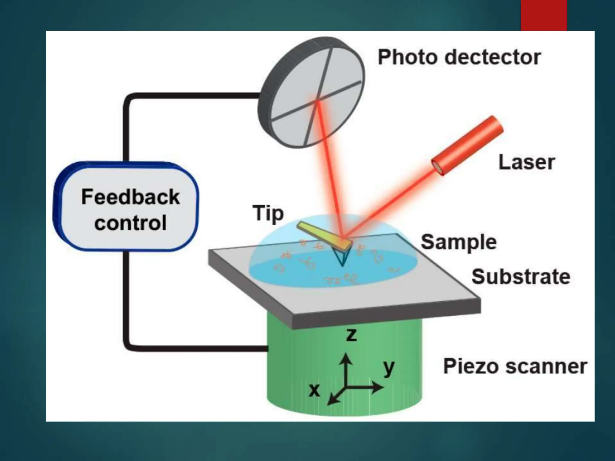
ATOMIC FORCE MICROSCOPY
(AFM) Analysis
History
 Atomic force microscopy (AFM) was developed
when people tried to extend STM technique to
investigate the electrically non-conductive
materials, like proteins.
 In 1986, Binnig and Quate demonstrated for the
first time the ideas of AFM, which used an ultra-
small probe tip at the end of a cantilever.
What is
AFM
 AFM is a type of scanning probe
microscopy (SPM), with demonstrated
resolution on the order of fractions of
a nanometer, more than 1000 times better than
the optical diffraction limit.
 The information is gathered by "feeling" or
"touching" the surface with a mechanical
probe
 AFM provides a 3D profile of the surface on a
nanoscale, by measuring forces between a sharp
probe and the surface.
 TheAFM has three major abilities:
force measurement, imaging, and manipulation.
 It is powerful because anAFM can generate
images at atomic resolution with angstrom scale
resolution height information, with minimum
sample preparation.
Principle Of AFM
 Surface Sensing
An AFM uses a cantilever with a very sharp tip to
scan over a sample surface. As the tip approaches
the surface, the close-range, attractive force
between the surface and the tip cause the
cantilever to deflect towards the surface.
However, as the cantilever is brought even closer
to the surface, such that the tip makes contact
with it, increasingly repulsive force takes over
and causes the cantilever to deflect away from the
surface.
 Detection Method
Alaser beam is used to detect cantilever
deflections towards or away from the surface.
By reflecting an incident beam off the flat top
of the cantilever, any cantilever deflection will
cause slight changes in the direction of the
reflected beam.Aposition-sensitive photo
diode (PSPD) can be used to track these
changes. Thus, if anAFM tip passes over a
raised surface feature, the resulting cantilever
deflection (and the subsequent change in
direction of reflected beam) is recorded by the
PSPD.
 Imaging
AnAFM images the topography of a
sample surface by scanning the
cantilever over a region of interest. The
raised and lowered features on the
sample surface influence the deflection
of the cantilever, which is monitored by
the PSPD. TheAFM can generate an
accurate topographic map of the surface
features.
How Are Force Measured
 The probe is placed on the end of a cantilever
(which one can think of as a spring).
 The amount of force between the probe and
surface is dependant on the spring constant
(stiffness of the cantilever and the distance
between th probe and the sample surface.
 This force can be described using Hooke’s
Law:
F= -k·x
F = Force
k = spring constant
x = cantilever deflection
• If the spring constant of
cantilever (typically ~ 0.1-1
N/m) is less than surface,
the cantilever bends and the
deflection is monitored.
•This typically results in
forces ranging from nN (10
) to µN (10-6) in the open
air.
WhatAre Probes Made Of?
They are generally made up
of Silicon or Si3N4.
Probes may be coated with
other materials for the
addiontal SPM application
such as CFM(Chemical
force Microscopy) and
MFM(Magnetic Force
Microscopy)
Why is different from optical
microscopy
 No lenses are needed.
 No need of light source to illuminate the
sample.
 No eyeplece to look through the sample.
 Itself imaging device technique which is able
to measure the very small forces b/w the atom
or molecules
Why AFM is better than STM
 It give information about the conducting
and non-conducting surfaces.
 3-D image of surfaces are obtained by
this techniques.
 Sample can be analyzed in openAir.
Applicatio
n
The AFM has been applied to problems in a wide range of
disciplines of the natural sciences, including solid-state
physics, semiconductor science and technology, molecular
engineering, polymer chemistry and physics, surface
chemistry, molecular biology, cell biology,
 and medicine.
It gives information about the toughness,
roughness and smoothness value of surface.
 Applications in the field of solid state physics
include (a) the identification of atoms at a
surface, (b) the evaluation of interactions
between a specific atom and its neighboring
atoms.
 In molecular biology,AFM can be used to
study the structure and mechanical properties
of protein complexes and assemblies. For
example, AFM has been used to
image microtubules and measure their
stiffness.
 In cellular biology,AFM can be used to
attempt to distinguish cancer cells and normal
cells based on a hardness of cells, and to
evaluate interactions between a specific cell
and its neighboring cells in a competitive
culture system.
 Soft surfaces are analyzed by this technique
without damaging it like Lipids.
 Covalent bond strength is measured by this
Technique
References
 http://www.parkafm.com
 www.nanoscience.com
 http://www.nanoscience.gatech.edu
 MikroMasch
http://www.spmtips.com/products/cantilevers/datashe
ets/sc11/ (Accessed 11/06/06).
 Nanosensors Homepage http://www.nanosensors.com
(Accessed 11/06/06).
 MikroMasch
http://www.spmtips.com/products/cantilevers/datashe
ets/hi-res- w/ (Accessed 1/26/07).
 NanoWizardAFM Handbook
 The Hessian BlobAlgorithm: Precise Particle
Detection inAtomic Force Microscopy
Imagery.
 Atomic-resolution three-dimensional
hydration structures on a heterogeneously
charged surface

ATOMIC FORCE MICROSCOPY (AFM) Analysis.pptx

  • 1.
  • 2.
    History  Atomic forcemicroscopy (AFM) was developed when people tried to extend STM technique to investigate the electrically non-conductive materials, like proteins.  In 1986, Binnig and Quate demonstrated for the first time the ideas of AFM, which used an ultra- small probe tip at the end of a cantilever.
  • 3.
    What is AFM  AFMis a type of scanning probe microscopy (SPM), with demonstrated resolution on the order of fractions of a nanometer, more than 1000 times better than the optical diffraction limit.  The information is gathered by "feeling" or "touching" the surface with a mechanical probe
  • 4.
     AFM providesa 3D profile of the surface on a nanoscale, by measuring forces between a sharp probe and the surface.  TheAFM has three major abilities: force measurement, imaging, and manipulation.  It is powerful because anAFM can generate images at atomic resolution with angstrom scale resolution height information, with minimum sample preparation.
  • 5.
    Principle Of AFM Surface Sensing An AFM uses a cantilever with a very sharp tip to scan over a sample surface. As the tip approaches the surface, the close-range, attractive force between the surface and the tip cause the cantilever to deflect towards the surface. However, as the cantilever is brought even closer to the surface, such that the tip makes contact with it, increasingly repulsive force takes over and causes the cantilever to deflect away from the surface.
  • 6.
     Detection Method Alaserbeam is used to detect cantilever deflections towards or away from the surface. By reflecting an incident beam off the flat top of the cantilever, any cantilever deflection will cause slight changes in the direction of the reflected beam.Aposition-sensitive photo diode (PSPD) can be used to track these changes. Thus, if anAFM tip passes over a raised surface feature, the resulting cantilever deflection (and the subsequent change in direction of reflected beam) is recorded by the PSPD.
  • 8.
     Imaging AnAFM imagesthe topography of a sample surface by scanning the cantilever over a region of interest. The raised and lowered features on the sample surface influence the deflection of the cantilever, which is monitored by the PSPD. TheAFM can generate an accurate topographic map of the surface features.
  • 9.
    How Are ForceMeasured  The probe is placed on the end of a cantilever (which one can think of as a spring).  The amount of force between the probe and surface is dependant on the spring constant (stiffness of the cantilever and the distance between th probe and the sample surface.  This force can be described using Hooke’s Law: F= -k·x
  • 10.
    F = Force k= spring constant x = cantilever deflection • If the spring constant of cantilever (typically ~ 0.1-1 N/m) is less than surface, the cantilever bends and the deflection is monitored. •This typically results in forces ranging from nN (10 ) to µN (10-6) in the open air.
  • 12.
    WhatAre Probes MadeOf? They are generally made up of Silicon or Si3N4. Probes may be coated with other materials for the addiontal SPM application such as CFM(Chemical force Microscopy) and MFM(Magnetic Force Microscopy)
  • 13.
    Why is differentfrom optical microscopy  No lenses are needed.  No need of light source to illuminate the sample.  No eyeplece to look through the sample.  Itself imaging device technique which is able to measure the very small forces b/w the atom or molecules
  • 14.
    Why AFM isbetter than STM  It give information about the conducting and non-conducting surfaces.  3-D image of surfaces are obtained by this techniques.  Sample can be analyzed in openAir.
  • 15.
    Applicatio n The AFM hasbeen applied to problems in a wide range of disciplines of the natural sciences, including solid-state physics, semiconductor science and technology, molecular engineering, polymer chemistry and physics, surface chemistry, molecular biology, cell biology,  and medicine. It gives information about the toughness, roughness and smoothness value of surface.
  • 16.
     Applications inthe field of solid state physics include (a) the identification of atoms at a surface, (b) the evaluation of interactions between a specific atom and its neighboring atoms.  In molecular biology,AFM can be used to study the structure and mechanical properties of protein complexes and assemblies. For example, AFM has been used to image microtubules and measure their stiffness.
  • 17.
     In cellularbiology,AFM can be used to attempt to distinguish cancer cells and normal cells based on a hardness of cells, and to evaluate interactions between a specific cell and its neighboring cells in a competitive culture system.  Soft surfaces are analyzed by this technique without damaging it like Lipids.  Covalent bond strength is measured by this Technique
  • 18.
    References  http://www.parkafm.com  www.nanoscience.com http://www.nanoscience.gatech.edu  MikroMasch http://www.spmtips.com/products/cantilevers/datashe ets/sc11/ (Accessed 11/06/06).  Nanosensors Homepage http://www.nanosensors.com (Accessed 11/06/06).  MikroMasch http://www.spmtips.com/products/cantilevers/datashe ets/hi-res- w/ (Accessed 1/26/07).  NanoWizardAFM Handbook
  • 19.
     The HessianBlobAlgorithm: Precise Particle Detection inAtomic Force Microscopy Imagery.  Atomic-resolution three-dimensional hydration structures on a heterogeneously charged surface