Downloaded 218 times

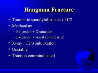
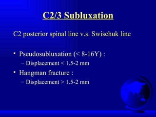
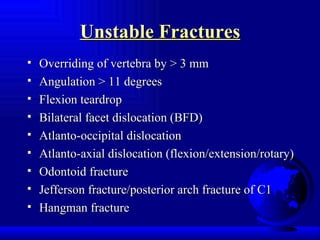
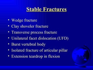

This document provides an overview of cervical spine injuries and key considerations for their radiological evaluation. It discusses important anatomical structures, mechanisms of injury, classifications of common fractures like Jefferson fractures and Hangman's fractures, criteria for stability versus instability, and special considerations like SCIWORA in pediatric populations. A wide range of cervical spine pathologies are addressed.


