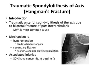
3. Traumatic Spondylolithesis of Axis
(Hangman's Fracture)
• Introduction
• Traumatic anterior spondylolithesis of the axis due
to bilateral fracture of pars interarticularis
– MVA is most common cause
• Mechanism is
– hyperextension
• leads to fracture of pars
– secondary flexion
• tears PLL and disc allowing subluxation
• Associated injuries
– 30% have concomitant c-spine fx
7. Treatment
• Nonoperative
– rigid cervical collar 4-6 weeks
• indications
– Type I fractures (< 3mm horizontal displacement)
– closed reduction followed by halo immobilization for 8-12
weeks
• indications
– Type II with 3-5 mm displacement
– Type IIA
• reduction technique
– Type II
» cervical axial traction combined with extension
– Type IIA
» hyperextension (avoid axial traction in Type IIA)
8. Treatment
• Operative
– reduction with surgical stabilization
• indications
– Type II with > 5 mm displacement and severe angulation
– Type III (facet dislocations)
• technique
– anterior C2-3 interbody fusion
– posterior C1-3 fusion
– bilateral C2 pars screw osteosynthesis
9. Closed Cervical Traction
• Indications
– subaxial cervical fractures with malalignment
– unilateral and bilateral facet dislocations
– displaced odontoid fractures
– select hangman's fractures
– C1-2 rotatory subluxation
• Contraindications
– patient who is not awake, alert, and cooperative
– presence of a skull fracture may be a contraindication
10.
11. Patient position
• Preferred setting
– emergency room, operating room, ICU for close
observation and frequent flouroscopy/radiographs
• Patient position
– supine with reverse trendelenburg or use of arm and leg
weights and can help prevent patient migration to the top
of the bed with addition of weights.
• Sedation
– small doses of diazepam can be administered to aid in
muscle relaxation
– however patient must remain awake and able to converse
12. Pin Placement
• Pin placement (Garner-Wells pins)
– pin placement is 1 cm above pinna in line with external
auditory meatus and below the equator of the skull.
• Pin tightness
– On Gardner-Wells tons pins are tightened until spring
loaded indicator protrudes 1 mm above surface
– over tightening of the pins can result in penetration fo the
inner table of the calvarium
• Pin strength
– stainless steel pins have higher failure loads than titanium
and MRI-compatible graphite and should be used with
traction of > 50lbs.
13. Reduction with Serial Traction
• Serial traction
– an intital 10lbs is added.
– weights are increased at 10lb increments every 20 minutes
– serial exams and radiographs are taken after each weight is placed
– maximal weight is controversial
• some authors recommend weight limits of 70 lbs
• recent studies report up to 140 lbs is safe
• Reduction maneuvers
– reduction of a unilateral facet dislocation
• reduction maneuver performed after facet is distracted to a perched position
• maintain axial load and rotate head 30-40 degree past midline in the direction
of the dislocation
• stop once resistance is felt and confirm with radiographs
– reduction of bilateral facet dislocation
• reduction maneuver performed after facet is distracted to a perched position
• palpate the stepoff in the spinal process posteriorly and apply an anterior
directed force caudal to the level of the dislocation
• rotate the head 40 degree beyond midline in one direction, and then rotate 40
degreee in the other direction while axial traction is maintained.
15. Halo Orthosis Immobilization
• Fixes skull relative to torso
– provides most rigid form of cervical spine external
immobilization
– ideal for upper C-spine injury
• Allows intercalated paradoxical motion in the
subaxial cervical spine
– therefore not ideal for lower cervical spine
injuries (lateral bending least controlled)
• "snaking phenomenon"
– recumbent lateral radiograph shows focal kyphosis in midcervical
spine
– yet, upright lateral radiograph shows maintained lordosis in
midcervical spine
16.
17. Indications
• Indications
• definitive treatment of cervical spine trauma including
– occipital condyle fx
– occiptiocervical dislocation
– stable Type II Atlas fx (stable Jefferson fx)
– type II odontoid fractures in young patients
– type II and IIA hangman’s fractures
• adjunctive postoperative stabilization following cervical spine surgery
• Contraindications
– absolute
• cranial fractures
• infection
• severe soft-tissue injury
– relative
• polytrauma
• severe chest trauma
• barrel-shaped chest
• obesity
• advanced age
• CT scan prior to halo application
– indications
• clinical suspicion for cranial fracture
• children younger than 10 to determine thickness of bone