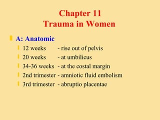
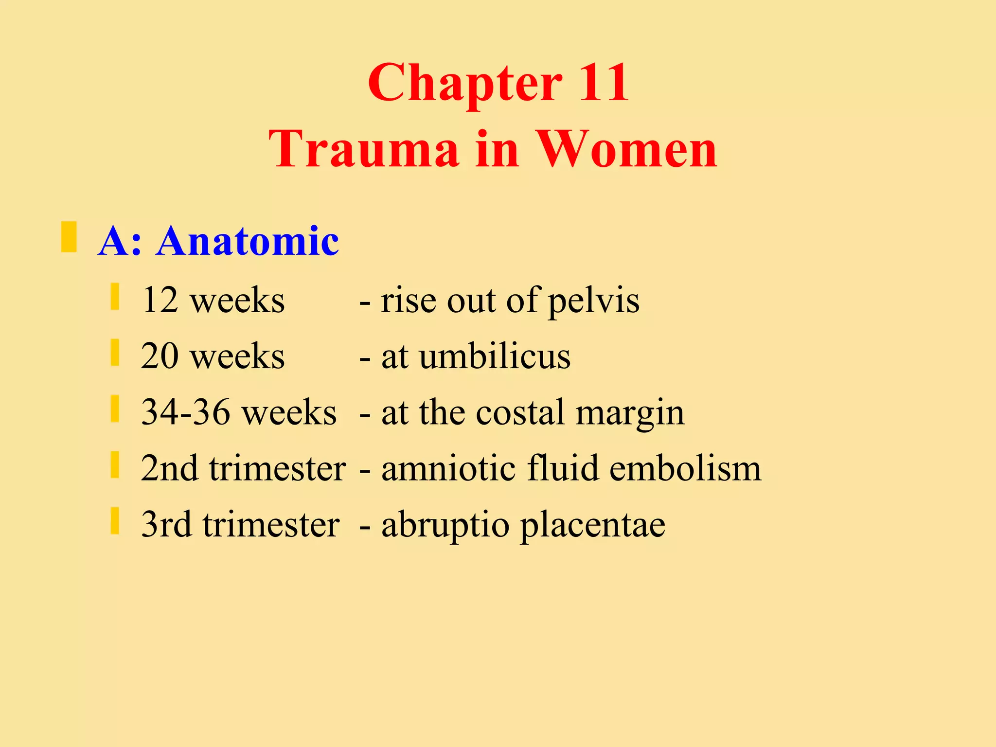
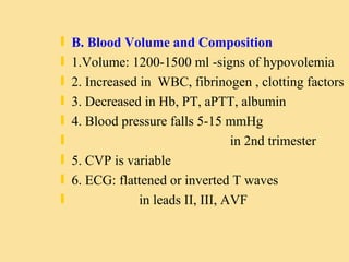
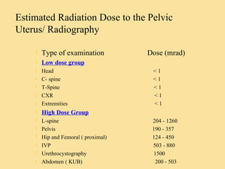
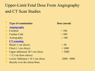
Chapter 11  Trauma in Women A: Anatomic 12 weeks  - rise out of pelvis 20 weeks  - at umbilicus 34-36 weeks  - at the costal margin 2nd trimester - amniotic fluid embolism 3rd trimester - abruptio placentae
B. Blood Volume and Composition 1.Volume: 1200-1500 ml -signs of hypovolemia 2. Increased in  WBC, fibrinogen , clotting factors 3. Decreased in Hb, PT, aPTT, albumin 4. Blood pressure falls 5-15 mmHg  in 2nd trimester 5. CVP is variable 6. ECG: flattened or inverted T waves  in leads II, III, AVF
C. respiratory Increased in tidal volume Decreased in residual volume Hypocapnea ( Pco2 of 30 mmHG)  in late pregnancy D. Musculoskeletal 7th months: the symphysis pubis widens (4-8mm) The sacroiliac-joint space increased
Mechanism of Injury A. Blunt Injury 1. Direct Injury 2. Indirect Injury  Abrutio Placentae  & Uterine Rupture Seat belt: forward flexion and uterine  compression B. Penetrating Injury Dense uterine musculature & Amnion Low incidence of maternal visceral injury
Assessment and Management A: Primary Survey and Resuscitation 1. Maternal: Hyperventilation 4 - 6 inches elevation of right buttock Fetus may be in shock before  maternal hypovolemia shock signs  Vasopressors - fetal hypoxia B: Fetus: Uterine rupture Abruptio placentae Continued fetal heart tones  20 -24 wks  of gestation
B. Adjuncts to primary survey Maternal: Monitor on her left side after physical examination Monitor of the CVP response to fluid Maternal bicarbonate is usually low Fetus: 20-24 wks heart tones: 120- 160 beats / min Continous monitor with cardiotocodynamometry Consultation if abnormal fetal heart rates
C. Secondary Assessment 1. DPL:  perform above the umbilicus Presence of uterine contractions 2.Vaginal Examination : Amniotic fluid with PH of 7 - 7.5 :  ruptured of chorioamniotic membrane Bleeding in 3rd trimester: disruption of placenta  impending fetal death The fetus may be in jeopardy even with apparent,  minor maternal injury
D. Definite Care Uterine rupture: shock or no s/s Placental abruptio: leading cause of fetal death 30% no vaginal bleeding All pregnant Rh-negative trauma patient should considered for RH immunoglobulin therapy .  Initial management is directed at resuscitation and stabilization of the pregnant patient. Perimortem c/s may be successful if it is done within 4-5 mins arrest.
Radiography in Pregnant Women No fetus risk: 5 - 10 rad. The maximum risk attributable to 10 rad of exposure is approx. 0.1 % After 20th weeks of gestation: cause no fetal abnormalities. Routine C-spine, CXR, Pelvis obtained with shielding: negligible fetal exposure CT beam in direct line to fetus: 3 - 9 rad. CT scan above uterus: < 3 rad to fetus.
Radiography to fetus varies: 1. The type of study 2. The size of patient 3. Position of the fetus 4. Type of machine 5. Method of shielding 6. The number of section obtained 7. Fetal/uterine size 8. Coned x-ray beam aimed  > 10 cm away from  fetus are not dangerous.
Estimated Radiation Dose to the Pelvic Uterus/ Radiography Type of examination  Dose (mrad) Low dose group Head  < 1 C- spine  < 1 T-Spine  < 1 CXR  < 1 Extremities  < 1 High Dose Group L-spine  204 - 1260 Pelvis  190 - 357 Hip and Femoral ( proximal)  124 - 450 IVP  503 - 880 Urethrocystography  1500 Abdomen ( KUB)  200 - 503
Upper-Limit Fetal Dose From Angiography and CT Scan Studies   Type of examination  Dose (mrad) Angiography Cerebral  < 100 Cardiac Cath  < 500 Aortography  < 100 CT scanning Head ( 1 cm slices)  < 50 Chest ( 1 cm slices)  < 1000 Upper abdomen( 20 1-cm slices  < 3000 > 2.5 cm from uterus) Lower Abdomen ( 10 1-cm slices  3000 - 9000 directly over the uterus/fetus

Trauma In Women

  • 1.
    Chapter 11 Trauma in Women A: Anatomic 12 weeks - rise out of pelvis 20 weeks - at umbilicus 34-36 weeks - at the costal margin 2nd trimester - amniotic fluid embolism 3rd trimester - abruptio placentae
  • 2.
    B. Blood Volumeand Composition 1.Volume: 1200-1500 ml -signs of hypovolemia 2. Increased in WBC, fibrinogen , clotting factors 3. Decreased in Hb, PT, aPTT, albumin 4. Blood pressure falls 5-15 mmHg in 2nd trimester 5. CVP is variable 6. ECG: flattened or inverted T waves in leads II, III, AVF
  • 3.
    C. respiratory Increasedin tidal volume Decreased in residual volume Hypocapnea ( Pco2 of 30 mmHG) in late pregnancy D. Musculoskeletal 7th months: the symphysis pubis widens (4-8mm) The sacroiliac-joint space increased
  • 4.
    Mechanism of InjuryA. Blunt Injury 1. Direct Injury 2. Indirect Injury Abrutio Placentae & Uterine Rupture Seat belt: forward flexion and uterine compression B. Penetrating Injury Dense uterine musculature & Amnion Low incidence of maternal visceral injury
  • 5.
    Assessment and ManagementA: Primary Survey and Resuscitation 1. Maternal: Hyperventilation 4 - 6 inches elevation of right buttock Fetus may be in shock before maternal hypovolemia shock signs Vasopressors - fetal hypoxia B: Fetus: Uterine rupture Abruptio placentae Continued fetal heart tones 20 -24 wks of gestation
  • 6.
    B. Adjuncts toprimary survey Maternal: Monitor on her left side after physical examination Monitor of the CVP response to fluid Maternal bicarbonate is usually low Fetus: 20-24 wks heart tones: 120- 160 beats / min Continous monitor with cardiotocodynamometry Consultation if abnormal fetal heart rates
  • 7.
    C. Secondary Assessment1. DPL: perform above the umbilicus Presence of uterine contractions 2.Vaginal Examination : Amniotic fluid with PH of 7 - 7.5 : ruptured of chorioamniotic membrane Bleeding in 3rd trimester: disruption of placenta impending fetal death The fetus may be in jeopardy even with apparent, minor maternal injury
  • 8.
    D. Definite CareUterine rupture: shock or no s/s Placental abruptio: leading cause of fetal death 30% no vaginal bleeding All pregnant Rh-negative trauma patient should considered for RH immunoglobulin therapy . Initial management is directed at resuscitation and stabilization of the pregnant patient. Perimortem c/s may be successful if it is done within 4-5 mins arrest.
  • 9.
    Radiography in PregnantWomen No fetus risk: 5 - 10 rad. The maximum risk attributable to 10 rad of exposure is approx. 0.1 % After 20th weeks of gestation: cause no fetal abnormalities. Routine C-spine, CXR, Pelvis obtained with shielding: negligible fetal exposure CT beam in direct line to fetus: 3 - 9 rad. CT scan above uterus: < 3 rad to fetus.
  • 10.
    Radiography to fetusvaries: 1. The type of study 2. The size of patient 3. Position of the fetus 4. Type of machine 5. Method of shielding 6. The number of section obtained 7. Fetal/uterine size 8. Coned x-ray beam aimed > 10 cm away from fetus are not dangerous.
  • 11.
    Estimated Radiation Doseto the Pelvic Uterus/ Radiography Type of examination Dose (mrad) Low dose group Head < 1 C- spine < 1 T-Spine < 1 CXR < 1 Extremities < 1 High Dose Group L-spine 204 - 1260 Pelvis 190 - 357 Hip and Femoral ( proximal) 124 - 450 IVP 503 - 880 Urethrocystography 1500 Abdomen ( KUB) 200 - 503
  • 12.
    Upper-Limit Fetal DoseFrom Angiography and CT Scan Studies Type of examination Dose (mrad) Angiography Cerebral < 100 Cardiac Cath < 500 Aortography < 100 CT scanning Head ( 1 cm slices) < 50 Chest ( 1 cm slices) < 1000 Upper abdomen( 20 1-cm slices < 3000 > 2.5 cm from uterus) Lower Abdomen ( 10 1-cm slices 3000 - 9000 directly over the uterus/fetus