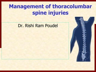
Management of thoracolumbar 
spine injuries 
Dr. Rishi Ram Poudel
Cause of injury.. Indian 
Scenario..!! 
fall from height RTA Violence Gun shot 
injuries 
52.3 
38.4 
5 4.3 
Upendra B, Khandwal P, Chowdhury B, Jayaswal A: Correlation of outcome measures with 
epidemiological factors in thoracolumbar spinal trauma. Indain J Orthop 2007 
Oct;41(4):290-4.
Why dorsolumbar junction more 
succeptible?? 
Location between the stiff kyphotic dorsal 
spine and more mobile lordotic lumbar spine
Dennis three column concept..
Middle column providing 
greatest mechanical stability
Compression fractures.. Denis 
Failure under compression of anterior column. The middle column is intact 
and acts as a hinge.
Burst fractures.. Denis 
Both end plates Superior end plate Inferior end plate 
Burst rotation Burst lateral flexion 
Failure under axial load of both the anterior and middle column originating at the 
level of one or both end plates of the same vertebrae
Burst fractures.. 
Lateral film: 
• Fracture of posterior wall cortex 
• Loss of height of posterior vertebral body 
• Retropulsion of fragment into canal 
AP film 
Increase in interpediculate distance
Unstable burst fractures.. 
• Loss of height >50% 
• Kyphotic deformity >30 degrees 
• Substantial posterior column injury 
• Progessive deformity 
• Progessive neurological deficit
Reason for Kyphotic deformity..biomechanics..!!
Seat-Belt type injuries.. 
Denis 
Chance fracture 
Failure of both posterior and middle columns under tension forces 
generated by flexion with its axis placed in the anterior column
Fracture dislocations.. 
Flexion- rotation 
Denis 
Slice fracture
Fracture dislocations.. 
Shear type fracture dislocation 
Denis 
Postero-anterior shear injury with 
floating lamina 
Anterio-posterior shear injury
Fracture dislocations.. 
Flexion-distraction 
Denis 
Tear of anterior annulus 
fibrosus and stripping of 
ALL during subluxation
Fracture classification 
Denis
AXIAL-COMPRESSION MECHANISM 
Morphologic Anatomic Neurologic Therapeutic 
Compression 
Anterior column compressed Intact Orthosis 
fracture 
Stable burst Retropulsed middle column 
Posterior ligament intact 
Intact 
Compromised 
Orthosis 
Anterior 
decompression/stabilization
FLEXION-COMPRESSION MECHANISM 
Morphologic Anatomic Neurologic Therapeutic 
Wedge fracture Anterior column collapse 
(< 40% VB height) 
Kyphosis >30º 
Intact 
Intact 
Orthosis 
Posterior instrumentation 
Unstable burst Kyphosis 20-30º 
<50% canal compromise 
Kyphosis > 30º 
>50% loss of anterior VB 
height 
>70%canal compromise 
Marked vertebral body 
comminution 
Intact 
Intact/Compromised 
Orthosis vs posterior 
segmental 
instrumentation in 
distraction and extension 
Anterior 
decompression/stabilization 
Consider combined anterior 
and posterior stabilization
FLEXION-DISTRACTION MECHANISM 
Morphologic Anatomic Neurologic Therapeutic 
TRUE CHANCE Tensile injury through bone Intact Orthosis if 
anatomically reduced 
LIGAMENTOUS 
CHANCE 
Tensile injury through 
ligament 
Intact/Compromised Posterior reduction 
and compression 
instrumentation
EXTENSION MECHANISM 
Morphologic Anatomic Neurologic Therapeutic 
ANTERIOR OPENING 
OF DISC 
Tensile failure of ALL 
and anterior annulus 
Intact Orthosis 
FRACTURE 
DISLOCATION 
Laminar fracture 
Anterior and middle 
column ligament 
disruption 
Posterior vertebral body 
translation 
Intact/Compromised 
Posterior 
decompression, 
reduction and 
instrumentation
ROTATION AND SHEAR MECHANISM 
Morphologic Anatomic Neurologic Therapeutic 
FRACTURE 
DISLOCATION 
Facet fractures 
Three column disruption 
Vertebral translation 
Intact/Compromised 
Posterior reduction 
and instrumentation
AO/Magrel classification..!!
McCormack load shearing classification 
A. Comminution/Involvement of vertebral body 
Low scores (3-6) can be managed with 
short segment posterior stabilization only 
B. Displacement/ Apposition of fracture parts 
High scores (7-9) require additional anterior 
stabilization to prevent failure of posterior 
implant 
C. Deformity correction[(A+B)/2-C]
ASIA impairment scale 
A COMPLETE: No motor or sensory function is preserved in the sacral 
segments S4-S5 
B INCOMPLETE: Sensory but not motor function is preserved below the 
neurologic level and includes sacral segments S4-S5 
C INCOMPLETE: Motor function is preserved below the neurological 
level and more than half of key muscles below neurologic level have a 
muscle grade less than 3 
D INCOMPLETE: Motor function is preserved below the neurological 
level and at least half of the key muscles below the neurologic level 
have a muscle grade more than 3 
E NORMAL motor and sensory functions
Thoracolumbar Injury Classification and 
Severity Score(TLICS)
Evaluating PLC..!! 
Clinical signs: 
1. Palpable 
interspinous 
defect 
2. Posterior 
tenderness 
X ray: 
1. Kyphosis >30 
degrees 
2. > 50% 
compression of 
anterior 
vertebral body 
3. interspinous 
spacing greater 
than 7 mm than 
adjacent 
vertebrae 
CT scan: 
1. Diastasis of facet 
joints 
2. Spinous process 
avulsion 
MRI: 
1. Edema in region of 
PLC (T2) 
2. Disruption of PLC 
components (T1) 
(SSL,ISL,LF,Capsule)
Initial management… 
ATLS protocol 
• Injuries impairing respiratory and circulatory 
function treated with priority 
• Log roll technique for manipulating the 
patient 
• C-spine immobilization
Systematic Approach 
1 
2 
3 
4 
5
Systematic Approach 
Miss a Step and you are nowhere..!!
Examination 
Trauma Bay 
E.R. 
• Information 
• Mechanism 
– energy, energy 
• Direction of Impact 
• Associated Injuries 
Starts in the….
Does “unexaminable” mean no 
examination? 
NO! 
• Inspect for bruising or ecchymosis 
• Palpate for step-off or deformity 
• Rectal Tone 
• Reflex exam 
– Bulbocavernosus 
– Clonus/Babinski
Thoracolumbar spine..!! 
Lateral view..: 
• Vetebral Body heights 
• Alignment of bodies/Angulation of spine 
• Contour of bodies 
• Presence of disc spaces 
• Encroachment of body on canal 
• Loss of vertebral body height 
• Kyphosis measurement – COBB angle
AP view: 
• Alignment 
• Symmetry/ Shape of 
pedicles 
• Interpedicular distance 
• Position of spinous 
process 
• Contour of bodies
Spinal shock..!! 
Commonly used but poorly understood 
term 
Loss of spinal reflexes caudal to a spinal cord 
injury. 
First phase of response to spinal cord to injury 
associated with initial flaccid paralysis below 
the lesion
Spinal shock..! 
The return of bulbocavernosus reflex marks 
the resolution of spinal shock 
Reflex is not always initially lost and may take 
longer to recover making assessment 
confusing
Spinal shock: Pathophysiology..!! 
After resolution of shock ,variable preserved 
EDEMA 
functions below the injury level 
1.Residual axons with sprouting collaterals 
2. Denervation hypersensitivity 
VENOUS 
CONGESTION 
DAMAGE TO BLOOD 
VESSELS 
MICROHAEMMORRHAGE 
SPINAL SHOCK 
If no motor or sensory function below the level of injury can 
be documented when spinal shock ends, a complete spinal 
cord injury is present, and the prognosis is poor for recovery 
of distal motor or sensory function
Neurogenic shock..!! 
Loss of symphatetic outflow related to spinal 
shock 
Vasodilation of the viscera and peripheries 
resulting in hypotension without 
TACHYCARDIA 
Fluid administration Pulmonary edema
SCI: Complete Vs Incomplete 
Complete 
• No function below level of injury 
• Absence of sensation and voluntary movement 
in S4/5 distribution 
The difference between a complete and 
incomplete spinal cord injury is the PRESENCE 
OF SACRAL SPARING identified by the 
presence Incomplete 
of ANAL SENSATION 
Preservation of sensation in S4/5 distribution and 
voluntary control of anal sphincter
Role of steroid..!! 
NASCIS II & III 
High doses of methyl-prednisolone 
Closed, blunt spinal cord trauma presenting 
within 8 hours 
• Loading dose of 30 mg/kg given as bolus i/v 
• Continue infusion 5.4mg /kg x 24 hours if the 
patient presented within 3 hours of injury 
• Continue infusion x 48 hours if the patient 
presented 3 to 8 hours after injury
Polytraumatized spine patients..! 
Perioperative and post-opeartive mortality and 
morbidity were not increased by emergent stabilization 
Neurologic improvement was increased and life 
threatening complications were reduced 
Mc Lain RF, Benson DR:Urgent surgical stabilization of spine 
fractures in polytraumatized patients Spine 1999;24:1646
Managing Thoracolumbar Spine Fractures..! 
NEUROLOGICAL DEFICIT 
YES 
COMPLETE INCOMPLETE 
POSTERIOR 
FUSION ANTERIOR 
DECOMPRESSION & FUSION 
POSTERIOR 
TRANSPEDICULAR 
DECOMPRESSION AND 
FUSION 
CIRCUMFERENTIAL 
PROCEDURES 
NO 
UNSTABLE STABLE CONSERVATIVE 
PLC disrupted PLC intact 
Posterior fusion 
± 
Minimally 
invasive(VBA) 
Circumferential 
fusion 
Anterior fusion 
Posterior fusion 
with indirect 
reduction ± 
Minimally 
invasive(VBA) 
Stable fracture 
Involving less than 2 
columns 
Kyphosis < 25 degrees 
Compression < 50% of 
anterior column 
Canal compromise < 50%
TLICS guidelines..!!
Non-operative management 
• Fractures with <10% vertebral height loss do not need external 
support. 
• Fractures with < 40% height loss and < 25 degrees kyphosis can be 
treated with a Jewett brace for 6 to 8 weeks. 
• In fractures below T5, a plaster jacket or TLSO can be used. 
• In higher fractures (above T5), a cervical component should be 
added to the brace. 
Burst fractures in brace should regularly be assessed in standing radiographs 
with orthosis 
Bony chance fracture if anatomically reduced can be treated with bracing
Burst fracture: non-operative vs operative treatment 
Operative management is related with better kyphosis 
correction but with similar pain and functional outcomes 4 
years post-operatively
Laminectomy: posterior direct 
decompression 
Indications: 
• Comminuted posterior elements causing direct neural 
compression 
• Epidural hematoma requiring evacuation 
• Repair of dural tear associated with burst and laminar 
fractures during posterior instrumentation and fusion
Posterior instrumentation:distraction and ligamentotaxis 
Contraindications: 
• Canal compromise >67% 
• Delay in operative treatment for > 4 days 
• Where pedicle screw insertion is not feasible 
(atypical morphology, small dimension or traumatic 
fracture) 
Requires intact PLC
Indications: 
• Retropulsed fragments occupying >67% canal area 
ANTERIOR DECOMPRESSION AND FUSION 
• Extensive vertebral body comminution with 
significant kyphotic deformity 
Greater neurologic improvement as compared to 
posterior or posterolateral decompression 
• Delay in operative treatment for 4 days 
• Return of normal bowel and bladder control achieved 
more frequently 
• Traumatic disc herniations causing symptomatic 
cord or root compression 
• Even in cases of long-standing compression after fracture 
modest recovery 
Patients with incomplete deficits (spinal cord or cauda equina) are 
ideal candidates because they have greater chances for neural 
recovery
Combined anterior and posterior 
methods 
• When canal is compromised circumferentially 
• Severe coronal or sagittal plane deformity 
(>40 degrees) 
• Structural augmentation is deemed 
necessary(multiple contiguous levels of 
injury, poor bone quality or osteoporosis)
Kyphosis even in absence of neurological 
deficit likely to progress. 
>30 degrees : late neurological deficit 
Burst fracture in thoracolumbar region (T11-L2) with 
neurological deficit from a retropulsed fragment should 
undergo anterior decompression and fusion as a solitary 
procedure or in combination with a posterior approach 
However, relative indications and contraindications depending 
Isolated upon (LMNOPS) 
anterior procedures L2 and below to 
be Location 
avoided: Pseudo-arthosis and vascular 
concern 
Mechanism 
Neurology 
Open vs closed 
Stability 
Posterior element disruption and osteoporotic 
bone: additional posterior intrumentation
Vertebroplasty and Kyphoplasty..!! 
Indications: 
• Osteoporotic VCF not responding to conservative management 
• Spinal metastatic lesions & fractures 
• Hemangiomas 
Goal of vertebroplasty is to improve strength and stability 
Goal of Kyphoplasty is to restore vertebral body height and stability. The 
use of baloon creates a void for cement placement under lower pressure 
and thus results in lower incidence of cement extravasation
Can be safely done in patients with refractory pain to 
conservative treatments.
Vertebroplasty technique..!!
Dangers..Needle injury..! 
Canal breach Lateral passage and 
aortic damage
Complications ..!! 
Transient increase in pain in the injected level 
Cement leakage 
Pulmonary embolism(marrow,cement,air) 
Infection
Vertebroplasty vs kyphoplasty: Debate 
continues…. 
Kyphoplasty : more controlled procedure with 
height restoration and less chances of cement 
extravasation 
Vertebroplasty: faster, more straightforward, 
cheaper that has not shown to give inferior results. 
Increased rates of cement migration doesn’t 
significantly results in increased morbidity
DAMAGE CONTROL SURGERY : 3 PHASE APPROACH 
Establish rapid 
control of 
hemorrhage 
Identify major 
injuries 
1 
Stabilize major 
fractures 
Reduce 
dislocated joints 
Decontaminate 
open wounds 
2 
Once normal 
physiology is 
restored , 
definitive surgical 
repairs 
3
Posterior internal 
stabilization of 
thoracic or lumbar 
trauma 
Anterior decompression or 
complex stabilization 
procedures as allowed by 
patient’s physiologic 
condition 
Window of opportunity 
Definitive surgery electively scheduled for 
experienced spine surgeon 
General care: ventilatory care, pressure care , bowel 
bladder management, thromboembolic prophylaxis, 
control of infections . Minimize systemic insult 
to the patient
The ability to provide good internal fixation with minimal soft 
tissue disruption is the key point of the AO principles of 
treatment of extremity fractures. SPINAL FRACTURES ARE NO 
DIFFERENT. 
Primary role: restore or augment posterior tension band 
APPLICATIONS: 
1.Axial compression injuries (compression/burst #) 
Supplemenatal posterior fixation of anterior corpecectomy when required for decompression or 
anterior column support. 
As a primary procedure : MIS decompression and radiculopathy 
2.Flexion-distraction injuries 
Pure bony Chance Fractures allowing direct osteosynthesis rather than spinal fusion 
3.Damage control Orthopaedics 
Temporary stabilization in patients with multiple traumatic injuries who might not be 
physiologically able to tolerate definitive open procedure early in their hospitalization 
4.When bracing of stable fractures is difficult or contraindicated 
As an internal splint for patients associated with chest trauma, significant respiratory 
compromise , morbid obesity. 
Concept of MIS procedures: 
• Avoid need for large surgical dissection resulting in less 
denervation and muscle atrophy as well as less damage to stabilizing 
structures such as facet joints. 
• Reduce the morbidity associated with standard open procedures
Surgical technique..!! 
Properly aligned AP and Lateral images 
Flat supeior end plate (only one end 
plate shadow) 
Pedicles just below superior end plate 
and spinous processes centered 
between the pedicles 
Superior end plate flat 
Single posterior vertebral body shadow 
Superimposed pedicles
Surgical technique…!! 
1 cm vertical incision lateral to pedicle 
Insertion of Jamshidi needle: 
Starting point: supero-lateral quadrant of pedicle , advanced 
down untill it appears to be at posterior border of vertebral 
body on lateral image. 
Tip of needle within medial half of pedicle on AP image 
Guide wire 
Dilators: Largest dilator as a working tube 
Cannulated pedicle screw over guide wire
Surgical technique..!!
VIDEO-ASSISTED THORACIC SURGERY(VATS) 
Despite a long learning curve and technical 
demands it has several advantages 
• Better cosmesis 
• Adequate exposure from T2-L1 
• Less morbidity 
• Better illumination, magnification 
Contraindications: 
• Inability to tolerate single lung ventilation 
• Emphysema, acute respiratory insufficiency 
• Previous thoracotomy
Short segment fixation plus 
transpedicular augmentation..!!
Transpedicular augmentation:better final 
restoration of vertebral height 
Allen li et al: Indian J Orthop. 2007 Oct-Dec; 41(4): 362–367.
Rehabilitation..!! 
Bladder dysfunction: 
• Intermittent catheterization 
• Supra-pubic catheterization in penile ulceration 
• Valsalva, Crede method 
Bowel dysfunction: 
• Regular intake of high fluids and dietary fibers 
• Pulse water irrigation of rectum 
• Electrical stimulation of abdominal musculature 
• Prokinetic agents: cisapride 
• Suppositories
Rehabilitation..!! 
Spasticity 
• Physical therapy: rhythmic passive movements, muscular 
stretching exercises 
• Direct muscle electrical stimulation 
• Oral baclofen 
Pain 
• Non-pharmacologic massage and heat, TENS 
• Pharmacologic: Gabapentin 
DVT prevention 
• Compression stockings 
• LMWH
Rehabilitation..!! 
Bed sore prevention 
• Posture change every 2 hourly 
• Air mattress 
• Use of pillows and foam wedges at bony 
prominences 
High protein diet 
Debridement of established sore to fasten 
healing
Rehabilitation..!! 
Postural hypotension 
• Clonidine 
• Elastic stockings, abdominal binders 
Prevention of respiratory infections 
Chest physiotherapy, steam inhalation, 
incentive spirometry 
Manual assisted coughing
Rehabilitation..!! 
Community re-intregation is must.
Thank You..!!!!!

thoracolumbar spinal trauma

  • 1.
    Management of thoracolumbar spine injuries Dr. Rishi Ram Poudel
  • 3.
    Cause of injury..Indian Scenario..!! fall from height RTA Violence Gun shot injuries 52.3 38.4 5 4.3 Upendra B, Khandwal P, Chowdhury B, Jayaswal A: Correlation of outcome measures with epidemiological factors in thoracolumbar spinal trauma. Indain J Orthop 2007 Oct;41(4):290-4.
  • 4.
    Why dorsolumbar junctionmore succeptible?? Location between the stiff kyphotic dorsal spine and more mobile lordotic lumbar spine
  • 5.
  • 6.
    Middle column providing greatest mechanical stability
  • 7.
    Compression fractures.. Denis Failure under compression of anterior column. The middle column is intact and acts as a hinge.
  • 8.
    Burst fractures.. Denis Both end plates Superior end plate Inferior end plate Burst rotation Burst lateral flexion Failure under axial load of both the anterior and middle column originating at the level of one or both end plates of the same vertebrae
  • 9.
    Burst fractures.. Lateralfilm: • Fracture of posterior wall cortex • Loss of height of posterior vertebral body • Retropulsion of fragment into canal AP film Increase in interpediculate distance
  • 10.
    Unstable burst fractures.. • Loss of height >50% • Kyphotic deformity >30 degrees • Substantial posterior column injury • Progessive deformity • Progessive neurological deficit
  • 11.
    Reason for Kyphoticdeformity..biomechanics..!!
  • 12.
    Seat-Belt type injuries.. Denis Chance fracture Failure of both posterior and middle columns under tension forces generated by flexion with its axis placed in the anterior column
  • 13.
    Fracture dislocations.. Flexion-rotation Denis Slice fracture
  • 14.
    Fracture dislocations.. Sheartype fracture dislocation Denis Postero-anterior shear injury with floating lamina Anterio-posterior shear injury
  • 15.
    Fracture dislocations.. Flexion-distraction Denis Tear of anterior annulus fibrosus and stripping of ALL during subluxation
  • 16.
  • 17.
    AXIAL-COMPRESSION MECHANISM MorphologicAnatomic Neurologic Therapeutic Compression Anterior column compressed Intact Orthosis fracture Stable burst Retropulsed middle column Posterior ligament intact Intact Compromised Orthosis Anterior decompression/stabilization
  • 18.
    FLEXION-COMPRESSION MECHANISM MorphologicAnatomic Neurologic Therapeutic Wedge fracture Anterior column collapse (< 40% VB height) Kyphosis >30º Intact Intact Orthosis Posterior instrumentation Unstable burst Kyphosis 20-30º <50% canal compromise Kyphosis > 30º >50% loss of anterior VB height >70%canal compromise Marked vertebral body comminution Intact Intact/Compromised Orthosis vs posterior segmental instrumentation in distraction and extension Anterior decompression/stabilization Consider combined anterior and posterior stabilization
  • 19.
    FLEXION-DISTRACTION MECHANISM MorphologicAnatomic Neurologic Therapeutic TRUE CHANCE Tensile injury through bone Intact Orthosis if anatomically reduced LIGAMENTOUS CHANCE Tensile injury through ligament Intact/Compromised Posterior reduction and compression instrumentation
  • 20.
    EXTENSION MECHANISM MorphologicAnatomic Neurologic Therapeutic ANTERIOR OPENING OF DISC Tensile failure of ALL and anterior annulus Intact Orthosis FRACTURE DISLOCATION Laminar fracture Anterior and middle column ligament disruption Posterior vertebral body translation Intact/Compromised Posterior decompression, reduction and instrumentation
  • 21.
    ROTATION AND SHEARMECHANISM Morphologic Anatomic Neurologic Therapeutic FRACTURE DISLOCATION Facet fractures Three column disruption Vertebral translation Intact/Compromised Posterior reduction and instrumentation
  • 22.
  • 23.
    McCormack load shearingclassification A. Comminution/Involvement of vertebral body Low scores (3-6) can be managed with short segment posterior stabilization only B. Displacement/ Apposition of fracture parts High scores (7-9) require additional anterior stabilization to prevent failure of posterior implant C. Deformity correction[(A+B)/2-C]
  • 24.
    ASIA impairment scale A COMPLETE: No motor or sensory function is preserved in the sacral segments S4-S5 B INCOMPLETE: Sensory but not motor function is preserved below the neurologic level and includes sacral segments S4-S5 C INCOMPLETE: Motor function is preserved below the neurological level and more than half of key muscles below neurologic level have a muscle grade less than 3 D INCOMPLETE: Motor function is preserved below the neurological level and at least half of the key muscles below the neurologic level have a muscle grade more than 3 E NORMAL motor and sensory functions
  • 25.
    Thoracolumbar Injury Classificationand Severity Score(TLICS)
  • 26.
    Evaluating PLC..!! Clinicalsigns: 1. Palpable interspinous defect 2. Posterior tenderness X ray: 1. Kyphosis >30 degrees 2. > 50% compression of anterior vertebral body 3. interspinous spacing greater than 7 mm than adjacent vertebrae CT scan: 1. Diastasis of facet joints 2. Spinous process avulsion MRI: 1. Edema in region of PLC (T2) 2. Disruption of PLC components (T1) (SSL,ISL,LF,Capsule)
  • 27.
    Initial management… ATLSprotocol • Injuries impairing respiratory and circulatory function treated with priority • Log roll technique for manipulating the patient • C-spine immobilization
  • 28.
  • 29.
    Systematic Approach Missa Step and you are nowhere..!!
  • 30.
    Examination Trauma Bay E.R. • Information • Mechanism – energy, energy • Direction of Impact • Associated Injuries Starts in the….
  • 31.
    Does “unexaminable” meanno examination? NO! • Inspect for bruising or ecchymosis • Palpate for step-off or deformity • Rectal Tone • Reflex exam – Bulbocavernosus – Clonus/Babinski
  • 32.
    Thoracolumbar spine..!! Lateralview..: • Vetebral Body heights • Alignment of bodies/Angulation of spine • Contour of bodies • Presence of disc spaces • Encroachment of body on canal • Loss of vertebral body height • Kyphosis measurement – COBB angle
  • 33.
    AP view: •Alignment • Symmetry/ Shape of pedicles • Interpedicular distance • Position of spinous process • Contour of bodies
  • 34.
    Spinal shock..!! Commonlyused but poorly understood term Loss of spinal reflexes caudal to a spinal cord injury. First phase of response to spinal cord to injury associated with initial flaccid paralysis below the lesion
  • 35.
    Spinal shock..! Thereturn of bulbocavernosus reflex marks the resolution of spinal shock Reflex is not always initially lost and may take longer to recover making assessment confusing
  • 36.
    Spinal shock: Pathophysiology..!! After resolution of shock ,variable preserved EDEMA functions below the injury level 1.Residual axons with sprouting collaterals 2. Denervation hypersensitivity VENOUS CONGESTION DAMAGE TO BLOOD VESSELS MICROHAEMMORRHAGE SPINAL SHOCK If no motor or sensory function below the level of injury can be documented when spinal shock ends, a complete spinal cord injury is present, and the prognosis is poor for recovery of distal motor or sensory function
  • 37.
    Neurogenic shock..!! Lossof symphatetic outflow related to spinal shock Vasodilation of the viscera and peripheries resulting in hypotension without TACHYCARDIA Fluid administration Pulmonary edema
  • 38.
    SCI: Complete VsIncomplete Complete • No function below level of injury • Absence of sensation and voluntary movement in S4/5 distribution The difference between a complete and incomplete spinal cord injury is the PRESENCE OF SACRAL SPARING identified by the presence Incomplete of ANAL SENSATION Preservation of sensation in S4/5 distribution and voluntary control of anal sphincter
  • 39.
    Role of steroid..!! NASCIS II & III High doses of methyl-prednisolone Closed, blunt spinal cord trauma presenting within 8 hours • Loading dose of 30 mg/kg given as bolus i/v • Continue infusion 5.4mg /kg x 24 hours if the patient presented within 3 hours of injury • Continue infusion x 48 hours if the patient presented 3 to 8 hours after injury
  • 40.
    Polytraumatized spine patients..! Perioperative and post-opeartive mortality and morbidity were not increased by emergent stabilization Neurologic improvement was increased and life threatening complications were reduced Mc Lain RF, Benson DR:Urgent surgical stabilization of spine fractures in polytraumatized patients Spine 1999;24:1646
  • 41.
    Managing Thoracolumbar SpineFractures..! NEUROLOGICAL DEFICIT YES COMPLETE INCOMPLETE POSTERIOR FUSION ANTERIOR DECOMPRESSION & FUSION POSTERIOR TRANSPEDICULAR DECOMPRESSION AND FUSION CIRCUMFERENTIAL PROCEDURES NO UNSTABLE STABLE CONSERVATIVE PLC disrupted PLC intact Posterior fusion ± Minimally invasive(VBA) Circumferential fusion Anterior fusion Posterior fusion with indirect reduction ± Minimally invasive(VBA) Stable fracture Involving less than 2 columns Kyphosis < 25 degrees Compression < 50% of anterior column Canal compromise < 50%
  • 42.
  • 43.
    Non-operative management •Fractures with <10% vertebral height loss do not need external support. • Fractures with < 40% height loss and < 25 degrees kyphosis can be treated with a Jewett brace for 6 to 8 weeks. • In fractures below T5, a plaster jacket or TLSO can be used. • In higher fractures (above T5), a cervical component should be added to the brace. Burst fractures in brace should regularly be assessed in standing radiographs with orthosis Bony chance fracture if anatomically reduced can be treated with bracing
  • 44.
    Burst fracture: non-operativevs operative treatment Operative management is related with better kyphosis correction but with similar pain and functional outcomes 4 years post-operatively
  • 45.
    Laminectomy: posterior direct decompression Indications: • Comminuted posterior elements causing direct neural compression • Epidural hematoma requiring evacuation • Repair of dural tear associated with burst and laminar fractures during posterior instrumentation and fusion
  • 46.
    Posterior instrumentation:distraction andligamentotaxis Contraindications: • Canal compromise >67% • Delay in operative treatment for > 4 days • Where pedicle screw insertion is not feasible (atypical morphology, small dimension or traumatic fracture) Requires intact PLC
  • 47.
    Indications: • Retropulsedfragments occupying >67% canal area ANTERIOR DECOMPRESSION AND FUSION • Extensive vertebral body comminution with significant kyphotic deformity Greater neurologic improvement as compared to posterior or posterolateral decompression • Delay in operative treatment for 4 days • Return of normal bowel and bladder control achieved more frequently • Traumatic disc herniations causing symptomatic cord or root compression • Even in cases of long-standing compression after fracture modest recovery Patients with incomplete deficits (spinal cord or cauda equina) are ideal candidates because they have greater chances for neural recovery
  • 48.
    Combined anterior andposterior methods • When canal is compromised circumferentially • Severe coronal or sagittal plane deformity (>40 degrees) • Structural augmentation is deemed necessary(multiple contiguous levels of injury, poor bone quality or osteoporosis)
  • 49.
    Kyphosis even inabsence of neurological deficit likely to progress. >30 degrees : late neurological deficit Burst fracture in thoracolumbar region (T11-L2) with neurological deficit from a retropulsed fragment should undergo anterior decompression and fusion as a solitary procedure or in combination with a posterior approach However, relative indications and contraindications depending Isolated upon (LMNOPS) anterior procedures L2 and below to be Location avoided: Pseudo-arthosis and vascular concern Mechanism Neurology Open vs closed Stability Posterior element disruption and osteoporotic bone: additional posterior intrumentation
  • 50.
    Vertebroplasty and Kyphoplasty..!! Indications: • Osteoporotic VCF not responding to conservative management • Spinal metastatic lesions & fractures • Hemangiomas Goal of vertebroplasty is to improve strength and stability Goal of Kyphoplasty is to restore vertebral body height and stability. The use of baloon creates a void for cement placement under lower pressure and thus results in lower incidence of cement extravasation
  • 51.
    Can be safelydone in patients with refractory pain to conservative treatments.
  • 54.
  • 55.
    Dangers..Needle injury..! Canalbreach Lateral passage and aortic damage
  • 56.
    Complications ..!! Transientincrease in pain in the injected level Cement leakage Pulmonary embolism(marrow,cement,air) Infection
  • 57.
    Vertebroplasty vs kyphoplasty:Debate continues…. Kyphoplasty : more controlled procedure with height restoration and less chances of cement extravasation Vertebroplasty: faster, more straightforward, cheaper that has not shown to give inferior results. Increased rates of cement migration doesn’t significantly results in increased morbidity
  • 58.
    DAMAGE CONTROL SURGERY: 3 PHASE APPROACH Establish rapid control of hemorrhage Identify major injuries 1 Stabilize major fractures Reduce dislocated joints Decontaminate open wounds 2 Once normal physiology is restored , definitive surgical repairs 3
  • 59.
    Posterior internal stabilizationof thoracic or lumbar trauma Anterior decompression or complex stabilization procedures as allowed by patient’s physiologic condition Window of opportunity Definitive surgery electively scheduled for experienced spine surgeon General care: ventilatory care, pressure care , bowel bladder management, thromboembolic prophylaxis, control of infections . Minimize systemic insult to the patient
  • 60.
    The ability toprovide good internal fixation with minimal soft tissue disruption is the key point of the AO principles of treatment of extremity fractures. SPINAL FRACTURES ARE NO DIFFERENT. Primary role: restore or augment posterior tension band APPLICATIONS: 1.Axial compression injuries (compression/burst #) Supplemenatal posterior fixation of anterior corpecectomy when required for decompression or anterior column support. As a primary procedure : MIS decompression and radiculopathy 2.Flexion-distraction injuries Pure bony Chance Fractures allowing direct osteosynthesis rather than spinal fusion 3.Damage control Orthopaedics Temporary stabilization in patients with multiple traumatic injuries who might not be physiologically able to tolerate definitive open procedure early in their hospitalization 4.When bracing of stable fractures is difficult or contraindicated As an internal splint for patients associated with chest trauma, significant respiratory compromise , morbid obesity. Concept of MIS procedures: • Avoid need for large surgical dissection resulting in less denervation and muscle atrophy as well as less damage to stabilizing structures such as facet joints. • Reduce the morbidity associated with standard open procedures
  • 61.
    Surgical technique..!! Properlyaligned AP and Lateral images Flat supeior end plate (only one end plate shadow) Pedicles just below superior end plate and spinous processes centered between the pedicles Superior end plate flat Single posterior vertebral body shadow Superimposed pedicles
  • 62.
    Surgical technique…!! 1cm vertical incision lateral to pedicle Insertion of Jamshidi needle: Starting point: supero-lateral quadrant of pedicle , advanced down untill it appears to be at posterior border of vertebral body on lateral image. Tip of needle within medial half of pedicle on AP image Guide wire Dilators: Largest dilator as a working tube Cannulated pedicle screw over guide wire
  • 63.
  • 64.
    VIDEO-ASSISTED THORACIC SURGERY(VATS) Despite a long learning curve and technical demands it has several advantages • Better cosmesis • Adequate exposure from T2-L1 • Less morbidity • Better illumination, magnification Contraindications: • Inability to tolerate single lung ventilation • Emphysema, acute respiratory insufficiency • Previous thoracotomy
  • 66.
    Short segment fixationplus transpedicular augmentation..!!
  • 67.
    Transpedicular augmentation:better final restoration of vertebral height Allen li et al: Indian J Orthop. 2007 Oct-Dec; 41(4): 362–367.
  • 68.
    Rehabilitation..!! Bladder dysfunction: • Intermittent catheterization • Supra-pubic catheterization in penile ulceration • Valsalva, Crede method Bowel dysfunction: • Regular intake of high fluids and dietary fibers • Pulse water irrigation of rectum • Electrical stimulation of abdominal musculature • Prokinetic agents: cisapride • Suppositories
  • 69.
    Rehabilitation..!! Spasticity •Physical therapy: rhythmic passive movements, muscular stretching exercises • Direct muscle electrical stimulation • Oral baclofen Pain • Non-pharmacologic massage and heat, TENS • Pharmacologic: Gabapentin DVT prevention • Compression stockings • LMWH
  • 70.
    Rehabilitation..!! Bed soreprevention • Posture change every 2 hourly • Air mattress • Use of pillows and foam wedges at bony prominences High protein diet Debridement of established sore to fasten healing
  • 71.
    Rehabilitation..!! Postural hypotension • Clonidine • Elastic stockings, abdominal binders Prevention of respiratory infections Chest physiotherapy, steam inhalation, incentive spirometry Manual assisted coughing
  • 72.
  • 73.