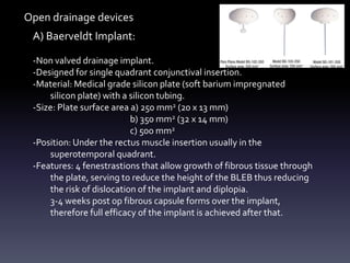
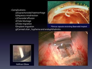
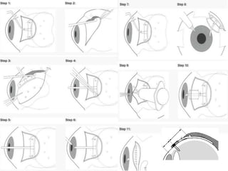
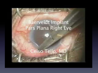
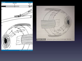
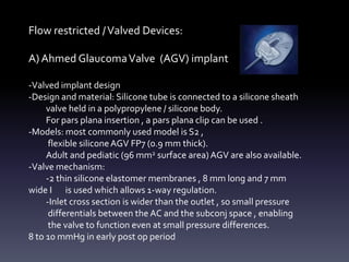
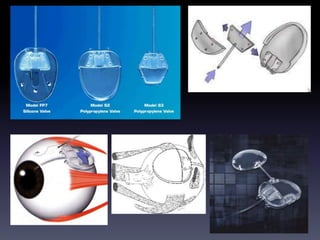
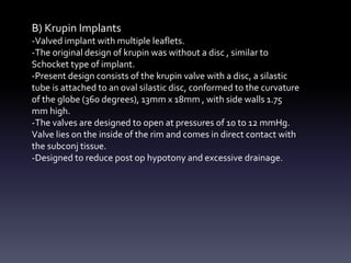
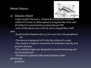
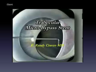
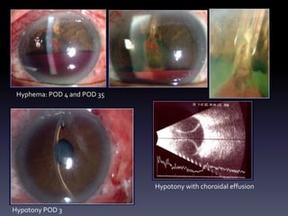
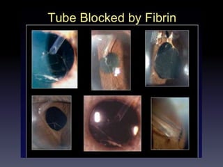
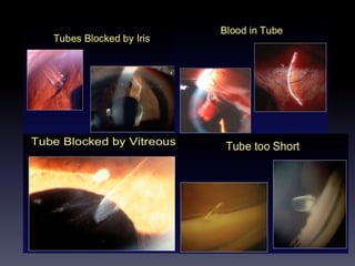
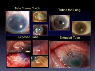
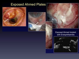
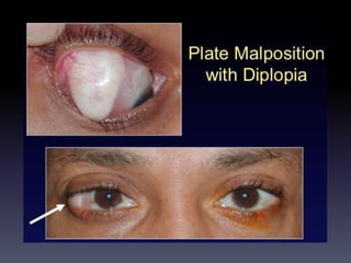
SlideShare a Scribd company logo
GLAUCOMA DRAINGE-
DEVICE SURGERY
Dr. Nilay Patel
Dept of Ophthalmology
Dr. D .Y. Patil University,School of Medicine
Physiology of Drainage Implants
• A silicone tube that extends from the AC to a plate, disc, or encircling
element beneath the conjunctiva and tenon capsule. External plate has a
ridge through which the tube enters on to its surface.The ridge decreases
the risk of obstruction with the surrounding tissue and fibrous capsule and
forms a filtering bleb posteriorly near the equator.
• A fibrous capsule forms a filtering bleb around the external portion of the
draining device.
• After insertion, a thin collagenous capsule, surrounded by granulomatous
reaction is present which subsides after 4 months.The capsule thickness
remains stable and collagen stroma becomes less compact.
• There is a direct relation between surface area of the implants and the
filtering capacity of their surrounding capsule.
• Reduction in bleb diameter decreases surface tension, capsular fibrosis and
thickness thus increasing the effectiveness of the filtering surface.
• All drainage devices develop an elevated IOP weeks to months after
implantation due to capsule formation.This is termed as Hypertensive Phase
The indications for GDD implantation include the following:
• Neovascular glaucoma
• PKP with glaucoma
• Retinal detachment surgery with glaucoma
• ICE syndrome
• Traumatic glaucoma
• Uveitic glaucoma
• Open-angle glaucoma with failed trabeculectomy
• Epithelial downgrowth
• Refractory infantile glaucoma
• Contact lens wearers who need glaucoma filtration surgery
Implant designs:
A) Open-Tube Drainage Devices
-Baerveldt Implant:a) 250 mm2 area
b) 350 mm2 area
c) 500 mm2 area
d) 35o mm2 area pars plana
-Molteno Implant
-SchocketTube Shunt
B) Flow Restricted/Valved Devices
-Ahmed GlaucomaValve
-Krupin Implants.
C) Newer Devices
-iStent
-Ex-PRESS Device
-SolxGold Shunt
-OptiMed glaucoma Pressure
Regulator
-SusannaGlaucoma Implant
A) Baerveldt Implant:
-Non valved drainage implant.
-Designed for single quadrant conjunctival insertion.
-Material: Medical grade silicon plate (soft barium impregnated
silicon plate) with a silicon tubing.
-Size: Plate surface area a) 250 mm2 (20 x 13 mm)
b) 350 mm2 (32 x 14 mm)
c) 500 mm2
-Position: Under the rectus muscle insertion usually in the
superotemporal quadrant.
-Features: 4 fenestrastions that allow growth of fibrous tissue through
the plate, serving to reduce the height of the BLEB thus reducing
the risk of dislocation of the implant and diplopia.
3-4 weeks post op fibrous capsule forms over the implant,
therefore full efficacy of the implant is achieved after that.
Open drainage devices
-Complications:
a)Suprachoroidal haemorrhage
b)Aqueous misdirection
c)Choroidal effusion
d)Tube blockage
e)Tube migration
f)Implant migration
g)Corneal ulcer , hyphema and endophthalmitis.
B) Molteno Implant:
-Non valved drainage implant introduced in 1969.
-Original design had a single plate ( 13 mm diameter and 135mm2
surface area ).To increase the efficacy second plate was added.
3Rd generation plate was developed, it has a bowl shaped
structure ( it is designed to function as a biological valve by l
imiting the available area of filtration during times of low
aqueous production.)
-Material:Acrylic plate with silicon tubing
-Size: Surface area of single plate is 135mm2 and 13 mm diameter
Double plate is 270 mm2 , 175 mm2 and 230 mm2.
Silicon tubing had an external diameter of 0.62mm and internal
diameter of 0.30mm.
-Position: Under the rectus muscle insertion usually in the
superotemporal quadrant.
-Features:V-shaped pressure ridge on the upper surface encases
10.5mm2 around the tube opening to deal with the problem of
Hypotony and excessive filtration in post op period.
C) SchocketTube Shunt:
-Non valved drainage implant.
-A Silicon or Silastic tube is extended from the AC to a 360 degree
encircling silicon band which functioned in developing the
reservoir for aqueous drainage.
-Material: Silicon or Silastic tubing.
-Position: 90 degree insertion of the silicon tube originating from
the AC to the encircling band beneath 2 or more rectus muscles
-Features: Its provides a larger surface area of reservoir compared
to molteno implant.
-Procedure: A 30-mm long silastic lacrimal tube with an internal
diameter of 0.30 mm is sutured inside the groove of a #20 silicone band,
leaving 15 mm of the tube extending from the band.The encircling band
is circumferentially sutured to the sclera, 10–12 mm from the limbus
beneath the four rectus muscles, with 5-0 Supramid sutures.A square 3–
4mm scleral flap hinged at the limbus is elevated, and a slightly beveled
puncture wound is made into the anterior chamber.The end of the tube is
beveled and inserted into the anterior cham- ber so that a 3-mm length is
suspended between the iris and cor- nea with the bevel facing anteriorly.
The scleral flap is closed with 10-0 nylon, and the conjunctival flap is
sutured with 8-0Vicryl.
Flow restricted /Valved Devices:
A) Ahmed GlaucomaValve (AGV) implant
-Valved implant design
-Design and material: Silicone tube is connected to a silicone sheath
valve held in a polypropylene / silicone body.
For pars plana insertion , a pars plana clip can be used .
-Models: most commonly used model is S2 ,
flexible siliconeAGV FP7 (0.9 mm thick).
Adult and pediatic (96 mm2 surface area)AGV are also available.
-Valve mechanism:
-2 thin silicone elastomer membranes , 8 mm long and 7 mm
wide I is used which allows 1-way regulation.
-Inlet cross section is wider than the outlet , so small pressure
differentials between the AC and the subconj space , enabling
the valve to function even at small pressure differences.
8 to 10 mmHg in early post op period
-Venturi effect of the valve can be explained by Bernoulli’s equation of fluid
dynamics.
( flow rate of a fluid is inversely proportional to the pressure of the fluid)
-The aqueous humor flows slowly into the trapezoid chamber and increases
the pressure.On reaching the pre-set threshold value , the valve opens thus
decreasing the pressure.
-The fluid velocity has to increase as it leaves the chamber (due to rise in
pressure) through the drainage tube .The tension in the silicone membranes
helps reduce hypotony as the valves closes as pressure decreases.
-No-touch zone on the AVG is area of the chamber with silicone leaflets.
If the implant is grasped with the forceps along the centre line, it may separate
the valve cover from the implant leading to failure due to adhesion of the valve
membrane.
Ahmed GlaucomaValve
B) Krupin Implants
-Valved implant with multiple leaflets.
-The original design of krupin was without a disc , similar to
Schocket type of implant.
-Present design consists of the krupin valve with a disc, a silastic
tube is attached to an oval silastic disc, conformed to the curvature
of the globe (360 degrees), 13mm x 18mm , with side walls 1.75
mm high.
-The valves are designed to open at pressures of 10 to 12 mmHg.
Valve lies on the inside of the rim and comes in direct contact with
the subconj tissue.
-Designed to reduce post op hypotony and excessive drainage.
Newer Setons:
1) Glaukos iStent
-Light weightTitanium L-shaped device placed inside the
schlemm’s canal, to allow aqueous to bypass the inner wall
of schlemm’s canal and the juxtacanalicularTM
-1mm of the device sits in the SC and is shaped like a half
pipe.
-Small snorkel shaped tube 0.5 mm size sits in the peripheral
AC
-The device is designed to fit into the schlemm’s canal,
-The implant is heparin covered for thrombolytic activity and
prevent stenosis.
-The 3 barbed ridges are designed to prevent loosening and
secure placement.
-The device is placed in the nasal quadrant under
gonioscopic
guidance.
iStent
2) Ex-PRESS mini shunt:
- 400 micron wide x 3 mm long , stainless steel device.
- It has a beveled sharpened rounded tip, disclike flange (<1 mm2) at the
proximal end and a spurlike projection that prevents its extrusion.
- Thickness corresponds to the scleral thickness, the external flange and
the inner spur are angled to conform to the scleral anatomy.
- Inner diameter is 125 microns and outer diameter is 250 micron, thin
enough to fit into the scleral spur.
- Sterilization: by gamma radiation , stored at 15-30 degree C
- Immediate bleb formation on 1st or 2nd post op day.
Ex-PRESS mini shunt
3) Gold Micro-Shunt
- Deep Light Glaucoma tx:Titanium sapphire laser and photo titratable gold
micro shunt
- Laser emits microsecond infra red light pulses that passes through the
trabecular meshwork tissues to produce an opening for outflow.
- The 99.5% inert gold shunt is placed which have micro channels , which can
also be titrated post op using titanium sapphire laser.
- The biocompatible gold plate is 5.2 mm long, 2 mm wide and 60 micron thick
containing multiple microchannels
- It is implanted through a 3 mm clear corneal incision with a help of a pre
loaded injector.
- Thus the channels of the gold shunt form a bridge b/w AC and suprachoroidal
space.
4) OptiMed glaucoma Pressure Regulator:
- Silicon tube with a PMMA plate.
- Inner diameter of the tube is 0.38 mm and outer diameter is 0.76 mm.
- 5 mm PMMA tube is inserted into the silicone base with dimensions of
1 x 2 x 3 x 4 mm , base contains 180 to 200 microtubules.
- It functions when the pressure exceeds 10 mmHg , through the capillary
action, it works on the principle of Poiseuille’s formula.
5) Susanna Glaucoma Implant:
- A reservior body conforming to the shape of the globe at its equator and a
ridge in the end plate to protect the inner opening of the silicone tube from
blockage by fibrous tissue growth.
- Fenestrating end plate promotes fibrous tissue anchoring.(to avoid motion)
- The footplate measures 4 mm length allow easy fixation at 6 mm from the
limbus and yet allowing the plate to be placed at 10 mm from the limbus ( to
reduce extrusion)
COMPLICATIONS:
a) Intraoperative complications:
• Bleeding
• Misdirection of the silicone tube into the posterior chamber in
presence of peripheral anterior synechiae
• Loss ofAC: if port size is large
b) Early Post operative complication:
• Hypotony: maybe asso with Choroidal effusions.
use of valved implants or by occluding the tubing with a ligature
• Increased IOP: due to occlusion , with iris ,YAG ablation of the iris
• Tube-corneal contact: avoided by placing the tube properly
intraoperatively
• Early post op endophthalmitis
c) Late complications:
• Encysted bleb: in the hypertensive phase rise in IOP due to
development of a thick capsule around the plate 4 to 6 weeks postop
regimen consists of Diclofenac 75 mg , prednisolone 40 mg and
topical steroids.
1 cm3 can be deflated by needling
• Erosion of the silicon tube.
• Plate migration
• Eye movement limitations
• Endophthalmitis
• Epithelial ingrowth and invasion into the fibrous capsule
• Sterile hypopyon
• Irregular pupil due to adhesions of the iris roots
• Globe perforation
• RD, supra choroidal h’age,VH, choroidal effusion.
Hyphema: POD 4 and POD 35
Hypotony POD 3
Hypotony with choroidal effusion
THANK YOU

More Related Content

What's hot

Cataract Surgery: Small Pupil by Dr Joobin Khadamy
Cataract Surgery: Small Pupil by Dr Joobin KhadamyCataract Surgery: Small Pupil by Dr Joobin Khadamy
Cataract Surgery: Small Pupil by Dr Joobin Khadamy
Joobin Khadamy . MD
 
Piggyback iol
Piggyback iolPiggyback iol
Piggyback iol
Fahmida Hoque
 
Phakic iol ppt
Phakic iol pptPhakic iol ppt
Phakic iol ppt
DrArino John
 
Ophthalmic viscosurgical devices presentation
Ophthalmic viscosurgical devices presentationOphthalmic viscosurgical devices presentation
Ophthalmic viscosurgical devices presentation
Ahmed Yusuf
 
Surgical induced astigmatism
Surgical induced astigmatismSurgical induced astigmatism
Surgical induced astigmatism
Namrata Gupta
 
Role of oct in glaucoma
Role of oct in glaucomaRole of oct in glaucoma
Role of oct in glaucoma
Dr Samarth Mishra
 
Minimally invasive glaucoma surgery
Minimally invasive glaucoma surgery Minimally invasive glaucoma surgery
Minimally invasive glaucoma surgery
aditisingh77985
 
Ocular viscoelastic devices(OVD)
Ocular viscoelastic devices(OVD)Ocular viscoelastic devices(OVD)
Ocular viscoelastic devices(OVD)
Sivateja Challa
 
Minimally invasive Glaucoma surgery MIGS
Minimally invasive Glaucoma surgery MIGSMinimally invasive Glaucoma surgery MIGS
Minimally invasive Glaucoma surgery MIGS
ankita mahapatra
 
Premium intraocular lenses The past, present and-3.pptx
Premium intraocular lenses The past, present and-3.pptxPremium intraocular lenses The past, present and-3.pptx
Premium intraocular lenses The past, present and-3.pptx
Mushtaq Ahmad
 
ophthalmic viscosurgical devices
ophthalmic viscosurgical devicesophthalmic viscosurgical devices
ophthalmic viscosurgical devices
Amr mohamed
 
Complications of trabeculectomy
Complications of trabeculectomyComplications of trabeculectomy
Complications of trabeculectomy
Sumeet Agrawal
 
BASIC INFO ON FUDUS FLORESCENCE ANGIOGRAPHY
BASIC INFO ON FUDUS FLORESCENCE ANGIOGRAPHYBASIC INFO ON FUDUS FLORESCENCE ANGIOGRAPHY
BASIC INFO ON FUDUS FLORESCENCE ANGIOGRAPHYNalin Nayan
 
Limbal stem cell Deficiency; amniotic membrane transplantation
Limbal stem cell Deficiency; amniotic membrane transplantationLimbal stem cell Deficiency; amniotic membrane transplantation
Limbal stem cell Deficiency; amniotic membrane transplantation
Om Patel
 
Glaucoma drainage devices
Glaucoma drainage devicesGlaucoma drainage devices
Glaucoma drainage devices
vaishusmail
 
Pigment dispersion syndrome
Pigment dispersion syndromePigment dispersion syndrome
Pigment dispersion syndrome
SSSIHMS-PG
 
Diagnosis of pre perimetric glaucoma
Diagnosis of pre perimetric glaucomaDiagnosis of pre perimetric glaucoma
Diagnosis of pre perimetric glaucoma
Sadhwini Harish
 
Wound construction For Cataract Surgery
Wound construction For Cataract SurgeryWound construction For Cataract Surgery
Wound construction For Cataract SurgeryDevin Prabhakar
 
Vitreous substitutes in ophthalmology
 Vitreous substitutes in ophthalmology Vitreous substitutes in ophthalmology
Vitreous substitutes in ophthalmology
mdalbanuddin
 
Post keratoplasty Case
Post keratoplasty CasePost keratoplasty Case

What's hot (20)

Cataract Surgery: Small Pupil by Dr Joobin Khadamy
Cataract Surgery: Small Pupil by Dr Joobin KhadamyCataract Surgery: Small Pupil by Dr Joobin Khadamy
Cataract Surgery: Small Pupil by Dr Joobin Khadamy
 
Piggyback iol
Piggyback iolPiggyback iol
Piggyback iol
 
Phakic iol ppt
Phakic iol pptPhakic iol ppt
Phakic iol ppt
 
Ophthalmic viscosurgical devices presentation
Ophthalmic viscosurgical devices presentationOphthalmic viscosurgical devices presentation
Ophthalmic viscosurgical devices presentation
 
Surgical induced astigmatism
Surgical induced astigmatismSurgical induced astigmatism
Surgical induced astigmatism
 
Role of oct in glaucoma
Role of oct in glaucomaRole of oct in glaucoma
Role of oct in glaucoma
 
Minimally invasive glaucoma surgery
Minimally invasive glaucoma surgery Minimally invasive glaucoma surgery
Minimally invasive glaucoma surgery
 
Ocular viscoelastic devices(OVD)
Ocular viscoelastic devices(OVD)Ocular viscoelastic devices(OVD)
Ocular viscoelastic devices(OVD)
 
Minimally invasive Glaucoma surgery MIGS
Minimally invasive Glaucoma surgery MIGSMinimally invasive Glaucoma surgery MIGS
Minimally invasive Glaucoma surgery MIGS
 
Premium intraocular lenses The past, present and-3.pptx
Premium intraocular lenses The past, present and-3.pptxPremium intraocular lenses The past, present and-3.pptx
Premium intraocular lenses The past, present and-3.pptx
 
ophthalmic viscosurgical devices
ophthalmic viscosurgical devicesophthalmic viscosurgical devices
ophthalmic viscosurgical devices
 
Complications of trabeculectomy
Complications of trabeculectomyComplications of trabeculectomy
Complications of trabeculectomy
 
BASIC INFO ON FUDUS FLORESCENCE ANGIOGRAPHY
BASIC INFO ON FUDUS FLORESCENCE ANGIOGRAPHYBASIC INFO ON FUDUS FLORESCENCE ANGIOGRAPHY
BASIC INFO ON FUDUS FLORESCENCE ANGIOGRAPHY
 
Limbal stem cell Deficiency; amniotic membrane transplantation
Limbal stem cell Deficiency; amniotic membrane transplantationLimbal stem cell Deficiency; amniotic membrane transplantation
Limbal stem cell Deficiency; amniotic membrane transplantation
 
Glaucoma drainage devices
Glaucoma drainage devicesGlaucoma drainage devices
Glaucoma drainage devices
 
Pigment dispersion syndrome
Pigment dispersion syndromePigment dispersion syndrome
Pigment dispersion syndrome
 
Diagnosis of pre perimetric glaucoma
Diagnosis of pre perimetric glaucomaDiagnosis of pre perimetric glaucoma
Diagnosis of pre perimetric glaucoma
 
Wound construction For Cataract Surgery
Wound construction For Cataract SurgeryWound construction For Cataract Surgery
Wound construction For Cataract Surgery
 
Vitreous substitutes in ophthalmology
 Vitreous substitutes in ophthalmology Vitreous substitutes in ophthalmology
Vitreous substitutes in ophthalmology
 
Post keratoplasty Case
Post keratoplasty CasePost keratoplasty Case
Post keratoplasty Case
 

Similar to Gdd

Glaucoma drainage devices[1]
Glaucoma drainage devices[1]Glaucoma drainage devices[1]
Glaucoma drainage devices[1]
paresh nichlani
 
ADVANCES IN GLAUCOMA SURGERY - MIGS
ADVANCES IN GLAUCOMA SURGERY - MIGSADVANCES IN GLAUCOMA SURGERY - MIGS
ADVANCES IN GLAUCOMA SURGERY - MIGS
Priyanka Raj
 
Minimally Invasive Glaucoma Surgery (MIGS)
Minimally Invasive Glaucoma Surgery (MIGS)Minimally Invasive Glaucoma Surgery (MIGS)
Minimally Invasive Glaucoma Surgery (MIGS)
Meironi Waimir
 
Phacoemulsification part 3
Phacoemulsification part 3Phacoemulsification part 3
Phacoemulsification part 3
Priyanka Raj
 
Mivs
MivsMivs
Casting procedures/cosmetic dentistry course by Indian dental academy
Casting procedures/cosmetic dentistry course by Indian dental academyCasting procedures/cosmetic dentistry course by Indian dental academy
Casting procedures/cosmetic dentistry course by Indian dental academy
Indian dental academy
 
Fluid control.pptx
Fluid control.pptxFluid control.pptx
Fluid control.pptx
Royal Dental College Library
 
article.smst2004.Vascular filter with improved strength and flexibility
article.smst2004.Vascular filter with improved strength and flexibilityarticle.smst2004.Vascular filter with improved strength and flexibility
article.smst2004.Vascular filter with improved strength and flexibilityPetrus Besselink
 
Obturation of Root Canal - An Endodontic Overview
Obturation of Root Canal - An Endodontic OverviewObturation of Root Canal - An Endodontic Overview
Obturation of Root Canal - An Endodontic Overview
Iraqi Dental Academy
 
MIVS 2018
MIVS 2018   MIVS 2018
MIVS 2018
Anurag Shandil
 
Gingival tissue management
Gingival tissue management Gingival tissue management
Gingival tissue management
Abhijeet Pallewar
 
Recent advances in dcr
Recent advances in dcrRecent advances in dcr
Recent advances in dcr
Dinesh Madduri
 
Phacoemulsification
PhacoemulsificationPhacoemulsification
Phacoemulsification
DR. ROHIT AGRAWAL
 
Management of tracheostomy
Management of tracheostomyManagement of tracheostomy
Management of tracheostomy
mohamed abuelnaga
 
ET TUBES presentation by Dr. Animesh Aman Singh
ET TUBES presentation  by Dr. Animesh Aman SinghET TUBES presentation  by Dr. Animesh Aman Singh
ET TUBES presentation by Dr. Animesh Aman Singh
19anisingh
 
Debulking device
Debulking deviceDebulking device
Debulking device
Ashish Golwara
 
BMV balloons- FINAL.pptx
BMV balloons- FINAL.pptxBMV balloons- FINAL.pptx
BMV balloons- FINAL.pptx
RohitWalse2
 
Clinical management of edentulous maxillectomy /prosthodontic courses
Clinical management of edentulous maxillectomy /prosthodontic coursesClinical management of edentulous maxillectomy /prosthodontic courses
Clinical management of edentulous maxillectomy /prosthodontic courses
Indian dental academy
 

Similar to Gdd (20)

Glaucoma drainage devices[1]
Glaucoma drainage devices[1]Glaucoma drainage devices[1]
Glaucoma drainage devices[1]
 
ADVANCES IN GLAUCOMA SURGERY - MIGS
ADVANCES IN GLAUCOMA SURGERY - MIGSADVANCES IN GLAUCOMA SURGERY - MIGS
ADVANCES IN GLAUCOMA SURGERY - MIGS
 
Minimally Invasive Glaucoma Surgery (MIGS)
Minimally Invasive Glaucoma Surgery (MIGS)Minimally Invasive Glaucoma Surgery (MIGS)
Minimally Invasive Glaucoma Surgery (MIGS)
 
Micro valves
Micro valvesMicro valves
Micro valves
 
Phacoemulsification part 3
Phacoemulsification part 3Phacoemulsification part 3
Phacoemulsification part 3
 
Mivs
MivsMivs
Mivs
 
Casting procedures/cosmetic dentistry course by Indian dental academy
Casting procedures/cosmetic dentistry course by Indian dental academyCasting procedures/cosmetic dentistry course by Indian dental academy
Casting procedures/cosmetic dentistry course by Indian dental academy
 
Fluid control.pptx
Fluid control.pptxFluid control.pptx
Fluid control.pptx
 
article.smst2004.Vascular filter with improved strength and flexibility
article.smst2004.Vascular filter with improved strength and flexibilityarticle.smst2004.Vascular filter with improved strength and flexibility
article.smst2004.Vascular filter with improved strength and flexibility
 
Obturation of Root Canal - An Endodontic Overview
Obturation of Root Canal - An Endodontic OverviewObturation of Root Canal - An Endodontic Overview
Obturation of Root Canal - An Endodontic Overview
 
MIVS 2018
MIVS 2018   MIVS 2018
MIVS 2018
 
Gingival tissue management
Gingival tissue management Gingival tissue management
Gingival tissue management
 
Recent advances in dcr
Recent advances in dcrRecent advances in dcr
Recent advances in dcr
 
Phacoemulsification
PhacoemulsificationPhacoemulsification
Phacoemulsification
 
Management of tracheostomy
Management of tracheostomyManagement of tracheostomy
Management of tracheostomy
 
ET TUBES presentation by Dr. Animesh Aman Singh
ET TUBES presentation  by Dr. Animesh Aman SinghET TUBES presentation  by Dr. Animesh Aman Singh
ET TUBES presentation by Dr. Animesh Aman Singh
 
Dental implants
Dental implants Dental implants
Dental implants
 
Debulking device
Debulking deviceDebulking device
Debulking device
 
BMV balloons- FINAL.pptx
BMV balloons- FINAL.pptxBMV balloons- FINAL.pptx
BMV balloons- FINAL.pptx
 
Clinical management of edentulous maxillectomy /prosthodontic courses
Clinical management of edentulous maxillectomy /prosthodontic coursesClinical management of edentulous maxillectomy /prosthodontic courses
Clinical management of edentulous maxillectomy /prosthodontic courses
 

Recently uploaded

Alcohol_Dr. Jeenal Mistry MD Pharmacology.pdf
Alcohol_Dr. Jeenal Mistry MD Pharmacology.pdfAlcohol_Dr. Jeenal Mistry MD Pharmacology.pdf
Alcohol_Dr. Jeenal Mistry MD Pharmacology.pdf
Dr Jeenal Mistry
 
Evaluation of antidepressant activity of clitoris ternatea in animals
Evaluation of antidepressant activity of clitoris ternatea in animalsEvaluation of antidepressant activity of clitoris ternatea in animals
Evaluation of antidepressant activity of clitoris ternatea in animals
Shweta
 
The Normal Electrocardiogram - Part I of II
The Normal Electrocardiogram - Part I of IIThe Normal Electrocardiogram - Part I of II
The Normal Electrocardiogram - Part I of II
MedicoseAcademics
 
For Better Surat #ℂall #Girl Service ❤85270-49040❤ Surat #ℂall #Girls
For Better Surat #ℂall #Girl Service ❤85270-49040❤ Surat #ℂall #GirlsFor Better Surat #ℂall #Girl Service ❤85270-49040❤ Surat #ℂall #Girls
For Better Surat #ℂall #Girl Service ❤85270-49040❤ Surat #ℂall #Girls
Savita Shen $i11
 
NVBDCP.pptx Nation vector borne disease control program
NVBDCP.pptx Nation vector borne disease control programNVBDCP.pptx Nation vector borne disease control program
NVBDCP.pptx Nation vector borne disease control program
Sapna Thakur
 
Report Back from SGO 2024: What’s the Latest in Cervical Cancer?
Report Back from SGO 2024: What’s the Latest in Cervical Cancer?Report Back from SGO 2024: What’s the Latest in Cervical Cancer?
Report Back from SGO 2024: What’s the Latest in Cervical Cancer?
bkling
 
Novas diretrizes da OMS para os cuidados perinatais de mais qualidade
Novas diretrizes da OMS para os cuidados perinatais de mais qualidadeNovas diretrizes da OMS para os cuidados perinatais de mais qualidade
Novas diretrizes da OMS para os cuidados perinatais de mais qualidade
Prof. Marcus Renato de Carvalho
 
Superficial & Deep Fascia of the NECK.pptx
Superficial & Deep Fascia of the NECK.pptxSuperficial & Deep Fascia of the NECK.pptx
Superficial & Deep Fascia of the NECK.pptx
Dr. Rabia Inam Gandapore
 
POST OPERATIVE OLIGURIA and its management
POST OPERATIVE OLIGURIA and its managementPOST OPERATIVE OLIGURIA and its management
POST OPERATIVE OLIGURIA and its management
touseefaziz1
 
Surat @ℂall @Girls ꧁❤8527049040❤꧂@ℂall @Girls Service Vip Top Model Safe
Surat @ℂall @Girls ꧁❤8527049040❤꧂@ℂall @Girls Service Vip Top Model SafeSurat @ℂall @Girls ꧁❤8527049040❤꧂@ℂall @Girls Service Vip Top Model Safe
Surat @ℂall @Girls ꧁❤8527049040❤꧂@ℂall @Girls Service Vip Top Model Safe
Savita Shen $i11
 
New Directions in Targeted Therapeutic Approaches for Older Adults With Mantl...
New Directions in Targeted Therapeutic Approaches for Older Adults With Mantl...New Directions in Targeted Therapeutic Approaches for Older Adults With Mantl...
New Directions in Targeted Therapeutic Approaches for Older Adults With Mantl...
i3 Health
 
Hemodialysis: Chapter 3, Dialysis Water Unit - Dr.Gawad
Hemodialysis: Chapter 3, Dialysis Water Unit - Dr.GawadHemodialysis: Chapter 3, Dialysis Water Unit - Dr.Gawad
Hemodialysis: Chapter 3, Dialysis Water Unit - Dr.Gawad
NephroTube - Dr.Gawad
 
heat stroke and heat exhaustion in children
heat stroke and heat exhaustion in childrenheat stroke and heat exhaustion in children
heat stroke and heat exhaustion in children
SumeraAhmad5
 
Pharynx and Clinical Correlations BY Dr.Rabia Inam Gandapore.pptx
Pharynx and Clinical Correlations BY Dr.Rabia Inam Gandapore.pptxPharynx and Clinical Correlations BY Dr.Rabia Inam Gandapore.pptx
Pharynx and Clinical Correlations BY Dr.Rabia Inam Gandapore.pptx
Dr. Rabia Inam Gandapore
 
ARTHROLOGY PPT NCISM SYLLABUS AYURVEDA STUDENTS
ARTHROLOGY PPT NCISM SYLLABUS AYURVEDA STUDENTSARTHROLOGY PPT NCISM SYLLABUS AYURVEDA STUDENTS
ARTHROLOGY PPT NCISM SYLLABUS AYURVEDA STUDENTS
Dr. Vinay Pareek
 
BENIGN PROSTATIC HYPERPLASIA.BPH. BPHpdf
BENIGN PROSTATIC HYPERPLASIA.BPH. BPHpdfBENIGN PROSTATIC HYPERPLASIA.BPH. BPHpdf
BENIGN PROSTATIC HYPERPLASIA.BPH. BPHpdf
DR SETH JOTHAM
 
Are There Any Natural Remedies To Treat Syphilis.pdf
Are There Any Natural Remedies To Treat Syphilis.pdfAre There Any Natural Remedies To Treat Syphilis.pdf
Are There Any Natural Remedies To Treat Syphilis.pdf
Little Cross Family Clinic
 
KDIGO 2024 guidelines for diabetologists
KDIGO 2024 guidelines for diabetologistsKDIGO 2024 guidelines for diabetologists
KDIGO 2024 guidelines for diabetologists
د.محمود نجيب
 
ANATOMY AND PHYSIOLOGY OF URINARY SYSTEM.pptx
ANATOMY AND PHYSIOLOGY OF URINARY SYSTEM.pptxANATOMY AND PHYSIOLOGY OF URINARY SYSTEM.pptx
ANATOMY AND PHYSIOLOGY OF URINARY SYSTEM.pptx
Swetaba Besh
 
Knee anatomy and clinical tests 2024.pdf
Knee anatomy and clinical tests 2024.pdfKnee anatomy and clinical tests 2024.pdf
Knee anatomy and clinical tests 2024.pdf
vimalpl1234
 

Recently uploaded (20)

Alcohol_Dr. Jeenal Mistry MD Pharmacology.pdf
Alcohol_Dr. Jeenal Mistry MD Pharmacology.pdfAlcohol_Dr. Jeenal Mistry MD Pharmacology.pdf
Alcohol_Dr. Jeenal Mistry MD Pharmacology.pdf
 
Evaluation of antidepressant activity of clitoris ternatea in animals
Evaluation of antidepressant activity of clitoris ternatea in animalsEvaluation of antidepressant activity of clitoris ternatea in animals
Evaluation of antidepressant activity of clitoris ternatea in animals
 
The Normal Electrocardiogram - Part I of II
The Normal Electrocardiogram - Part I of IIThe Normal Electrocardiogram - Part I of II
The Normal Electrocardiogram - Part I of II
 
For Better Surat #ℂall #Girl Service ❤85270-49040❤ Surat #ℂall #Girls
For Better Surat #ℂall #Girl Service ❤85270-49040❤ Surat #ℂall #GirlsFor Better Surat #ℂall #Girl Service ❤85270-49040❤ Surat #ℂall #Girls
For Better Surat #ℂall #Girl Service ❤85270-49040❤ Surat #ℂall #Girls
 
NVBDCP.pptx Nation vector borne disease control program
NVBDCP.pptx Nation vector borne disease control programNVBDCP.pptx Nation vector borne disease control program
NVBDCP.pptx Nation vector borne disease control program
 
Report Back from SGO 2024: What’s the Latest in Cervical Cancer?
Report Back from SGO 2024: What’s the Latest in Cervical Cancer?Report Back from SGO 2024: What’s the Latest in Cervical Cancer?
Report Back from SGO 2024: What’s the Latest in Cervical Cancer?
 
Novas diretrizes da OMS para os cuidados perinatais de mais qualidade
Novas diretrizes da OMS para os cuidados perinatais de mais qualidadeNovas diretrizes da OMS para os cuidados perinatais de mais qualidade
Novas diretrizes da OMS para os cuidados perinatais de mais qualidade
 
Superficial & Deep Fascia of the NECK.pptx
Superficial & Deep Fascia of the NECK.pptxSuperficial & Deep Fascia of the NECK.pptx
Superficial & Deep Fascia of the NECK.pptx
 
POST OPERATIVE OLIGURIA and its management
POST OPERATIVE OLIGURIA and its managementPOST OPERATIVE OLIGURIA and its management
POST OPERATIVE OLIGURIA and its management
 
Surat @ℂall @Girls ꧁❤8527049040❤꧂@ℂall @Girls Service Vip Top Model Safe
Surat @ℂall @Girls ꧁❤8527049040❤꧂@ℂall @Girls Service Vip Top Model SafeSurat @ℂall @Girls ꧁❤8527049040❤꧂@ℂall @Girls Service Vip Top Model Safe
Surat @ℂall @Girls ꧁❤8527049040❤꧂@ℂall @Girls Service Vip Top Model Safe
 
New Directions in Targeted Therapeutic Approaches for Older Adults With Mantl...
New Directions in Targeted Therapeutic Approaches for Older Adults With Mantl...New Directions in Targeted Therapeutic Approaches for Older Adults With Mantl...
New Directions in Targeted Therapeutic Approaches for Older Adults With Mantl...
 
Hemodialysis: Chapter 3, Dialysis Water Unit - Dr.Gawad
Hemodialysis: Chapter 3, Dialysis Water Unit - Dr.GawadHemodialysis: Chapter 3, Dialysis Water Unit - Dr.Gawad
Hemodialysis: Chapter 3, Dialysis Water Unit - Dr.Gawad
 
heat stroke and heat exhaustion in children
heat stroke and heat exhaustion in childrenheat stroke and heat exhaustion in children
heat stroke and heat exhaustion in children
 
Pharynx and Clinical Correlations BY Dr.Rabia Inam Gandapore.pptx
Pharynx and Clinical Correlations BY Dr.Rabia Inam Gandapore.pptxPharynx and Clinical Correlations BY Dr.Rabia Inam Gandapore.pptx
Pharynx and Clinical Correlations BY Dr.Rabia Inam Gandapore.pptx
 
ARTHROLOGY PPT NCISM SYLLABUS AYURVEDA STUDENTS
ARTHROLOGY PPT NCISM SYLLABUS AYURVEDA STUDENTSARTHROLOGY PPT NCISM SYLLABUS AYURVEDA STUDENTS
ARTHROLOGY PPT NCISM SYLLABUS AYURVEDA STUDENTS
 
BENIGN PROSTATIC HYPERPLASIA.BPH. BPHpdf
BENIGN PROSTATIC HYPERPLASIA.BPH. BPHpdfBENIGN PROSTATIC HYPERPLASIA.BPH. BPHpdf
BENIGN PROSTATIC HYPERPLASIA.BPH. BPHpdf
 
Are There Any Natural Remedies To Treat Syphilis.pdf
Are There Any Natural Remedies To Treat Syphilis.pdfAre There Any Natural Remedies To Treat Syphilis.pdf
Are There Any Natural Remedies To Treat Syphilis.pdf
 
KDIGO 2024 guidelines for diabetologists
KDIGO 2024 guidelines for diabetologistsKDIGO 2024 guidelines for diabetologists
KDIGO 2024 guidelines for diabetologists
 
ANATOMY AND PHYSIOLOGY OF URINARY SYSTEM.pptx
ANATOMY AND PHYSIOLOGY OF URINARY SYSTEM.pptxANATOMY AND PHYSIOLOGY OF URINARY SYSTEM.pptx
ANATOMY AND PHYSIOLOGY OF URINARY SYSTEM.pptx
 
Knee anatomy and clinical tests 2024.pdf
Knee anatomy and clinical tests 2024.pdfKnee anatomy and clinical tests 2024.pdf
Knee anatomy and clinical tests 2024.pdf
 

Gdd

  • 1. GLAUCOMA DRAINGE- DEVICE SURGERY Dr. Nilay Patel Dept of Ophthalmology Dr. D .Y. Patil University,School of Medicine
  • 2. Physiology of Drainage Implants • A silicone tube that extends from the AC to a plate, disc, or encircling element beneath the conjunctiva and tenon capsule. External plate has a ridge through which the tube enters on to its surface.The ridge decreases the risk of obstruction with the surrounding tissue and fibrous capsule and forms a filtering bleb posteriorly near the equator. • A fibrous capsule forms a filtering bleb around the external portion of the draining device. • After insertion, a thin collagenous capsule, surrounded by granulomatous reaction is present which subsides after 4 months.The capsule thickness remains stable and collagen stroma becomes less compact. • There is a direct relation between surface area of the implants and the filtering capacity of their surrounding capsule. • Reduction in bleb diameter decreases surface tension, capsular fibrosis and thickness thus increasing the effectiveness of the filtering surface.
  • 3. • All drainage devices develop an elevated IOP weeks to months after implantation due to capsule formation.This is termed as Hypertensive Phase The indications for GDD implantation include the following: • Neovascular glaucoma • PKP with glaucoma • Retinal detachment surgery with glaucoma • ICE syndrome • Traumatic glaucoma • Uveitic glaucoma • Open-angle glaucoma with failed trabeculectomy • Epithelial downgrowth • Refractory infantile glaucoma • Contact lens wearers who need glaucoma filtration surgery
  • 4. Implant designs: A) Open-Tube Drainage Devices -Baerveldt Implant:a) 250 mm2 area b) 350 mm2 area c) 500 mm2 area d) 35o mm2 area pars plana -Molteno Implant -SchocketTube Shunt B) Flow Restricted/Valved Devices -Ahmed GlaucomaValve -Krupin Implants. C) Newer Devices -iStent -Ex-PRESS Device -SolxGold Shunt -OptiMed glaucoma Pressure Regulator -SusannaGlaucoma Implant
  • 5. A) Baerveldt Implant: -Non valved drainage implant. -Designed for single quadrant conjunctival insertion. -Material: Medical grade silicon plate (soft barium impregnated silicon plate) with a silicon tubing. -Size: Plate surface area a) 250 mm2 (20 x 13 mm) b) 350 mm2 (32 x 14 mm) c) 500 mm2 -Position: Under the rectus muscle insertion usually in the superotemporal quadrant. -Features: 4 fenestrastions that allow growth of fibrous tissue through the plate, serving to reduce the height of the BLEB thus reducing the risk of dislocation of the implant and diplopia. 3-4 weeks post op fibrous capsule forms over the implant, therefore full efficacy of the implant is achieved after that. Open drainage devices
  • 6. -Complications: a)Suprachoroidal haemorrhage b)Aqueous misdirection c)Choroidal effusion d)Tube blockage e)Tube migration f)Implant migration g)Corneal ulcer , hyphema and endophthalmitis.
  • 7.
  • 8.
  • 9. B) Molteno Implant: -Non valved drainage implant introduced in 1969. -Original design had a single plate ( 13 mm diameter and 135mm2 surface area ).To increase the efficacy second plate was added. 3Rd generation plate was developed, it has a bowl shaped structure ( it is designed to function as a biological valve by l imiting the available area of filtration during times of low aqueous production.) -Material:Acrylic plate with silicon tubing -Size: Surface area of single plate is 135mm2 and 13 mm diameter Double plate is 270 mm2 , 175 mm2 and 230 mm2. Silicon tubing had an external diameter of 0.62mm and internal diameter of 0.30mm. -Position: Under the rectus muscle insertion usually in the superotemporal quadrant. -Features:V-shaped pressure ridge on the upper surface encases 10.5mm2 around the tube opening to deal with the problem of Hypotony and excessive filtration in post op period.
  • 10.
  • 11.
  • 12. C) SchocketTube Shunt: -Non valved drainage implant. -A Silicon or Silastic tube is extended from the AC to a 360 degree encircling silicon band which functioned in developing the reservoir for aqueous drainage. -Material: Silicon or Silastic tubing. -Position: 90 degree insertion of the silicon tube originating from the AC to the encircling band beneath 2 or more rectus muscles -Features: Its provides a larger surface area of reservoir compared to molteno implant. -Procedure: A 30-mm long silastic lacrimal tube with an internal diameter of 0.30 mm is sutured inside the groove of a #20 silicone band, leaving 15 mm of the tube extending from the band.The encircling band is circumferentially sutured to the sclera, 10–12 mm from the limbus beneath the four rectus muscles, with 5-0 Supramid sutures.A square 3– 4mm scleral flap hinged at the limbus is elevated, and a slightly beveled puncture wound is made into the anterior chamber.The end of the tube is beveled and inserted into the anterior cham- ber so that a 3-mm length is suspended between the iris and cor- nea with the bevel facing anteriorly. The scleral flap is closed with 10-0 nylon, and the conjunctival flap is sutured with 8-0Vicryl.
  • 13.
  • 14. Flow restricted /Valved Devices: A) Ahmed GlaucomaValve (AGV) implant -Valved implant design -Design and material: Silicone tube is connected to a silicone sheath valve held in a polypropylene / silicone body. For pars plana insertion , a pars plana clip can be used . -Models: most commonly used model is S2 , flexible siliconeAGV FP7 (0.9 mm thick). Adult and pediatic (96 mm2 surface area)AGV are also available. -Valve mechanism: -2 thin silicone elastomer membranes , 8 mm long and 7 mm wide I is used which allows 1-way regulation. -Inlet cross section is wider than the outlet , so small pressure differentials between the AC and the subconj space , enabling the valve to function even at small pressure differences. 8 to 10 mmHg in early post op period
  • 15. -Venturi effect of the valve can be explained by Bernoulli’s equation of fluid dynamics. ( flow rate of a fluid is inversely proportional to the pressure of the fluid) -The aqueous humor flows slowly into the trapezoid chamber and increases the pressure.On reaching the pre-set threshold value , the valve opens thus decreasing the pressure. -The fluid velocity has to increase as it leaves the chamber (due to rise in pressure) through the drainage tube .The tension in the silicone membranes helps reduce hypotony as the valves closes as pressure decreases. -No-touch zone on the AVG is area of the chamber with silicone leaflets. If the implant is grasped with the forceps along the centre line, it may separate the valve cover from the implant leading to failure due to adhesion of the valve membrane.
  • 16.
  • 18. B) Krupin Implants -Valved implant with multiple leaflets. -The original design of krupin was without a disc , similar to Schocket type of implant. -Present design consists of the krupin valve with a disc, a silastic tube is attached to an oval silastic disc, conformed to the curvature of the globe (360 degrees), 13mm x 18mm , with side walls 1.75 mm high. -The valves are designed to open at pressures of 10 to 12 mmHg. Valve lies on the inside of the rim and comes in direct contact with the subconj tissue. -Designed to reduce post op hypotony and excessive drainage.
  • 19. Newer Setons: 1) Glaukos iStent -Light weightTitanium L-shaped device placed inside the schlemm’s canal, to allow aqueous to bypass the inner wall of schlemm’s canal and the juxtacanalicularTM -1mm of the device sits in the SC and is shaped like a half pipe. -Small snorkel shaped tube 0.5 mm size sits in the peripheral AC -The device is designed to fit into the schlemm’s canal, -The implant is heparin covered for thrombolytic activity and prevent stenosis. -The 3 barbed ridges are designed to prevent loosening and secure placement. -The device is placed in the nasal quadrant under gonioscopic guidance.
  • 21. 2) Ex-PRESS mini shunt: - 400 micron wide x 3 mm long , stainless steel device. - It has a beveled sharpened rounded tip, disclike flange (<1 mm2) at the proximal end and a spurlike projection that prevents its extrusion. - Thickness corresponds to the scleral thickness, the external flange and the inner spur are angled to conform to the scleral anatomy. - Inner diameter is 125 microns and outer diameter is 250 micron, thin enough to fit into the scleral spur. - Sterilization: by gamma radiation , stored at 15-30 degree C - Immediate bleb formation on 1st or 2nd post op day.
  • 23. 3) Gold Micro-Shunt - Deep Light Glaucoma tx:Titanium sapphire laser and photo titratable gold micro shunt - Laser emits microsecond infra red light pulses that passes through the trabecular meshwork tissues to produce an opening for outflow. - The 99.5% inert gold shunt is placed which have micro channels , which can also be titrated post op using titanium sapphire laser. - The biocompatible gold plate is 5.2 mm long, 2 mm wide and 60 micron thick containing multiple microchannels - It is implanted through a 3 mm clear corneal incision with a help of a pre loaded injector. - Thus the channels of the gold shunt form a bridge b/w AC and suprachoroidal space.
  • 24. 4) OptiMed glaucoma Pressure Regulator: - Silicon tube with a PMMA plate. - Inner diameter of the tube is 0.38 mm and outer diameter is 0.76 mm. - 5 mm PMMA tube is inserted into the silicone base with dimensions of 1 x 2 x 3 x 4 mm , base contains 180 to 200 microtubules. - It functions when the pressure exceeds 10 mmHg , through the capillary action, it works on the principle of Poiseuille’s formula.
  • 25. 5) Susanna Glaucoma Implant: - A reservior body conforming to the shape of the globe at its equator and a ridge in the end plate to protect the inner opening of the silicone tube from blockage by fibrous tissue growth. - Fenestrating end plate promotes fibrous tissue anchoring.(to avoid motion) - The footplate measures 4 mm length allow easy fixation at 6 mm from the limbus and yet allowing the plate to be placed at 10 mm from the limbus ( to reduce extrusion)
  • 26. COMPLICATIONS: a) Intraoperative complications: • Bleeding • Misdirection of the silicone tube into the posterior chamber in presence of peripheral anterior synechiae • Loss ofAC: if port size is large b) Early Post operative complication: • Hypotony: maybe asso with Choroidal effusions. use of valved implants or by occluding the tubing with a ligature • Increased IOP: due to occlusion , with iris ,YAG ablation of the iris • Tube-corneal contact: avoided by placing the tube properly intraoperatively • Early post op endophthalmitis
  • 27. c) Late complications: • Encysted bleb: in the hypertensive phase rise in IOP due to development of a thick capsule around the plate 4 to 6 weeks postop regimen consists of Diclofenac 75 mg , prednisolone 40 mg and topical steroids. 1 cm3 can be deflated by needling • Erosion of the silicon tube. • Plate migration • Eye movement limitations • Endophthalmitis • Epithelial ingrowth and invasion into the fibrous capsule • Sterile hypopyon • Irregular pupil due to adhesions of the iris roots • Globe perforation • RD, supra choroidal h’age,VH, choroidal effusion.
  • 28.
  • 29. Hyphema: POD 4 and POD 35 Hypotony POD 3 Hypotony with choroidal effusion
  • 30.
  • 31.
  • 32.
  • 33.
  • 34.