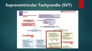
Approach to
tachy-arrhythmia
DR KTD PRIYADARSHANI
REGISTRAR IN EMERGENCY MEDICINE
PRIMARY CARE UNIT
TEACHING HOSPITAL- PERADENIYA
What is normal rhythm?
 HR 60-100 bpm
 Origin from SA node
 Propagate through normal conduction pathway
 With normal velocities
Causes of arrhythmias
 Abnormal rhythmicity of pacemaker
 Shift of the pacemaker from the sinus node to another place in the heart
 Blocks at different points in the impulse through the heart
 Abnormal pathways of impulse transmission through the heart
 Spontaneous generation of spurious impulses in almost any part of the
heart
Lets’ workout..
 7 step approach to ECG rhythm analysis
1. Rate
2. Pattern of QRS complexes
3. QRS morphology
4. P waves
5. Relationship between P waves and QRS complexes
6. Onset and termination
7. Response to vagal maneuvers
Resuscitation
 A, B- Start Oxygen
 C- IV access, hemodynamic stability
 D
 E
 If time allows- 12 lead ECG
 VBG- correct electrolyte imbalance
 2 main priorities
 Is the patient stable?
 What is nature of the arrhythmia?
Adverse features of arrhythmia
 Shock- SBP < 90mmHg, pallor, sweating, cold extremities, clammy,
confusion or impaired level of consciousness
 Syncope- transient loss of consciousness due to cerebral perfusion
 Myocardial ischemia- ischemic chest pain, evidence of ischemia on the ECG
 Heart failure- pulmonary edema and or raised JVP (+/- peripheral edema
and liver enlargement)
SINUS TACHYCARDIA
Sinus tachycardia
Sinus rhythm with resting heart rate (HR) > 100 bpm in adults, or above the normal range for age in
children
Normal heart rates
•Newborn: 110 – 150 bpm
•2 years: 85 – 125 bpm
•4 years: 75 – 115 bpm
•6 years+: 60 – 100 bpm
Management
 Correct Causes
 Hypoxia
 Hypovolemia
 Fever
 Thyrotoxicosis
 Anemia
 Pain
 Pharmacological
 Beta-agonists: adrenaline, isoprenaline, salbutamol, dobutamine
 Sympathomimetics: amphetamines, cocaine, methylphenidate
 Antimuscarinics: antihistamines, TCAs, carbamazepine, atropine
 Others: caffeine, theophylline, marijuana
Supraventricular Tachycardia (SVT)
SVT
• Regular tachycardia ~140-280 bpm
• Narrow QRS complexes (< 120ms)
• P waves if visible exhibit retrograde conduction
with P-wave inversion in leads II, III, aVF.
Management
 Unstable patient
 Unstable + adverse
features = synchronized
electrical cardio version.
 Sedate patient
 120-150J biphasic
 Repeat 3 times with
stepwise increase in joules
 Stable BP>100 mmHg
 Vagal maneuvers—Valsalva and/or carotid sinus massage.
 Adenosine = large vein + flush
 6 mg as a rapid IV bolus.
 12 mg and again and 18 mg if no response.
 transient sense of impending doom, chest discomfort and shortness of breth
 Consider verapamil 2.5–5 mg IV over 2 min (if adenosine is contraindicated)
 or verapamil (continuous slow infusion of 1 mg/min to a maximum of 20 mg or 5 mg IV slowly,
which may be repeated).
 Diltiazem infusion (2 mg/min to a maximum of 50 mg) is an alternativ
 Flecainide (2 mg/kg over 30–45 minutes) would be considered third-line therapy
 Catheter ablation may be considered in recurrent episodes not amenable to medical treatment.
Atrial flutter
Atrial flutter
• Narrow complex tachycardia
• Regular atrial activity at ~300 bpm
• “Saw-tooth” pattern of inverted flutter waves in leads II, III, aVF
• Upright flutter waves in V1 that may resemble P waves
• Loss of the isoelectric baseline
• Ventricular rate depends on AV conduction ratio
Handy Tips For Spotting Flutter
• Rapid Recognition
 Narrow complex tachycardia at 150 bpm
(range 130-170)? Yes -> Suspect flutter!
• Vagal Manoeuvres +/- Adenosine
 Atrial flutter will not usually cardiovert
with these techniques (unlike AVNRT)
• RR intervals
 In atrial flutter with variable block the R-
intervals will be multiples of the P-P
interval
Management
 administer an antiarrhythmic
drug;
 initiate DC cardioversion low
energy 50J; or
 initiate rapid atrial pacing to
terminate the atrial flutter
Atrial fibrillation
Atrial fibrillation
• Irregularly irregular rhythm
• No P waves
• Absence of an isoelectric baseline
• Variable ventricular rate
• QRS complexes usually < 120ms, unless pre-
existing bundle branch block, accessory
pathway, or rate-related aberrant conduction
• Fibrillatory waves may be present and can be
either fine (amplitude < 0.5mm) or coarse
(amplitude > 0.5mm)
• Fibrillatory waves may mimic P waves leading
to misdiagnosis
 Causes of Atrial Fibrillation
Cardiac causes Non-cardiac causes
Common:
• Ischaemic heart disease
• Rheumatic heart disease
• Hypertension
• Sick sinus syndrome
• Pre-excited syndromes (e.g.
WPW)
Less common:
• Cardiomyopathy
• Pericardial disease
• Atrial septal defect
• Atrial myxoma
• Acute infections, especially pneumonia
• Electrolyte abnormalities
• Lung carcinoma
• Other intrathoracic pathology (e.g. pleural
effusion)
• Pulmonary embolism
• Thyrotoxicosis
Post-surgery (especially cardiac)
• Excessive alcohol or caffeine
Management
 Diagnosis of atrial fibrillation
 Assessment of duration
 Assessment for anticoagulation
 Rate or rhythm control
 Treatment of underlying / associated diseases
 non-life-threatening hemodynamic instability, caused by AF, as:
 Ventricular rate >150 bpm
 Ongoing chest pain
 Critical perfusion
 AF not permanent—electrical cardioversion should be performed. Where there is a delay
in organizing electrical cardioversion, intravenous amiodarone should be used.
 AF permanent—pharmacological rate control strategy should be used. Treatment should
be with intravenous β-blockers or rate-limiting calcium antagonists. If these are
contraindicated, amiodarone should be used.
 AF in WPW—flecainide may be used as an alternative for attempting pharmacological
cardioversion. AV node-blocking agents (e.g. diltiazem, verapamil, or digoxin) should not
be used.
 In stable patients there are several treatment options:
• Rate control by drug therapy (e.g. beta-blocker or rate-limiting calcium
antagonist)
• Rhythm control by drug therapy (e.g. amiodarone or flecanide)
• Rhythm control by electrical cardioversion
• Anticoagulation to prevent thromboembolism (should be considered in all
patients)
 NICE recommend that all patients should initially be rate controlled unless:
 AF is thought to be reversible
 Presence of heart failure thought to be caused by AF
 New onset AF
 Atrial flutter thought to benefit from ablation
 Rhythm strategy thought to be more suitable based on clinical judgement
 Rate control is used in those with permanent AF and those with persistent AF, where
a rate control treatment strategy has been chosen.
 Rhythm control may be achieved via electrical or pharmacological cardioversion.
 It should be considered in patients who remain symptomatic despite rate control, or where
a rate control strategy has been unsuccessful.
 In patients without haemodynamic compromise, this can be performed electively.
 NICE recommends that a transthoracic echocardiogram is performed prior to elective
cardioversion.
 Factors which make a patient unsuitable for cardioversion:
 Contraindication to anticoagulation
 Structural heart disease that precludes long-term maintenance of sinus
rhythm
 Long duration of AF (>12 months)
 History of multiple failed attempts/relapses
 Ongoing reversible cause (e.g. thyrotoxicosis)
 THROMBOPROPHYLAXIS In AF
 CHA2DSVASC score 0,1-M, 2-F
VENTRICULAR TACHYCARDIA
 AV dissociation
 fusion beats or capture beats
 wide QRS complexes>140 ms
 rate>100 bpm: commonly 150–200 bpm
 rhythm regular
 constant QRS axis, often with marked left axis deviation or northwest
axis
 deep S wave with r/S ratio<1 in right bundle branch block (RBBB)
morphology VT
 Monomorphic
 most common
 associated with MI
 Polymorphic
 QRS at 200 beats/min or more which change amplitude and axis so they appear to twist around
the baseline
VT vs SVT
 Criteria for diagnosis of VT using the 4-step Brugada algorithm:
Management
 PULSELESS
 ACLS protocol
 Immediate unsynchronized defibrillation
 CPR with minimal interruption (30:2, with 2 minute cycles)
 Intubation
 O2
 IV access
 Adrenaline 1mg 3min
 Amiodarone 300mg (following 3rd shock)
 Exclude reversible causes (4 H’s and T’s)
 CLINICALLY COMPROMISED
 Haemodynamically unstable, chest pain, ischaemia, heart failure, VR > 150/min ->
synchronised shock (x 3)
 O2
 IV access
 Rapid exclusion of reversible factors (wire, PA catheter in RV, hypoK+ or Mg2+)
 Synchronised DC Shock (50J Bi, 100 Mono) (150 ->200J-> 360J)
 Amiodarone 5mg/kg over 20-60 min if patient stable -> infusion
 Consider: 2nd line drugs
 procainamide 50mg/min
 lignocaine 1mg/kg
 sotalol 1mg/kg
 Magnesium
 Inotrope = Dopamine infusion
 Overdrive pacing
 CLINICALLY STABLE- debate between cardioversion and pharmacological
treatment
 O2
 Amiodarone
 IV amiodarone 150 mg as a slow bolus over 10 minutes. This can be repeated a second time
if conversion has not been achieved
 IV procainamide 100 mg (where available) every 5 minutes to a maximum dose of 10–20
mg/kg body weight
 IV lignocaine 50–100 mg IV push at a rate not more than 50 mg/min. This can be repeated a
second time if conversion is not achieved.
 Sotolol 1 mg/kg is used as second-line agent.
 Cardioversion if medical therapy fails (quickly) – will need sedation
 Consider pacing if cardioversion no effective
 Evaluation and treatment of cause (usually IHD)
 If associated with long QT -> consider Mg2+
Premature ventricular complexes
• Broad QRS complex (≥ 120
ms) with abnormal
morphology
• Premature — i.e. occurs
earlier than would be
expected for the next sinus
impulse
• Discordant ST segment and
T wave changes.
• Usually followed by a full
compensatory pause
• Retrograde capture of the
atria may or may not occur
 Causes
 Frequent or symptomatic PVCs may be due to:
 Anxiety
 Sympathomimetics
 Beta-agonists
 Excess caffeine
 Hypokalaemia
 Hypomagnesaemia
 Digoxin toxicity
 Myocardial ischemia
Management
References
 Diagnosis and management in emergency medicine by Brown and
Cardagon 7th edition
 Text book of Adult emergency medicine 4th edition by peter cameron
 Life in the fast lane- ECG library
Questions?
Thank you!

Approach to tachyarrhythmia

  • 1.
    Approach to tachy-arrhythmia DR KTDPRIYADARSHANI REGISTRAR IN EMERGENCY MEDICINE PRIMARY CARE UNIT TEACHING HOSPITAL- PERADENIYA
  • 2.
    What is normalrhythm?  HR 60-100 bpm  Origin from SA node  Propagate through normal conduction pathway  With normal velocities
  • 3.
    Causes of arrhythmias Abnormal rhythmicity of pacemaker  Shift of the pacemaker from the sinus node to another place in the heart  Blocks at different points in the impulse through the heart  Abnormal pathways of impulse transmission through the heart  Spontaneous generation of spurious impulses in almost any part of the heart
  • 4.
    Lets’ workout..  7step approach to ECG rhythm analysis 1. Rate 2. Pattern of QRS complexes 3. QRS morphology 4. P waves 5. Relationship between P waves and QRS complexes 6. Onset and termination 7. Response to vagal maneuvers
  • 5.
    Resuscitation  A, B-Start Oxygen  C- IV access, hemodynamic stability  D  E  If time allows- 12 lead ECG  VBG- correct electrolyte imbalance  2 main priorities  Is the patient stable?  What is nature of the arrhythmia?
  • 6.
    Adverse features ofarrhythmia  Shock- SBP < 90mmHg, pallor, sweating, cold extremities, clammy, confusion or impaired level of consciousness  Syncope- transient loss of consciousness due to cerebral perfusion  Myocardial ischemia- ischemic chest pain, evidence of ischemia on the ECG  Heart failure- pulmonary edema and or raised JVP (+/- peripheral edema and liver enlargement)
  • 8.
  • 9.
    Sinus tachycardia Sinus rhythmwith resting heart rate (HR) > 100 bpm in adults, or above the normal range for age in children Normal heart rates •Newborn: 110 – 150 bpm •2 years: 85 – 125 bpm •4 years: 75 – 115 bpm •6 years+: 60 – 100 bpm
  • 10.
    Management  Correct Causes Hypoxia  Hypovolemia  Fever  Thyrotoxicosis  Anemia  Pain  Pharmacological  Beta-agonists: adrenaline, isoprenaline, salbutamol, dobutamine  Sympathomimetics: amphetamines, cocaine, methylphenidate  Antimuscarinics: antihistamines, TCAs, carbamazepine, atropine  Others: caffeine, theophylline, marijuana
  • 11.
  • 12.
    SVT • Regular tachycardia~140-280 bpm • Narrow QRS complexes (< 120ms) • P waves if visible exhibit retrograde conduction with P-wave inversion in leads II, III, aVF.
  • 13.
    Management  Unstable patient Unstable + adverse features = synchronized electrical cardio version.  Sedate patient  120-150J biphasic  Repeat 3 times with stepwise increase in joules
  • 14.
     Stable BP>100mmHg  Vagal maneuvers—Valsalva and/or carotid sinus massage.  Adenosine = large vein + flush  6 mg as a rapid IV bolus.  12 mg and again and 18 mg if no response.  transient sense of impending doom, chest discomfort and shortness of breth  Consider verapamil 2.5–5 mg IV over 2 min (if adenosine is contraindicated)  or verapamil (continuous slow infusion of 1 mg/min to a maximum of 20 mg or 5 mg IV slowly, which may be repeated).  Diltiazem infusion (2 mg/min to a maximum of 50 mg) is an alternativ  Flecainide (2 mg/kg over 30–45 minutes) would be considered third-line therapy  Catheter ablation may be considered in recurrent episodes not amenable to medical treatment.
  • 15.
  • 16.
    Atrial flutter • Narrowcomplex tachycardia • Regular atrial activity at ~300 bpm • “Saw-tooth” pattern of inverted flutter waves in leads II, III, aVF • Upright flutter waves in V1 that may resemble P waves • Loss of the isoelectric baseline • Ventricular rate depends on AV conduction ratio
  • 17.
    Handy Tips ForSpotting Flutter • Rapid Recognition  Narrow complex tachycardia at 150 bpm (range 130-170)? Yes -> Suspect flutter! • Vagal Manoeuvres +/- Adenosine  Atrial flutter will not usually cardiovert with these techniques (unlike AVNRT) • RR intervals  In atrial flutter with variable block the R- intervals will be multiples of the P-P interval
  • 18.
    Management  administer anantiarrhythmic drug;  initiate DC cardioversion low energy 50J; or  initiate rapid atrial pacing to terminate the atrial flutter
  • 19.
  • 20.
    Atrial fibrillation • Irregularlyirregular rhythm • No P waves • Absence of an isoelectric baseline • Variable ventricular rate • QRS complexes usually < 120ms, unless pre- existing bundle branch block, accessory pathway, or rate-related aberrant conduction • Fibrillatory waves may be present and can be either fine (amplitude < 0.5mm) or coarse (amplitude > 0.5mm) • Fibrillatory waves may mimic P waves leading to misdiagnosis
  • 21.
     Causes ofAtrial Fibrillation Cardiac causes Non-cardiac causes Common: • Ischaemic heart disease • Rheumatic heart disease • Hypertension • Sick sinus syndrome • Pre-excited syndromes (e.g. WPW) Less common: • Cardiomyopathy • Pericardial disease • Atrial septal defect • Atrial myxoma • Acute infections, especially pneumonia • Electrolyte abnormalities • Lung carcinoma • Other intrathoracic pathology (e.g. pleural effusion) • Pulmonary embolism • Thyrotoxicosis Post-surgery (especially cardiac) • Excessive alcohol or caffeine
  • 22.
    Management  Diagnosis ofatrial fibrillation  Assessment of duration  Assessment for anticoagulation  Rate or rhythm control  Treatment of underlying / associated diseases
  • 24.
     non-life-threatening hemodynamicinstability, caused by AF, as:  Ventricular rate >150 bpm  Ongoing chest pain  Critical perfusion  AF not permanent—electrical cardioversion should be performed. Where there is a delay in organizing electrical cardioversion, intravenous amiodarone should be used.  AF permanent—pharmacological rate control strategy should be used. Treatment should be with intravenous β-blockers or rate-limiting calcium antagonists. If these are contraindicated, amiodarone should be used.  AF in WPW—flecainide may be used as an alternative for attempting pharmacological cardioversion. AV node-blocking agents (e.g. diltiazem, verapamil, or digoxin) should not be used.
  • 25.
     In stablepatients there are several treatment options: • Rate control by drug therapy (e.g. beta-blocker or rate-limiting calcium antagonist) • Rhythm control by drug therapy (e.g. amiodarone or flecanide) • Rhythm control by electrical cardioversion • Anticoagulation to prevent thromboembolism (should be considered in all patients)  NICE recommend that all patients should initially be rate controlled unless:  AF is thought to be reversible  Presence of heart failure thought to be caused by AF  New onset AF  Atrial flutter thought to benefit from ablation  Rhythm strategy thought to be more suitable based on clinical judgement
  • 26.
     Rate controlis used in those with permanent AF and those with persistent AF, where a rate control treatment strategy has been chosen.  Rhythm control may be achieved via electrical or pharmacological cardioversion.  It should be considered in patients who remain symptomatic despite rate control, or where a rate control strategy has been unsuccessful.  In patients without haemodynamic compromise, this can be performed electively.  NICE recommends that a transthoracic echocardiogram is performed prior to elective cardioversion.
  • 27.
     Factors whichmake a patient unsuitable for cardioversion:  Contraindication to anticoagulation  Structural heart disease that precludes long-term maintenance of sinus rhythm  Long duration of AF (>12 months)  History of multiple failed attempts/relapses  Ongoing reversible cause (e.g. thyrotoxicosis)  THROMBOPROPHYLAXIS In AF  CHA2DSVASC score 0,1-M, 2-F
  • 28.
  • 29.
     AV dissociation fusion beats or capture beats  wide QRS complexes>140 ms  rate>100 bpm: commonly 150–200 bpm  rhythm regular  constant QRS axis, often with marked left axis deviation or northwest axis  deep S wave with r/S ratio<1 in right bundle branch block (RBBB) morphology VT  Monomorphic  most common  associated with MI  Polymorphic  QRS at 200 beats/min or more which change amplitude and axis so they appear to twist around the baseline
  • 30.
    VT vs SVT Criteria for diagnosis of VT using the 4-step Brugada algorithm:
  • 31.
    Management  PULSELESS  ACLSprotocol  Immediate unsynchronized defibrillation  CPR with minimal interruption (30:2, with 2 minute cycles)  Intubation  O2  IV access  Adrenaline 1mg 3min  Amiodarone 300mg (following 3rd shock)  Exclude reversible causes (4 H’s and T’s)
  • 32.
     CLINICALLY COMPROMISED Haemodynamically unstable, chest pain, ischaemia, heart failure, VR > 150/min -> synchronised shock (x 3)  O2  IV access  Rapid exclusion of reversible factors (wire, PA catheter in RV, hypoK+ or Mg2+)  Synchronised DC Shock (50J Bi, 100 Mono) (150 ->200J-> 360J)  Amiodarone 5mg/kg over 20-60 min if patient stable -> infusion  Consider: 2nd line drugs  procainamide 50mg/min  lignocaine 1mg/kg  sotalol 1mg/kg  Magnesium  Inotrope = Dopamine infusion  Overdrive pacing
  • 33.
     CLINICALLY STABLE-debate between cardioversion and pharmacological treatment  O2  Amiodarone  IV amiodarone 150 mg as a slow bolus over 10 minutes. This can be repeated a second time if conversion has not been achieved  IV procainamide 100 mg (where available) every 5 minutes to a maximum dose of 10–20 mg/kg body weight  IV lignocaine 50–100 mg IV push at a rate not more than 50 mg/min. This can be repeated a second time if conversion is not achieved.  Sotolol 1 mg/kg is used as second-line agent.  Cardioversion if medical therapy fails (quickly) – will need sedation  Consider pacing if cardioversion no effective  Evaluation and treatment of cause (usually IHD)  If associated with long QT -> consider Mg2+
  • 35.
    Premature ventricular complexes •Broad QRS complex (≥ 120 ms) with abnormal morphology • Premature — i.e. occurs earlier than would be expected for the next sinus impulse • Discordant ST segment and T wave changes. • Usually followed by a full compensatory pause • Retrograde capture of the atria may or may not occur
  • 36.
     Causes  Frequentor symptomatic PVCs may be due to:  Anxiety  Sympathomimetics  Beta-agonists  Excess caffeine  Hypokalaemia  Hypomagnesaemia  Digoxin toxicity  Myocardial ischemia
  • 37.
  • 38.
    References  Diagnosis andmanagement in emergency medicine by Brown and Cardagon 7th edition  Text book of Adult emergency medicine 4th edition by peter cameron  Life in the fast lane- ECG library
  • 39.
  • 40.