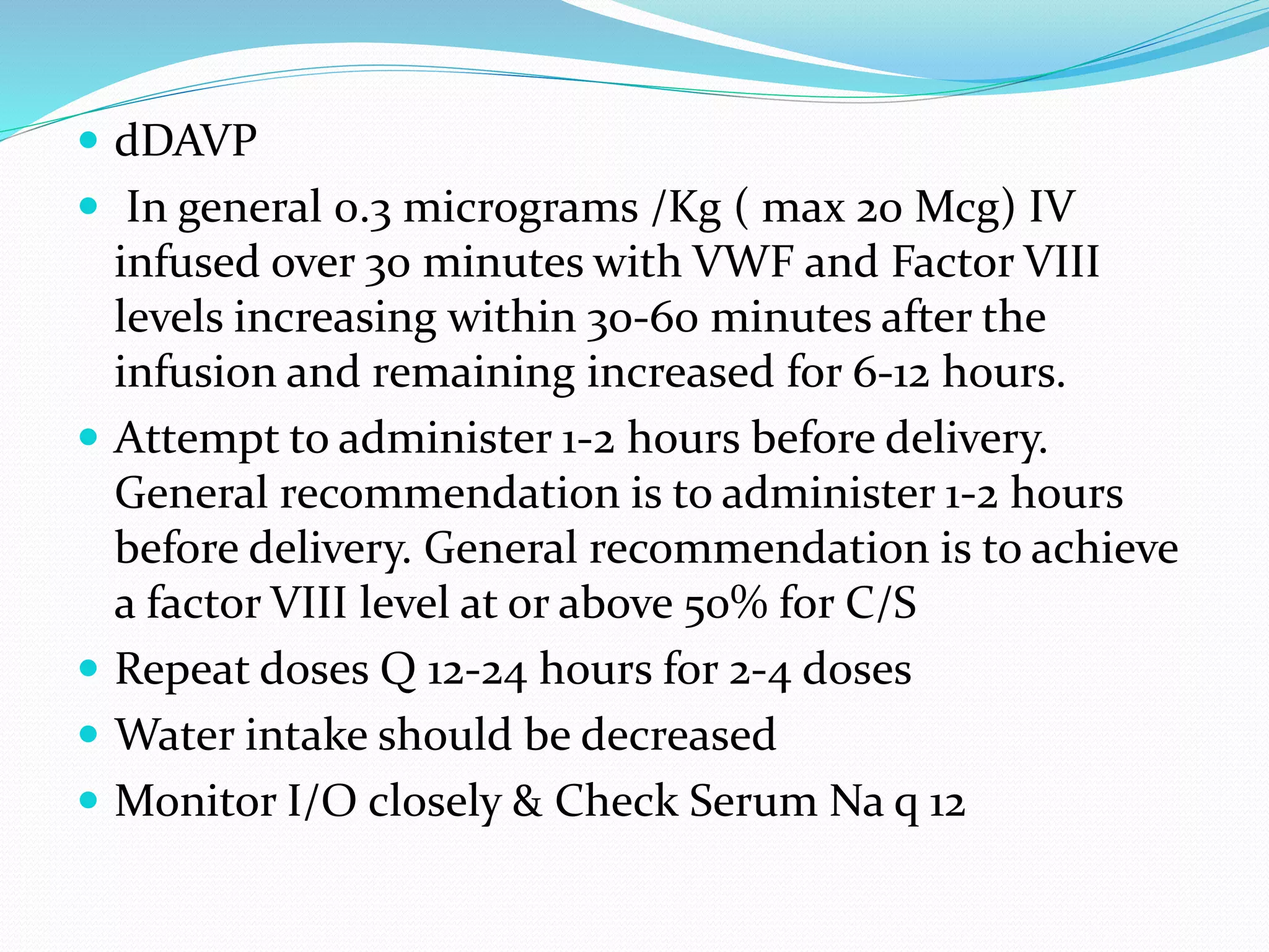
Chair Person:Dr.Praveen K
Student:Dr.Chetan R
The Platelet
 Platelets are released from the megakaryocyte
 Normal blood platelet count is 150,000–450,000/L.
 The major regulator of platelet production is the
hormone thrombopoietin (TPO), which is synthesized
in the liver.
 Synthesis is increased with inflammation and
specifically by interleukin 6.
 Thus a reduction in platelet and megakaryocyte mass
increases the level of TPO, which then stimulates
platelet production.
The Platelet
 Platelets circulate with an average life span of 7 to 10
days.
 Approximately one-third of the platelets reside in the
spleen, and this number increases in proportion to
splenic size, although the platelet count rarely
decreases to <40,000/L as the spleen enlarges.
 Platelets are physiologically very active, but are
anucleate, and thus have limited capacity to synthesize
new proteins
The Platelet
 Normal vascular endothelium contributes to prevent
thrombosis
 When vascular endothelium is injured, these inhibitory
effects are overcome, and platelets adhere to the exposed
intimal surface primarily through VWF, a protein present
in:
-Plasma
-The extracellular matrix of the subendothelial vessel wall.
 Platelet adhesion results in the generation of intracellular
signals that lead to activation of the platelet glycoprotein
(Gp) IIb/IIIa receptor and resultant platelet aggregation.
The Vessel Wall
 The endothelium normally presents an antithrombotic
surface but rapidly becomes prothrombotic when
stimulated, which promotes:
-Coagulation
-Inhibits fibrinolysis
-Activates platelets.
 In many cases, endothelium-derived vasodilators are
also platelet inhibitors (e.g., nitric oxide) and,
conversely, endothelium-derived vasoconstrictors
(e.g., endothelin) can also be platelet activators.
Hemostasis & Thrombosis
 Normal hemostasis comprises a series of regulated
processes that maintain blood in a fluid, clot-free state
in normal vessels while rapidly forming a localized
hemostatic plug at the site of vascular injury.
 The pathologic counterpart of hemostasis is
thrombosis, the formation of blood clot (thrombus)
within intact vessels. Both hemostasis and thrombosis
involve three elements: the vascular wall, platelets, and
the coagulation cascade
The Platelet
 Activated platelets undergo release of their granule
contents:
-Nucleotides,
-Adhesive proteins,
-Growth factors,
-Procoagulants
Thrombocytopenia
 Thrombocytopenia results from one or more of three
processes:
(1) decreased bone marrow production: ○ Inherited
○ Acquired
(2) sequestration, usually in an enlarged spleen
(3) increased platelet destruction.
Thrombocytopenia…
 A key step is to rule out "pseudothrombocytopenia”
(1) Is an in vitro artifact resulting from platelet
agglutination via antibodies (usually IgG, but also IgM
and IgA) when the calcium content is decreased by
blood collection in ethylenediamine tetraacetic (EDTA)
[the anticoagulant present in tubes (purple top)
(2) A blood smear should be evaluated and a platelet
count determined in blood collected into sodium citrate
(blue top tube) or heparin (green top tube)
A.Normal Peripheral Blood
B.Platelet Clumping in Pseudothrombocytopenia
Infection-Induced
Thrombocytopenia
 Many viral and bacterial infections result in
thrombocytopenia and are the most common
noniatrogenic cause of thrombocytopenia.
 This may or may not be associated with disseminated
intravascular coagulation (DIC), which is most
commonly seen in patients with systemic infections.
 Infections can affect both platelet production and
platelet survival.
Drug-Induced Thrombocytopenia
 Many drugs have been associated with
thrombocytopenia
 Classic drug-dependent antibodies are antibodies that
react with specific platelet surface antigens, and result
in thrombocytopenia only when the drug is present
(more common with quinine and sulfonamides).
Drug-Induced Thrombocytopenia
 Drug-dependent antibody binding can be
demonstrated by laboratory assays, showing antibody
binding in the presence of, but not without, the drug
present in the assay.
 The thrombocytopenia typically occurs after a period
of initial exposure (median length 21 days), or upon
reexposure, and usually resolves in 7–10 days after drug
withdrawal.
Heparin-Induced
Thrombocytopenia
 Drug-induced thrombocytopenia due to heparin differs from
that seen with other drugs in two major ways.
(1) The thrombocytopenia is not usually severe, with nadir
counts rarely <20,000/L.
(2) Heparin-induced thrombocytopenia (HIT) is not associated
with bleeding and, in fact, markedly increases the risk of
thrombosis.
• HIT results from antibody formation to a complex of the
platelet-specific protein platelet factor 4 (PF4) and heparin. The
antiheparin/PF4 antibody can activate platelets.
○ A fraction of those who develop antibodies will develop HIT,
and a portion of those (up to 50%) will develop thrombosis
(HITT).
Heparin-Induced
Thrombocytopenia
 HIT results from antibody formation to a complex of
the platelet-specific protein platelet factor 4 (PF4) and
heparin. The antiheparin/PF4 antibody can activate
platelets.
○ A fraction of those who develop antibodies will
develop HIT, and a portion of those (up to 50%) will
develop thrombosis (HITT).
Heparin-Induced
Thrombocytopenia
 HIT can occur after exposure to low-molecular-weight
heparin (LMWH) as well as unfractionated heparin (UFH),
although it is about 10 times more common with the latter.
 Most patients develop HIT after exposure to heparin for 5–
14 days.
 It occurs before 5 days in those who were exposed to
heparin in the prior few weeks or months (<~100 days) and
have circulating antiheparin/PF4 antibodies. Rarely,
thrombocytopenia and thrombosis begin several days after
all heparin has been stopped (termed delayed- onset HIT).
Heparin-Induced
Thrombocytopenia
 HIT (anti-heparin/PF4) antibodies can be detected
using two types of assays.
(1) (ELISA) with PF4/polyanion complex as the
antigen.
(2) Platelet activation assay
Heparin-Induced
Thrombocytopenia
Treatment:
• Prompt discontinuation of heparin and use of
alternative anticoagulants (at least 1 month).
○ The direct thrombin inhibitors (DTIs): argatroban,
lepirudin, bivalirudin
○ Antithrombin drugs: fondaparinux
○ HIT antibodies cross-react with LMWH, and these
preparations should not be used in the treatment of HIT
Immune Thrombocytopenic
Purpura (ITP)
 Is an acquired disorder in which there is immune-
mediated destruction of platelets
 In children, it is usually an acute disease, most
commonly following an infection, and with a self-
limited course.
 In adults, it usually runs a more chronic course.
 ITP is termed secondary if it is associated with an
underlying disorder; autoimmune disorders,
particularly systemic lupus erythematosus (SLE), and
infections, such as HIV and hepatitis C, are common
causes.
Immune Thrombocytopenic
Purpura (ITP)
 ITP is characterized by:
- Mucocutaneous bleeding
-A low, often very low, platelet count
-Normal peripheral blood cells and smear.
-Ecchymoses and petechiae
Immune Thrombocytopenic
Purpura (ITP)
 Antibodies (serologic testing) is usually not helpful
due to the low sensitivity and specificity of the current
tests.
 Evaluate for secondary causes of ITP:
-HIV, hepatitis C
-SLE
-Autoinmmune diseases
Immune Thrombocytopenic
Purpura (ITP): Treatment
 Prednisone at 1 mg/kg
 Rh0(D) immune globulin therapy (WinRho SDF), at 50–75
g/kg (only in Rh-positive patients!)
 Intravenous gamma globulin (IVIgG), is dosed at 2 g/kg
total, given in divided doses over 2–5 days.
 Rituximab, an anti-CD20 (B cell) antibody, in refractory
ITP.
 Splenectomy has been used for treatment of patients who
relapse after glucocorticoids are tapered
 Thrombopoietin receptor agonists are now available:
○ Romiplostim and eltrombopag
Inherited Thrombocytopenia
 Thrombocytopenia is rarely inherited
-May be inherited in an autosomal dominant,
autosomal recessive, or X-linked pattern.
-Autosomal dominant thrombocytopenia:
○ May-Hegglin anomaly, and Sebastian, Epstein's,
and Fechtner syndromes,
○ A common feature of these disorders is large
platelets (Fig. 115-1C).
C. Abnormal Large Platelet in Autosomal Dominant
Macrothrombocytopenia
Inherited Thrombocytopenia
 Autosomal recessive disorders:
-Congenital amegakaryocytic thrombocytopenia
-Thrombocytopenia with absent radii
-Bernard Soulier syndrome: due to absence of GPIb-
IX-V, the VWF adhesion receptor.
 X-linked disorders
-Include Wiskott-Aldrich syndrome and a
dyshematopoietic syndrome
Thrombotic Thrombocytopenic Purpura
and Hemolytic Uremic Syndrome
 Thrombotic thrombocytopenic microangiopathies:
-Are a group of disorders characterized by
thrombocytopenia, a microangiopathic hemolytic
anemia evident by fragmented RBCs and laboratory
evidence of hemolysis, and microvascular thrombosis.
-They include thrombotic thrombocytopenic purpura
(TTP) and hemolytic uremic syndrome (HUS)
D.Schistocytes and decreased platelets in Microangiopathic
Hemolytic Anemia
Thrombotic Thrombocytopenic
Purpura (TTP)
 TTP has a pentad of findings:
- Microangiopathic hemolytic anemia
-Thrombocytopenia
-Renal failure
-Neurologic findings,
-Fever.
Thrombotic Thrombocytopenic
Purpura (TTP)
 Antibodies to the metalloprotease ADAMTS13, that
cleaves VWF.
-The persistence of ultra-large VWF molecules
contribute to pathogenic platelet adhesion and
aggregation
 The level of ADAMTS13 activity, as well as antibodies,
can now be detected by laboratory assays.
Thrombotic Thrombocytopenic
Purpura (TTP)
 Antibodies to the metalloprotease ADAMTS13, that
cleaves VWF.
- The persistence of ultra-large VWF molecules
contribute to pathogenic platelet adhesion and
aggregation
 The level of ADAMTS13 activity, as well as antibodies,
can now be detected by laboratory assays
Thrombotic Thrombocytopenic
Purpura (TTP)
 Inherited TTP: Upshaw-Schulman syndrome
 Secondary TTP:
-HIV infection
-Pregnant women.
-Medication-related: ticlopidine, clopidogrel,
cyclosporine, mitomycin C, tacrolimus, quinine)
Thrombotic Thrombocytopenic
Purpura (TTP): Treatment
 TTP is a devastating disease if not diagnosed and
treated promptly.
 Plasma exchange remains the mainstay of treatment of
TTP.
 Glucocorticoids as an adjunct therapy
 Rituximab: under investigation
Hemolytic Uremic Syndrome
 HUS is a syndrome characterized by:
-Acute renal failure
-Microangiopathic hemolytic anemia
-Thrombocytopenia.
 It is seen predominantly in children and in most cases
is preceded by an episode of diarrhea, often
hemorrhagic in nature. Escherichia coli O157:H7 is the
most frequent, although not only, etiologic serotype
Hemolytic Uremic Syndrome
Treatment of HUS is primarily supportive.
 Dialysis
 Plasma exchange has not been shown to alter the
overall course.
Thromobcytosis
 Thrombocytosis is almost always due either to
(1) Iron deficiency;
(2) Inflammation, cancer, or infection (reactive
thrombocytosis);
(3) An underlying myeloproliferative process
[essential thrombocythemia or polycythemia vera)
(4) >1.5 million = myeloproliferative disorder
Qualitative Disorders of Platelet
Function
 Inherited:
• Relatively rare,
• Autosomal recessive disorders:
○ Glanzmann's thrombasthenia (absence of the platelet
GpIIbIIIa receptor)
○ Bernard Soulier syndrome (absence of the platelet GpIb-
IX-V receptor).
• Autosomal dominant disorders:
○ Platelet storage pool disorder (SPD): abnormalities of
platelet granule formation.
• Bleeding symptoms are usually mild in nature
Qualitative Disorders of Platelet
Function
 Treatment:
-Platelet transfusion if severe
-Milder bleeding symptoms frequently respond to
desmopressin (increases plasma VWF and FVIII levels; it
may also have a direct effect on platelet function)
Qualitative Disorders of Platelet
Function
 Acquired:
-Medications (antiplatelet therapy, penicillins)
-Uremia.
-Cardiopulmonary bypass due to the effect of the
artificial circuit on platelets
von Willebrand Disease
 VWD is the most common inherited bleeding disorder.
-Prevalence of 1%,
-VWF serves two roles:
○ (1) as the major adhesion molecule to subendothelium
○ (2) as the binding protein for FVIII, resulting in significant
prolongation of the FVIII half-life in circulation.
○ The platelet-adhesive function of VWF is critically dependent on
the presence of large VWF multimers, while FVIII binding is not.
○ Most of the symptoms of VWD are "platelet-like" except in more
severe VWD when the FVIII is low enough to produce symptoms
similar to those found in Factor VIII deficiency (hemophilia A).
von Willebrand Disease
 VWD has been classified into three major types, with
four subtypes of type 2
 The most common type of VWD is type 1 disease (80%
of cases).
-Patients have predominantly mucosal bleeding
symptoms
-Menorrhagia is a common manifestation of VWD.
Menstrual bleeding resulting in anemia should warrant
an evaluation for VWD and, if negative, functional
platelet disorders
Inherited VWD is classified into Three types:
 Type I is the most common form accounting for ~ 70%
of all patients with VWD. Caused by a variety of
mutations which all result in a quantitative deficiency
of VWF. AD inheritance
 Type II has 4 subtypes which in total account for ~ 25%
of all patients with VWD. Caused by a variety of
different mutations which in general adversely affect
the function of VWF not the amount. Type II is sub
classified into 4 subtypes of which the majority
manifest AD inheritance
Type II vWD has 4 subtypes
 Type IIA ~ 15 % of all VWD thereby making it the
second most common presentation for VWD. AD
Mutations result in a decrease in only large and
intermediate size VWF multimers causing decreased
function of VWF
 Type IIB ~ 5% of all VWD. AD inherited mutations
resulting in an overactive platelet binding site (GP1b)
that may result in thrombocytopenia mediated via
increased clearance of platelet aggregates
 Type IIM~ Rare AD mutation that results in reduced
binding to platelets
 Type IIN~ Rare AR mutation causing decreased
binding to Factor VIII resulting in Low factor VIII
 Type III is extremely rare (~1/1,000,000). AR
inheritance that results in extremely low VWF levels.
This is the most severe form of VWD due to very low
VWF levels resulting in decreased platelet aggregation
AND low Factor VIII levels
Clinical Presentation
 Bleeding Symptoms occur when an absolute decrease
in amount or function of VWF occurs. These
abnormalities result in decreased platelet plug
formation during the primary haemostatic response.
 Therefore many of the patients present with
Symptoms similar to those seen with platelet disorders
 i.e, Easy bruising, Skin bleeding, and prolonged
bleeding form the Gums/GI tract/Uterus
 The exception to this presentation is seen with Type
IIN and Type III (most severe form) VWD patients
who have low Factor VIII levels and present with soft
tissue, joint , and GU bleeding which are classic for
hemophilia.
 These Symptoms and the low factor VIII levels may
result in a misdiagnosis of Hemophilia A
Clinical Presentation for Type III and Type IIN
 Symptoms are generally severe and present at an early
age with bleeding @ circumcision, when deciduous
teeth erupt, or when learning to walk and crawl.
 Soft tissue , joint, and GU bleeding are the rule in
addition to easy bruising, skin bleeding, and GI
bleeding.
Clinical presentation
 Most patients with Type I or Type II have mild to
moderate bleeding abnormalities.
 Classic history includes frequent nose bleeds as a
child, lifelong easy bruising , and bleeding with
invasive dental procedures or tooth extractions
 Exacerbation of bleeding with ASA or NSAID use
 Many females may be asymptomatic until their first
menses
Lab tests for vWD
 Plasma VWF antigen level (VWF:Ag)
 Plasma VWF activity (ristocetin Cofactor activity)
 Factor VIII Activity
 Platelet function analyzer assay
 VWF Multimer Gel Electrophoresis
 Ristocetin induced platelet aggregation
 Bleeding time
Treatment Options
 Desmopressin (dDAVP)
 VWF replacement (Humate P or as a last resort
Cryoprecipitate)
 Antifibrinolytic therapy ( Epsilon aminocaproic acid ie
EACA or Tranexamic acid )
 Topical Agents
 Recombinant Factor VIIA (emergent use)
 Adjuvant Platelet transfusion
Trial of dDAVP
 Trial of dDAVP is recommended for all patients with
type I and Most type II patients (caution with type IIB
as thrombocytopenia MAY worsen and aggravate
bleeding)
 Effective Response for most patients is validated with
an increase in VWF activity to at least 30IU/dL and
optimally to 50IU/ dL
 Once adequate response is documented dDAVP can be
utilized for surgery or vaginal delivery
dDAVP
 Synthetic analogue of antidiuretic hormone that
increases VWF and Factor VIII levels.
 Side Effects include : Vasodilatation resulting in facial
flushing, headache, and sometimes hypotension and
nausea.
 Tachyphylaxis occurs after repeated doses
 WATER RETENTION WITH HYPONATREMIA AND
SEIZURES exacerbated by NSAIDS/ASA
 dDAVP
 In general 0.3 micrograms /Kg ( max 20 Mcg) IV
infused over 30 minutes with VWF and Factor VIII
levels increasing within 30-60 minutes after the
infusion and remaining increased for 6-12 hours.
 Attempt to administer 1-2 hours before delivery.
General recommendation is to administer 1-2 hours
before delivery. General recommendation is to achieve
a factor VIII level at or above 50% for C/S
 Repeat doses Q 12-24 hours for 2-4 doses
 Water intake should be decreased
 Monitor I/O closely & Check Serum Na q 12
Nasal Spray dDAVP
 Hemophilia A and mild-to-moderate von Willebrand
disease (type 1):
Intranasal (using high concentration spray [1.5
mg/mL]): <50 kg 150 mcg (1 spray);
>50 kg: 300 mcg (1 spray each nostril);
Repeat use is determined by the patients clinical
condition and laboratory profile.
If using preoperatively, administer 2 hours before
surgery.
VWF replacement therapy
 Is reserved for dDAVP REFRACTORY PATIENTS
 Recommended as primary therapy for patients with
Type III VWD
For Unresponsive Refractory Bleeding
 Recombinant Factor VIIa which may “bypass” the need
for factor VIII and also binds to activated platelets
 Consider platelet transfusion
References
 HARRISON’s Princples of Internal Medicine 19th Edn.
 Robbin’s Basic Pathology, 9th edition
 Monogram: Von Willebrand Disease Douglas
Montgomery M.D
THANK YOU

Approach to patient with platelet disorders

  • 1.
  • 2.
    The Platelet  Plateletsare released from the megakaryocyte  Normal blood platelet count is 150,000–450,000/L.  The major regulator of platelet production is the hormone thrombopoietin (TPO), which is synthesized in the liver.  Synthesis is increased with inflammation and specifically by interleukin 6.  Thus a reduction in platelet and megakaryocyte mass increases the level of TPO, which then stimulates platelet production.
  • 3.
    The Platelet  Plateletscirculate with an average life span of 7 to 10 days.  Approximately one-third of the platelets reside in the spleen, and this number increases in proportion to splenic size, although the platelet count rarely decreases to <40,000/L as the spleen enlarges.  Platelets are physiologically very active, but are anucleate, and thus have limited capacity to synthesize new proteins
  • 4.
    The Platelet  Normalvascular endothelium contributes to prevent thrombosis  When vascular endothelium is injured, these inhibitory effects are overcome, and platelets adhere to the exposed intimal surface primarily through VWF, a protein present in: -Plasma -The extracellular matrix of the subendothelial vessel wall.  Platelet adhesion results in the generation of intracellular signals that lead to activation of the platelet glycoprotein (Gp) IIb/IIIa receptor and resultant platelet aggregation.
  • 5.
    The Vessel Wall The endothelium normally presents an antithrombotic surface but rapidly becomes prothrombotic when stimulated, which promotes: -Coagulation -Inhibits fibrinolysis -Activates platelets.  In many cases, endothelium-derived vasodilators are also platelet inhibitors (e.g., nitric oxide) and, conversely, endothelium-derived vasoconstrictors (e.g., endothelin) can also be platelet activators.
  • 7.
    Hemostasis & Thrombosis Normal hemostasis comprises a series of regulated processes that maintain blood in a fluid, clot-free state in normal vessels while rapidly forming a localized hemostatic plug at the site of vascular injury.  The pathologic counterpart of hemostasis is thrombosis, the formation of blood clot (thrombus) within intact vessels. Both hemostasis and thrombosis involve three elements: the vascular wall, platelets, and the coagulation cascade
  • 10.
    The Platelet  Activatedplatelets undergo release of their granule contents: -Nucleotides, -Adhesive proteins, -Growth factors, -Procoagulants
  • 12.
    Thrombocytopenia  Thrombocytopenia resultsfrom one or more of three processes: (1) decreased bone marrow production: ○ Inherited ○ Acquired (2) sequestration, usually in an enlarged spleen (3) increased platelet destruction.
  • 13.
    Thrombocytopenia…  A keystep is to rule out "pseudothrombocytopenia” (1) Is an in vitro artifact resulting from platelet agglutination via antibodies (usually IgG, but also IgM and IgA) when the calcium content is decreased by blood collection in ethylenediamine tetraacetic (EDTA) [the anticoagulant present in tubes (purple top) (2) A blood smear should be evaluated and a platelet count determined in blood collected into sodium citrate (blue top tube) or heparin (green top tube)
  • 14.
  • 15.
    B.Platelet Clumping inPseudothrombocytopenia
  • 17.
    Infection-Induced Thrombocytopenia  Many viraland bacterial infections result in thrombocytopenia and are the most common noniatrogenic cause of thrombocytopenia.  This may or may not be associated with disseminated intravascular coagulation (DIC), which is most commonly seen in patients with systemic infections.  Infections can affect both platelet production and platelet survival.
  • 18.
    Drug-Induced Thrombocytopenia  Manydrugs have been associated with thrombocytopenia  Classic drug-dependent antibodies are antibodies that react with specific platelet surface antigens, and result in thrombocytopenia only when the drug is present (more common with quinine and sulfonamides).
  • 19.
    Drug-Induced Thrombocytopenia  Drug-dependentantibody binding can be demonstrated by laboratory assays, showing antibody binding in the presence of, but not without, the drug present in the assay.  The thrombocytopenia typically occurs after a period of initial exposure (median length 21 days), or upon reexposure, and usually resolves in 7–10 days after drug withdrawal.
  • 21.
    Heparin-Induced Thrombocytopenia  Drug-induced thrombocytopeniadue to heparin differs from that seen with other drugs in two major ways. (1) The thrombocytopenia is not usually severe, with nadir counts rarely <20,000/L. (2) Heparin-induced thrombocytopenia (HIT) is not associated with bleeding and, in fact, markedly increases the risk of thrombosis. • HIT results from antibody formation to a complex of the platelet-specific protein platelet factor 4 (PF4) and heparin. The antiheparin/PF4 antibody can activate platelets. ○ A fraction of those who develop antibodies will develop HIT, and a portion of those (up to 50%) will develop thrombosis (HITT).
  • 22.
    Heparin-Induced Thrombocytopenia  HIT resultsfrom antibody formation to a complex of the platelet-specific protein platelet factor 4 (PF4) and heparin. The antiheparin/PF4 antibody can activate platelets. ○ A fraction of those who develop antibodies will develop HIT, and a portion of those (up to 50%) will develop thrombosis (HITT).
  • 23.
    Heparin-Induced Thrombocytopenia  HIT canoccur after exposure to low-molecular-weight heparin (LMWH) as well as unfractionated heparin (UFH), although it is about 10 times more common with the latter.  Most patients develop HIT after exposure to heparin for 5– 14 days.  It occurs before 5 days in those who were exposed to heparin in the prior few weeks or months (<~100 days) and have circulating antiheparin/PF4 antibodies. Rarely, thrombocytopenia and thrombosis begin several days after all heparin has been stopped (termed delayed- onset HIT).
  • 25.
    Heparin-Induced Thrombocytopenia  HIT (anti-heparin/PF4)antibodies can be detected using two types of assays. (1) (ELISA) with PF4/polyanion complex as the antigen. (2) Platelet activation assay
  • 26.
    Heparin-Induced Thrombocytopenia Treatment: • Prompt discontinuationof heparin and use of alternative anticoagulants (at least 1 month). ○ The direct thrombin inhibitors (DTIs): argatroban, lepirudin, bivalirudin ○ Antithrombin drugs: fondaparinux ○ HIT antibodies cross-react with LMWH, and these preparations should not be used in the treatment of HIT
  • 27.
    Immune Thrombocytopenic Purpura (ITP) Is an acquired disorder in which there is immune- mediated destruction of platelets  In children, it is usually an acute disease, most commonly following an infection, and with a self- limited course.  In adults, it usually runs a more chronic course.  ITP is termed secondary if it is associated with an underlying disorder; autoimmune disorders, particularly systemic lupus erythematosus (SLE), and infections, such as HIV and hepatitis C, are common causes.
  • 28.
    Immune Thrombocytopenic Purpura (ITP) ITP is characterized by: - Mucocutaneous bleeding -A low, often very low, platelet count -Normal peripheral blood cells and smear. -Ecchymoses and petechiae
  • 29.
    Immune Thrombocytopenic Purpura (ITP) Antibodies (serologic testing) is usually not helpful due to the low sensitivity and specificity of the current tests.  Evaluate for secondary causes of ITP: -HIV, hepatitis C -SLE -Autoinmmune diseases
  • 30.
    Immune Thrombocytopenic Purpura (ITP):Treatment  Prednisone at 1 mg/kg  Rh0(D) immune globulin therapy (WinRho SDF), at 50–75 g/kg (only in Rh-positive patients!)  Intravenous gamma globulin (IVIgG), is dosed at 2 g/kg total, given in divided doses over 2–5 days.  Rituximab, an anti-CD20 (B cell) antibody, in refractory ITP.  Splenectomy has been used for treatment of patients who relapse after glucocorticoids are tapered  Thrombopoietin receptor agonists are now available: ○ Romiplostim and eltrombopag
  • 31.
    Inherited Thrombocytopenia  Thrombocytopeniais rarely inherited -May be inherited in an autosomal dominant, autosomal recessive, or X-linked pattern. -Autosomal dominant thrombocytopenia: ○ May-Hegglin anomaly, and Sebastian, Epstein's, and Fechtner syndromes, ○ A common feature of these disorders is large platelets (Fig. 115-1C).
  • 32.
    C. Abnormal LargePlatelet in Autosomal Dominant Macrothrombocytopenia
  • 33.
    Inherited Thrombocytopenia  Autosomalrecessive disorders: -Congenital amegakaryocytic thrombocytopenia -Thrombocytopenia with absent radii -Bernard Soulier syndrome: due to absence of GPIb- IX-V, the VWF adhesion receptor.  X-linked disorders -Include Wiskott-Aldrich syndrome and a dyshematopoietic syndrome
  • 34.
    Thrombotic Thrombocytopenic Purpura andHemolytic Uremic Syndrome  Thrombotic thrombocytopenic microangiopathies: -Are a group of disorders characterized by thrombocytopenia, a microangiopathic hemolytic anemia evident by fragmented RBCs and laboratory evidence of hemolysis, and microvascular thrombosis. -They include thrombotic thrombocytopenic purpura (TTP) and hemolytic uremic syndrome (HUS)
  • 35.
    D.Schistocytes and decreasedplatelets in Microangiopathic Hemolytic Anemia
  • 36.
    Thrombotic Thrombocytopenic Purpura (TTP) TTP has a pentad of findings: - Microangiopathic hemolytic anemia -Thrombocytopenia -Renal failure -Neurologic findings, -Fever.
  • 37.
    Thrombotic Thrombocytopenic Purpura (TTP) Antibodies to the metalloprotease ADAMTS13, that cleaves VWF. -The persistence of ultra-large VWF molecules contribute to pathogenic platelet adhesion and aggregation  The level of ADAMTS13 activity, as well as antibodies, can now be detected by laboratory assays.
  • 39.
    Thrombotic Thrombocytopenic Purpura (TTP) Antibodies to the metalloprotease ADAMTS13, that cleaves VWF. - The persistence of ultra-large VWF molecules contribute to pathogenic platelet adhesion and aggregation  The level of ADAMTS13 activity, as well as antibodies, can now be detected by laboratory assays
  • 40.
    Thrombotic Thrombocytopenic Purpura (TTP) Inherited TTP: Upshaw-Schulman syndrome  Secondary TTP: -HIV infection -Pregnant women. -Medication-related: ticlopidine, clopidogrel, cyclosporine, mitomycin C, tacrolimus, quinine)
  • 41.
    Thrombotic Thrombocytopenic Purpura (TTP):Treatment  TTP is a devastating disease if not diagnosed and treated promptly.  Plasma exchange remains the mainstay of treatment of TTP.  Glucocorticoids as an adjunct therapy  Rituximab: under investigation
  • 42.
    Hemolytic Uremic Syndrome HUS is a syndrome characterized by: -Acute renal failure -Microangiopathic hemolytic anemia -Thrombocytopenia.  It is seen predominantly in children and in most cases is preceded by an episode of diarrhea, often hemorrhagic in nature. Escherichia coli O157:H7 is the most frequent, although not only, etiologic serotype
  • 43.
    Hemolytic Uremic Syndrome Treatmentof HUS is primarily supportive.  Dialysis  Plasma exchange has not been shown to alter the overall course.
  • 44.
    Thromobcytosis  Thrombocytosis isalmost always due either to (1) Iron deficiency; (2) Inflammation, cancer, or infection (reactive thrombocytosis); (3) An underlying myeloproliferative process [essential thrombocythemia or polycythemia vera) (4) >1.5 million = myeloproliferative disorder
  • 45.
    Qualitative Disorders ofPlatelet Function  Inherited: • Relatively rare, • Autosomal recessive disorders: ○ Glanzmann's thrombasthenia (absence of the platelet GpIIbIIIa receptor) ○ Bernard Soulier syndrome (absence of the platelet GpIb- IX-V receptor). • Autosomal dominant disorders: ○ Platelet storage pool disorder (SPD): abnormalities of platelet granule formation. • Bleeding symptoms are usually mild in nature
  • 46.
    Qualitative Disorders ofPlatelet Function  Treatment: -Platelet transfusion if severe -Milder bleeding symptoms frequently respond to desmopressin (increases plasma VWF and FVIII levels; it may also have a direct effect on platelet function)
  • 47.
    Qualitative Disorders ofPlatelet Function  Acquired: -Medications (antiplatelet therapy, penicillins) -Uremia. -Cardiopulmonary bypass due to the effect of the artificial circuit on platelets
  • 48.
    von Willebrand Disease VWD is the most common inherited bleeding disorder. -Prevalence of 1%, -VWF serves two roles: ○ (1) as the major adhesion molecule to subendothelium ○ (2) as the binding protein for FVIII, resulting in significant prolongation of the FVIII half-life in circulation. ○ The platelet-adhesive function of VWF is critically dependent on the presence of large VWF multimers, while FVIII binding is not. ○ Most of the symptoms of VWD are "platelet-like" except in more severe VWD when the FVIII is low enough to produce symptoms similar to those found in Factor VIII deficiency (hemophilia A).
  • 49.
    von Willebrand Disease VWD has been classified into three major types, with four subtypes of type 2  The most common type of VWD is type 1 disease (80% of cases). -Patients have predominantly mucosal bleeding symptoms -Menorrhagia is a common manifestation of VWD. Menstrual bleeding resulting in anemia should warrant an evaluation for VWD and, if negative, functional platelet disorders
  • 50.
    Inherited VWD isclassified into Three types:  Type I is the most common form accounting for ~ 70% of all patients with VWD. Caused by a variety of mutations which all result in a quantitative deficiency of VWF. AD inheritance  Type II has 4 subtypes which in total account for ~ 25% of all patients with VWD. Caused by a variety of different mutations which in general adversely affect the function of VWF not the amount. Type II is sub classified into 4 subtypes of which the majority manifest AD inheritance
  • 51.
    Type II vWDhas 4 subtypes  Type IIA ~ 15 % of all VWD thereby making it the second most common presentation for VWD. AD Mutations result in a decrease in only large and intermediate size VWF multimers causing decreased function of VWF  Type IIB ~ 5% of all VWD. AD inherited mutations resulting in an overactive platelet binding site (GP1b) that may result in thrombocytopenia mediated via increased clearance of platelet aggregates
  • 52.
     Type IIM~Rare AD mutation that results in reduced binding to platelets  Type IIN~ Rare AR mutation causing decreased binding to Factor VIII resulting in Low factor VIII
  • 53.
     Type IIIis extremely rare (~1/1,000,000). AR inheritance that results in extremely low VWF levels. This is the most severe form of VWD due to very low VWF levels resulting in decreased platelet aggregation AND low Factor VIII levels
  • 54.
    Clinical Presentation  BleedingSymptoms occur when an absolute decrease in amount or function of VWF occurs. These abnormalities result in decreased platelet plug formation during the primary haemostatic response.  Therefore many of the patients present with Symptoms similar to those seen with platelet disorders  i.e, Easy bruising, Skin bleeding, and prolonged bleeding form the Gums/GI tract/Uterus
  • 55.
     The exceptionto this presentation is seen with Type IIN and Type III (most severe form) VWD patients who have low Factor VIII levels and present with soft tissue, joint , and GU bleeding which are classic for hemophilia.  These Symptoms and the low factor VIII levels may result in a misdiagnosis of Hemophilia A
  • 56.
    Clinical Presentation forType III and Type IIN  Symptoms are generally severe and present at an early age with bleeding @ circumcision, when deciduous teeth erupt, or when learning to walk and crawl.  Soft tissue , joint, and GU bleeding are the rule in addition to easy bruising, skin bleeding, and GI bleeding.
  • 57.
    Clinical presentation  Mostpatients with Type I or Type II have mild to moderate bleeding abnormalities.  Classic history includes frequent nose bleeds as a child, lifelong easy bruising , and bleeding with invasive dental procedures or tooth extractions  Exacerbation of bleeding with ASA or NSAID use  Many females may be asymptomatic until their first menses
  • 58.
    Lab tests forvWD  Plasma VWF antigen level (VWF:Ag)  Plasma VWF activity (ristocetin Cofactor activity)  Factor VIII Activity  Platelet function analyzer assay  VWF Multimer Gel Electrophoresis  Ristocetin induced platelet aggregation  Bleeding time
  • 60.
    Treatment Options  Desmopressin(dDAVP)  VWF replacement (Humate P or as a last resort Cryoprecipitate)  Antifibrinolytic therapy ( Epsilon aminocaproic acid ie EACA or Tranexamic acid )  Topical Agents  Recombinant Factor VIIA (emergent use)  Adjuvant Platelet transfusion
  • 61.
    Trial of dDAVP Trial of dDAVP is recommended for all patients with type I and Most type II patients (caution with type IIB as thrombocytopenia MAY worsen and aggravate bleeding)  Effective Response for most patients is validated with an increase in VWF activity to at least 30IU/dL and optimally to 50IU/ dL  Once adequate response is documented dDAVP can be utilized for surgery or vaginal delivery
  • 62.
    dDAVP  Synthetic analogueof antidiuretic hormone that increases VWF and Factor VIII levels.  Side Effects include : Vasodilatation resulting in facial flushing, headache, and sometimes hypotension and nausea.  Tachyphylaxis occurs after repeated doses  WATER RETENTION WITH HYPONATREMIA AND SEIZURES exacerbated by NSAIDS/ASA
  • 63.
     dDAVP  Ingeneral 0.3 micrograms /Kg ( max 20 Mcg) IV infused over 30 minutes with VWF and Factor VIII levels increasing within 30-60 minutes after the infusion and remaining increased for 6-12 hours.  Attempt to administer 1-2 hours before delivery. General recommendation is to administer 1-2 hours before delivery. General recommendation is to achieve a factor VIII level at or above 50% for C/S  Repeat doses Q 12-24 hours for 2-4 doses  Water intake should be decreased  Monitor I/O closely & Check Serum Na q 12
  • 64.
    Nasal Spray dDAVP Hemophilia A and mild-to-moderate von Willebrand disease (type 1): Intranasal (using high concentration spray [1.5 mg/mL]): <50 kg 150 mcg (1 spray); >50 kg: 300 mcg (1 spray each nostril); Repeat use is determined by the patients clinical condition and laboratory profile. If using preoperatively, administer 2 hours before surgery.
  • 65.
    VWF replacement therapy Is reserved for dDAVP REFRACTORY PATIENTS  Recommended as primary therapy for patients with Type III VWD For Unresponsive Refractory Bleeding  Recombinant Factor VIIa which may “bypass” the need for factor VIII and also binds to activated platelets  Consider platelet transfusion
  • 66.
    References  HARRISON’s Princplesof Internal Medicine 19th Edn.  Robbin’s Basic Pathology, 9th edition  Monogram: Von Willebrand Disease Douglas Montgomery M.D
  • 67.