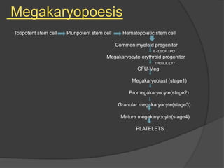
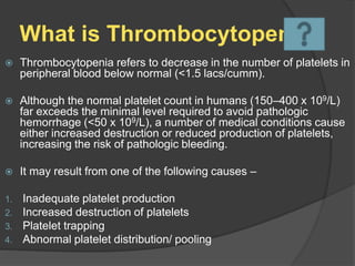
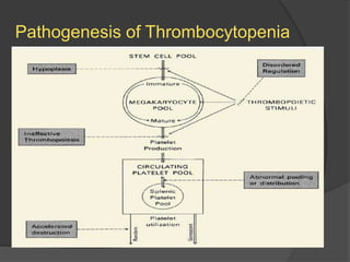
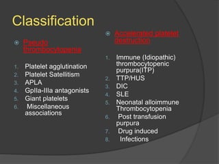
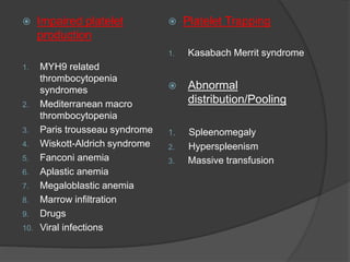
Malvika Tripathi
Department of Pathology
 Platelets were described by Addison in 1841 as
“extremely minute — granules” in clotting blood.
 They were termed platelets by Bizzozero, who also
observed their adhesive qualities as “increased
stickiness — when a vascular wall is damaged”.
 The same elements were identified by microscopic
examination of blood smears by Osler and Schaefer and
by Hayem in the late 19th century.
 Megakaryocytes have been recognized as rare marrow
cells for nearly two centuries, but it was the elegant
camera lucida studies of Howell in 1890 and his coining
of the term megakaryocyte that led to their broader
appreciation as distinct entities.
 In 1906, James Homer Wright put forth the hypothesis
that blood platelets are derived from the cytoplasm of
megakaryocytes and the basic elements of
thrombopoiesis were established.
 Platelet production begins in the yolk sac and, like the
remainder of hematopoiesis, shifts to the fetal liver and then to
the marrow at the time of gestation.
 Based on the adult blood volume (5 L), the number of platelets
per micro liter of blood (∼2 x 10⁵), and their circulatory half-life
(4-10 days), it can be calculated that each day an adult human
produces 1 x 10¹¹ platelets.
 The platelet count varies among the healthy population (1.5 to
4.5 x 10⁹ lakhs / cumm) but remains within a fairly narrow
range in any given individual.
 In times of increased demand, platelet production can rise 10-
fold or more.
Megakaryopoesis
Totipotent stem cell Pluripotent stem cell Hematopoietic stem cell
Common myeloid progenitor
IL-3,SCF,TPO
Megakaryocyte erythroid progenitor
TPO,IL6,IL11
CFU-Meg
Megakaryoblast (stage1)
Promegakaryocyte(stage2)
Granular megakaryocyte(stage3)
Mature megakaryocyte(stage4)
PLATELETS
Structure of platelets :-
Sol gel zone
Peripheral zone
Organelle zone
Platelet organelles & their
contents :-
1. Alpha granules :-
 Platelet specific protein – PF4, PDGF, thrombospondin, β
thromboglobulin.
 Coagulation specific protein- Fibrinogen, Factor V, vWF, High
molecular weight kinogen.
 Fibrinolytic system protein – a 2 antiplasmin , plasminogen , Platelet
aggregation inhibitor-1.
 Others – fibronectin, albumin.
2. Dense granules :-
 Anions –ATP, ADP, GTP, GDP.
 Cations - Serotonin, Calcium .
Role of platelets in
hemostasis :-
1. Adhesion :-
• Via GPIb on the surface of platelets.
• Congenital absence of GPIb results in Bernard’s
Soulier syndrome
• Congenital absence of vWF in plasma results in
VWD .
2. Release reaction ( secretion) :-
• Begins immediately after adhesion.
• ADP from dense granules promotes aggregation of
platelets.
• PF4 neutralises anticoagulant activity of Heparin .
• PDGF stimulates proliferation of vascular smooth muscles,
skin fibroblasts.
• TxA2 induces aggregation of other platelets & local
vasoconstriction.
3. Aggregation :-
• Binding of platelets to each other.
• Gp IIb & III a (fibrinogen receptors) exposed to
surface
• Binding of fibrinogen molecule causes aggregation of
platelets.
What is Thrombocytopenia
 Thrombocytopenia refers to decrease in the number of platelets in
peripheral blood below normal (<1.5 lacs/cumm).
 Although the normal platelet count in humans (150–400 x 109/L)
far exceeds the minimal level required to avoid pathologic
hemorrhage (<50 x 109/L), a number of medical conditions cause
either increased destruction or reduced production of platelets,
increasing the risk of pathologic bleeding.
 It may result from one of the following causes –
1. Inadequate platelet production
2. Increased destruction of platelets
3. Platelet trapping
4. Abnormal platelet distribution/ pooling
Pathogenesis of Thrombocytopenia
Classification
 Pseudo
thrombocytopenia
1. Platelet agglutination
2. Platelet Satellitism
3. APLA
4. GpIIa-IIIa antagonists
5. Giant platelets
6. Miscellaneous
associations
 Accelerated platelet
destruction
1. Immune (Idiopathic)
thrombocytopenic
purpura(ITP)
2. TTP/HUS
3. DIC
4. SLE
5. Neonatal alloimmune
Thrombocytopenia
6. Post transfusion
purpura
7. Drug induced
8. Infections
 Impaired platelet
production
1. MYH9 related
thrombocytopenia
syndromes
2. Mediterranean macro
thrombocytopenia
3. Paris trousseau syndrome
4. Wiskott-Aldrich syndrome
5. Fanconi anemia
6. Aplastic anemia
7. Megaloblastic anemia
8. Marrow infiltration
9. Drugs
10. Viral infections
 Platelet Trapping
1. Kasabach Merrit syndrome
 Abnormal
distribution/Pooling
1. Spleenomegaly
2. Hyperspleenism
3. Massive transfusion
Idiopathic Thrombocytopenic
purpura/Autoimmune
Thrombocytopenic purpura
 It is a common acquired autoimmune disorder
defined by a low platelet count secondary to
accelerated platelet destruction or impaired
thrombopoiesis by antiplatelet antibodies.
 It occurs in 2 forms – Acute and Chronic.
 Acute ITP occurs in children following viral
infection or vaccination. Sudden in onset.
 In acute ITP, Immune complexes bind to Fc
receptor on platelets that leads to immune
destruction of platelets by macrophages in spleen.
 Chronic ITP occurs predominantly in adult women
(20-40 years) and is not preceded by infection or
any underlying disease, Insidious onset, Self
limiting.
 In chronic ITP, antibodies directed against specific
glycoproteins IIb/IIIa or Ib/IX (Mainly IgG type). These
antibodies are also directed against megakaryocytes.
 These antibody coated platelets are recognized by Fc
receptors on macrophages and destroyed
mainly in spleen.
 GPIIb/IIIa are sites for fibrinogen binding during platelet
aggregation.
 These antibodies also block GPIIb/IIIa and cause
platelet dysfunction in addition to platelet destruction.
Parameters Acute ITP Chronic ITP
Age Childhood (2-4 years) Adults (15-40 years)
Sex No sex preference F>M
History of preceding viral
infections or vaccination
Common No history
Onset of bleeding Sudden Insidious
Type of bleeding Purpuric spots and
ecchymosis
Superficial
Site of bleeding Cutaneous and mucous
membranes (Gums,
Nose, GIT and
hematuria)
Skin and mucous
membrane
(Menorrahegia)
Degree of
thrombocytopenia
Severe Moderate
Spleen Just palpable Non palpable
Spontaneous remission Usual Need therapy
Recurrence Uncommon Common
Duration 1-6 months Months to year
Prognosis Very good Fair
 Blood loss may lead to anemia.
 Lymphocytes and Eosinophils are frequently increased
in acute ITP.
 Platelets are markedly reduced (<20,000/cumm) to
moderate and Macrothrombocytes are found.
 Number of large platelet is proportional to
megakaryocytes in marrow.
 Megakaryocytes are normal or increased in number in
bone marrow and frequently show morphological
changes such as hypo granularity of cytoplasm,
vacuolization and dense nuclear chromatin.
 If clinical features, complete blood counts and
blood smear are indicative of ITP then bone
marrow examination is not necessary for
diagnosis of ITP.
 Levels of platelet associated immunoglobulins
are raised in majority of patients with ITP.
Drug induced
thrombocytopenia
 Development of thrombocytopenia after quinine was
first described by Vipan in 1865, and since then a
large number of drugs have been found to cause
thrombocytopenia.
 Drug-induced thrombocytopenia generally affects
only a small percentage of patients taking a
particular drug, and is usually not severe, although it
can be fatal.
 Genetic or environmental factors both influence
susceptibility to drugs.
 Drugs may cause thrombocytopenia by different mechanisms.
 Dose-dependent myelosuppression and immune destruction of
the platelets are two well-known causes.
 One of the most severe and life-threatening immune
thrombocytopenias is heparin-induced thrombocytopenia (HIT),
an immune-mediated disorder caused by antibodies that
recognize a neoepitope in platelet factor 4 that is exposed when
platelet factor 4 binds heparin.
 The result is activation of platelets and the coagulation
cascade and, ultimately, thrombosis.
 HIT affects up to 5 percent of patients exposed to heparin.
 Other drugs causing drug induced
thrombocytopenia –
1. Sulfamethoxazole
2. Penicillin
3. Gold salts
4. Quinidine
5. Quinine
6. Diazepam
7. Lithium
8. Amiodarone
9. Acetazolamide
10. Amphotericin B
Neonatal Thrombocytopenia
 When the fetal platelets possessing paternally
derived antigens lacking in the mother enter
maternal circulation during gestation or delivery,
formation of alloantibody is stimulated.
 These maternal antibodies cross the placenta and
cause destruction of fetal platelets.
 The most common antigen against which
antibodies are formed is HPA-1a.
 The condition is usually resolved by 3 weeks
(maximum 3 months) after delivery.
 In severe cases purpura and hemorrhages are
evident at birth or manifest within few hours.
 Other causes of neonatal thrombocytopenia –
1. Thrombocytopenia with absent radius syndrome
2. Infections
3. Drug induced
4. Congenital megakaryocytic hyperplasia
5. Congenital leukemia
Post transfusion purpura
 Rare
 Sudden onset
 Bleeding occurs about 7-10 days after blood transfusion.
 Donor platelet (HPA-1a antigen) + Transfusion =
Destruction of patient’s platelets (already sensitized) =
Thrombocytopenia (severe).
 IV gamma globulins and plasmapheresis - treatment
modalities.
Disseminated intravascular
coagulation
 An acute, sub acute, or chronic
thrombohemorrhagic disorder, disseminated
intravascular coagulation (DIC) occurs as a
secondary complication in a variety of diseases.
 It is caused by the systemic activation of the
coagulation pathways, leading to the formation of
thrombi throughout the microcirculation.
 As a consequence of the widespread thromboses,
there is consumption of platelets and coagulation
factors and, secondarily, activation of fibrinolysis.
DIC is characterized by –
 Intravascular activation of extrinsic pathway of
coagulation with generation of thrombin and
fibrin.
 Reduction in level of endogenous
anticoagulants (antithrombin, protein C).
 Suppression of fibrinolytic system which causes
delayed and inadequate removal of fibrin.
These 3 factors in combination leads to generalized
deposition of fibrin in circulation and form micro
thrombi.
 2 types – Acute and chronic.
 Acute/ decompensated DIC –
Rapid and extensive activation of coagulation
leading to significant bleeding from consumption of
coagulation factors and widespread micrvascular
thrombosis with consequent end organ damage.
Ex – DIC due to sepsis or trauma.
 Sudden onset of spontaneous bleeding from
multiple sites like skin (petechie and ecchymosis),
GIT, urinary system, epistaxis, and oozing from
venepuncture sites.
 Chronic / Compensated DIC –
Slow activation of coagulation in small amount
with slow consumption of coagulation factors ;
Coagulation factors are normal or increased,
Clinical features are minimal or absent. Laboratory
abnormalities are the only evidence of DIC.
Ex – IUD, Liver diseases, giant hemangioma,
eclampsia, malignancy.
 Mild and protracted disease, manifests only with
venous thrombosis.
 Laboratory features –
 Acute DIC – Low platelet or falling platelets on
repeat testing, prolonged PT and aPTT, Low
fibrinogen or falling levels on repeat testing, Low
plasma level of coagulation inhibitors i.e. ATIII or
protein c, schistocytes on blood smear.
 Chronic DIC – Platelet count normal or slightly
reduced, PT and aPTT are normal.
Thrombotic thrombocytopenic
purpura
 2 types – Idiopathic and familial.
 Idiopathic –
Auto antibodies against ADAMTS13 lead to deficiency
of ADAMTS13 and accumulation of ultra large vWF
multimers that bind large number of platelets.
 Familial –
ADAMTS13 deficiency results from mutation in
ADAMTS 13 gene.
 ADAMTS13 – A disintegrin and metalloprotease with thrombospondin type 1 motif 13.
 Affects mainly young adults.
 More common in females.
 Pentad of manifestations include -
1. Microangiopathic hemolytic anemia
2. Bleeding manifestations secondary to severe
thrombocytopenia
3. Fluctuating neurological dysfunctions
4. Renal abnormalities
5. Fever
 These 5 features may not be present in all patients.
Hemolytic uraemic syndrome
 Characterized by triad of features :-
1) Acute renal failure
2) Thrombocytopenia
3) Microangiopathic hemolytic anaemia.
 Two types :- Typical and Atypical.
 Typical HUS –occur predominantly in children<5 yr
and is associated with Shiga toxin –producing E.coli
o157:H7.
 It is characterized by a prodrome of diarrhoeal
illness followed by Microangiopathic hemolytic
anaemia, Thrombocytopenia and renal failure.
 Atypical HUS- has similar clinical features but is
not preceded by diarrhoeal prodrome.
Pseudo thrombocytopenia/Spurious
thrombocytopenia
 Uncommon phenomenon caused by ex vivo
agglutination of platelets.
 This leads to platelet clumping.
 Due to this, platelet counts are reduced on
automated cell counters because they cannot
differentiate platelet clumps from individual cells.
 Causes –
1. Use of EDTA anticoagulant
2. With platelet cold agglutinins
3. Multiple myeloma.
Antibody induced platelet
agglutination
 Caused ex vivo either by anti platelet antibodies
or by activation of platelets during collection.
 Antibodies which cause platelet agglutination do
not appear in any pathologic process as they are
present in normal individual.
 These antibodies recognize the platelet
membrane glycoproteins which are modified or
exposed when calcium is chelated.
 These antibodies are typically of IgG type ; but
IgM and IgA are also described.
 This phenomenon is most common in presence
of EDTA as anticoagulant.
 But other anticoagulants can also cause antibody
induced agglutination of platelets such as –
1. Sodium citrate
2. Sodium oxalate
3. Acid citrate dextrose
4. Heparin
 Antibodies cause agglutination at room
temperature therefore the reaction can be
prevented if blood sample is kept at 37 Celsius.
 Clumping is usually evident in 60 minutes after
blood is drawn.
 In most cases antibodies are directed against
GPIIb/IIIa.
Platelet Satellitism
 Antibodies directed against GPIIb/IIIa react
simultaneously with Fc receptor III (FcRIII) of
leukocytes and attach platelets to neutrophils and
monocytes.
 Platelets form a rosette around periphery of
leukocytes.
 Neutrophils are most commonly involved.
 Monocytes can also be involved.
 These antibodies are naturally occurring and
their presence does not clearly correlate any
clinical situation, disease or drug.
 These antibodies fail to produce Satellitism in –
Platelets of patients with Glanzmann
Thrombasthenia (Absence of GPIIb/IIIa)
or
Patients with congenital absence of FCIII
receptor .
Antiphospholipid
antibodies(APLA)
 Some anti platelet antibodies from patients with
pseudo thrombocytopenia cross react with
negatively charged phospholipids and exhibit
anticardiolipin activity.
 Sera of these patients lose their ability to clump
platelets.
 Therefore antibodies against phospholipids can
bind to antigens modified by EDTA on platelet
membrane and cause platelet clumping.
 Antibodies from patients with thrombocytopenia
can induce platelet agglutination with donor
platelets in presence of EDTA.
 This agglutination can be prevented by –
1. Warming donor platelets to 37 celsius.
2. Pretreating donor platelets with aspirin,
Prostaglandin E1.
3. Monoclonal antibodies against GPIIb/IIIa.
MYH9 related
thrombocytopenia syndromes
 May-Hegglin anomaly, Fechtner syndrome,
Sebastian syndrome, and Epstein syndrome are
autosomal dominant macrothrombocytopenias
with mutations in the MYH9 gene.
 This gene is located on chromosome 22q12–13.
 This gene encodes NMMHC IIA , which is
expressed in platelets, kidney, leukocytes, and
the cochlea.
 In all cells in which the gene product is
expressed, except platelets and leukocytes, other
NMMHC isoforms (IIB and IIC) are also
expressed, and these can compensate
functionally for the defective IIA isoform,
restricting the most profound manifestations of
NMMHC-IIA deficiency to platelets and
leukocytes.
 The NMMHC-IIA protein appears to be an
important cytoskeletal contractile protein in
hematopoietic cells.
 One mutation in the MYH9 gene produced a
highly unstable protein with abnormal
organization of the megakaryocyte cytoskeleton.
 The defect in platelet number is likely a defect in
platelet maturation from proplatelets, as when
MYH9-deficient stem cells were differentiated to
megakaryocytes they produced proplatelets
normally.
 These syndromes include a triad of
Thrombocytopenia, Macrothrombocytes and
Dohle body like inclusions and other features.
 One hallmark feature of MYH9-related disorders
is revealed on the blood film, where neutrophilic
inclusions that appear blue with Wright- Giemsa
stain are noted.
 The inclusions correspond to cytoplasmic
aggregates of NMMHC-IIA, which are readily
detected by immunocytochemistry.
Mediterranean macro
thrombocytopenia
 Mild congenital thrombocytopenia with an autosomal
dominant pattern of inheritance.
 Many of the patients share clinical and molecular
features with the heterozygous Bernard-Soulier
syndrome phenotype.
 Linkage analyses reveal a heterozygous Ala156Val
missense substitution in the GPIb gene (also known
as the Bolzano mutation), which is also present in
patients with Bernard-Soulier syndrome.
 The clinical manifestations of Mediterranean
macro thrombocytopenia are variable, with the
severity of bleeding related to both platelet
number and function.
 A related syndrome with concomitant
stomatocytosis and hemolysis (Mediterranean
stomatocytosis/ macrothrombocytopenia) and
autosomal recessive transmission is caused by
mutations in the genes ABCG5 and ABCG8.
Paris Trousseau
syndrome/Jacobsen
Thrombopenia
 Congenital dysmorphology syndrome in which
affected individuals manifest trigonocephaly,
facial dysmorphism, heart defects, and mental
retardation.
 Result from deletion of the long arm of
chromosome 11 at 11q23, a region that includes
the FLI1 gene, the product of which is a
transcription factor involved in megakaryopoiesis.
 All affected patients have mild to moderate
thrombocytopenia and dysfunctional platelets.
 The blood film shows a subpopulation of platelets
containing giant granules.
 Marrow examination reveals two distinct
subpopulations of megakaryocytes with expansion of
immature megakaryocytic progenitors,
dysmegakaryopoiesis, and many
micromegakaryocytes.
 Pathologic bleeding usually is mild.
Kasabach-Merritt Syndrome
 Profound thrombocytopenia related to platelet
trapping within a vascular tumor, either a Kaposi-
like hemangioendothelioma or a tufted angioma.
 The syndrome presents predominantly during
infancy, but several adult cases have been
reported.
 These vascular tumors should be differentiated
from vascular malformations such as classic
benign hemangiomas.
 Thrombocytopenia in KMS usually is severe and
associated with DIC.
 Contributing factors include "platelet trapping" by
abnormally proliferating endothelium within the
hemangioma and platelet consumption associated
with DIC.
 Platelet trapping has been demonstrated by
immunohistochemical staining of the tumors with anti-
CD61 antibodies (a marker of platelets and
megakaryocytes)141 and by nuclear studies using
51Cr-labeled platelets and 111 In.
 The mainstay of treatment is eradication of the tumor.
Thrombocytopenia Associated
with HIV Infection
 Thrombocytopenia is common in patients infected
with HIV, with the prevalence of thrombocytopenia,
depending on the subpopulation of patients studied.
 Among HIV-infected drug users, the prevalence is
approximately 36.9 percent, compared to 8.7 percent
in drug users without HIV infection.
 In homosexual men, the prevalence is approximately
16 percent in the HIV-infected group and 3 percent in
the HIV-negative group.
 The high prevalence of thrombocytopenia in
homosexuals and intravenous drug users without
HIV probably results from the high frequency of
hepatitis in these populations.
 Causes –
1. Accelerated platelet destruction primarily related to immune
complexes.
2. Decreased platelet production specially in advanced disease.
3. Splenic sequestration.
4. Platelet consumption associated with thrombotic
thrombocytopenic purpura(TTP).
5. Medications, Concurrent infections such as hepatitis C,
Hemorrhagic malignancies may contribute to development of
thrombocytopenia.
 Platelet kinetic studies performed in HIV-positive
patients with thrombocytopenia demonstrate that
the mean platelet life span usually is very short, an
indication of accelerated platelet destruction.
 HIV appears capable of triggering a large
repertoire of immune complexes that participate in
the destruction of platelets.
 Immune complexes containing anti-F(ab')2
antibodies are found in homosexual individuals
with HIV and thrombocytopenia.
 Antibodies against CD4 and the CD4 receptor
gp120 have been found in HIV-positive patients
with and without thrombocytopenia.
 These antibodies are capable of forming
complexes through their specificity-determining
regions.
 These immune complexes can bind platelets and
have been postulated to play a role in the
thrombocytopenia associated with HIV.
 In addition to increased platelet destruction, reduced
platelet production appears to play a role in the
thrombocytopenia observed in HIV patients.
 A direct effect of viral infection on platelet production is
suggested by the observation that platelet production
increases when patients are treated with zidovudine.
 Infected patients with thrombocytopenia also have
increased levels of TPO, again supporting the notion of
ineffective platelet production in the origin of HIV-
associated thrombocytopenia.
 The defect appears to lie at the level of
megakaryopoiesis, as decreased levels of megakaryocyte
progenitors in marrow have been observed.
 The response to antiretroviral therapy suggests that
the virus infects megakaryocytes or their precursors in
the marrow.
 HIV-1 entry into cells requires sequential interaction of
the viral envelope gp120 with CD4 and a co receptor
on the host cell plasma membrane, either C-C
chemokine receptor-5 (CCR5) or CXC chemokine
receptor-4 (CXCR4).
 All of these receptors are expressed by
megakaryocytes.
 Patients with HIV are at higher risk for developing
thrombotic microangiopathies.
 Thrombocytopenic patients with HIV rarely
experience clinically important bleeding (except,
of course, those with hemophilia).
 The platelet counts rarely dip below 50 x 109/L,
and the thrombocytopenia often spontaneously
resolves.
Nutritional deficiencies and
alcohol induced
thrombocytopenia
 Thrombocytopenia may be seen in association
with vitamin B12 deficiency when the latter results
from auto antibodies against parietal cells or
intrinsic factor and is associated with immune
thrombocytopenia.
 Various other autoimmune disorders can coexist
with pernicious anemia, including autoimmune
vitiligo and autoimmune thyroiditis.
 Thrombocytopenia in alcoholic patients almost
always results from liver cirrhosis with relative TPO
deficiency (the liver is the primary origin of circulating
TPO levels), congestive Spleenomegaly, and/or from
folic acid deficiency.
 Suppression of platelet production sufficient to
produce thrombocytopenia requires consumption of
large quantities of ethanol over several days.
 Thrombocytopenia usually resolves in 5 to 21 days
with cessation of ethanol ingestion, sometimes with a
transient rebound thrombocytosis.
Myelodysplastic
syndrome(MDS)
 Myelodysplastic syndromes are clonal myeloid
disorders characterized by blood cytopenias in
combination with a hyper cellular marrow that often
exhibit dysplastic changes in any of the three
hematopoietic lineages.
 Thrombocytopenia is present in approximately 50
percent of patients and usually occurs in conjunction
with other cytopenias.
 Moreover, platelets from patients with MDS often
display functional abnormalities and, when present,
usually augment the thrombocytopenia-related
bleeding disorder.
 The presence of micromegakaryocytes or micro
mononuclear megakaryocytes in marrow from
MDS patients indicates altered megakaryopoiesis.
 Maturation of megakaryocytes is arrested in MDS,
as suggested by immunohistochemistry studies
showing an increased number of megakaryocytic
precursors in the marrow of affected patients.
 Besides maturation arrest, an increased rate of
apoptosis is seen in all subtypes of MDS.
 Abnormal megakaryopoiesis is due to
dysfunctional TPO receptor or an abnormality in
the downstream signaling pathway rather than
from diminished expression of receptor itself.
 Supportive therapy in addition to chemotherapy
is the treatment of choice.
Aplastic anemia
 Aplastic anemia is a pancytopenia that results
from failure of marrow hematopoiesis.
 However, some patients with MDS or
amegakaryocytic thrombocytopenia present with
low platelet counts and then progress to
pancytopenia and aplastic anemia.
 In aplastic anemia, blood testing reveals markedly
decreased cell counts, reticulocytopenia, and lack
of circulating blasts.
 A considerable amount of data support the
hypothesis that aplastic anemia results from an
autoimmune attack directed against HSC.
 Activated T-helper type 1 T cells that produce
interferon-, tumor necrosis factor, and IL-2 are
responsible for suppression of the hematopoietic
cell compartment.
 This cytotoxic activation leads to a Fas-mediated
cell-cycle arrest and death of CD4+T cells.
References
I. Williams hematology 8th edition
II. De Grutchy’s clinical hematology 6th
edition
III. Robbins basic pathology 8th edition
Thank You

Thrombocytopenia

  • 1.
  • 2.
     Platelets weredescribed by Addison in 1841 as “extremely minute — granules” in clotting blood.  They were termed platelets by Bizzozero, who also observed their adhesive qualities as “increased stickiness — when a vascular wall is damaged”.  The same elements were identified by microscopic examination of blood smears by Osler and Schaefer and by Hayem in the late 19th century.
  • 3.
     Megakaryocytes havebeen recognized as rare marrow cells for nearly two centuries, but it was the elegant camera lucida studies of Howell in 1890 and his coining of the term megakaryocyte that led to their broader appreciation as distinct entities.  In 1906, James Homer Wright put forth the hypothesis that blood platelets are derived from the cytoplasm of megakaryocytes and the basic elements of thrombopoiesis were established.
  • 4.
     Platelet productionbegins in the yolk sac and, like the remainder of hematopoiesis, shifts to the fetal liver and then to the marrow at the time of gestation.  Based on the adult blood volume (5 L), the number of platelets per micro liter of blood (∼2 x 10⁵), and their circulatory half-life (4-10 days), it can be calculated that each day an adult human produces 1 x 10¹¹ platelets.  The platelet count varies among the healthy population (1.5 to 4.5 x 10⁹ lakhs / cumm) but remains within a fairly narrow range in any given individual.  In times of increased demand, platelet production can rise 10- fold or more.
  • 5.
    Megakaryopoesis Totipotent stem cellPluripotent stem cell Hematopoietic stem cell Common myeloid progenitor IL-3,SCF,TPO Megakaryocyte erythroid progenitor TPO,IL6,IL11 CFU-Meg Megakaryoblast (stage1) Promegakaryocyte(stage2) Granular megakaryocyte(stage3) Mature megakaryocyte(stage4) PLATELETS
  • 6.
    Structure of platelets:- Sol gel zone Peripheral zone Organelle zone
  • 7.
    Platelet organelles &their contents :- 1. Alpha granules :-  Platelet specific protein – PF4, PDGF, thrombospondin, β thromboglobulin.  Coagulation specific protein- Fibrinogen, Factor V, vWF, High molecular weight kinogen.  Fibrinolytic system protein – a 2 antiplasmin , plasminogen , Platelet aggregation inhibitor-1.  Others – fibronectin, albumin. 2. Dense granules :-  Anions –ATP, ADP, GTP, GDP.  Cations - Serotonin, Calcium .
  • 8.
    Role of plateletsin hemostasis :-
  • 9.
    1. Adhesion :- •Via GPIb on the surface of platelets. • Congenital absence of GPIb results in Bernard’s Soulier syndrome • Congenital absence of vWF in plasma results in VWD .
  • 10.
    2. Release reaction( secretion) :- • Begins immediately after adhesion. • ADP from dense granules promotes aggregation of platelets. • PF4 neutralises anticoagulant activity of Heparin . • PDGF stimulates proliferation of vascular smooth muscles, skin fibroblasts. • TxA2 induces aggregation of other platelets & local vasoconstriction.
  • 11.
    3. Aggregation :- •Binding of platelets to each other. • Gp IIb & III a (fibrinogen receptors) exposed to surface • Binding of fibrinogen molecule causes aggregation of platelets.
  • 12.
    What is Thrombocytopenia Thrombocytopenia refers to decrease in the number of platelets in peripheral blood below normal (<1.5 lacs/cumm).  Although the normal platelet count in humans (150–400 x 109/L) far exceeds the minimal level required to avoid pathologic hemorrhage (<50 x 109/L), a number of medical conditions cause either increased destruction or reduced production of platelets, increasing the risk of pathologic bleeding.  It may result from one of the following causes – 1. Inadequate platelet production 2. Increased destruction of platelets 3. Platelet trapping 4. Abnormal platelet distribution/ pooling
  • 13.
  • 14.
    Classification  Pseudo thrombocytopenia 1. Plateletagglutination 2. Platelet Satellitism 3. APLA 4. GpIIa-IIIa antagonists 5. Giant platelets 6. Miscellaneous associations  Accelerated platelet destruction 1. Immune (Idiopathic) thrombocytopenic purpura(ITP) 2. TTP/HUS 3. DIC 4. SLE 5. Neonatal alloimmune Thrombocytopenia 6. Post transfusion purpura 7. Drug induced 8. Infections
  • 15.
     Impaired platelet production 1.MYH9 related thrombocytopenia syndromes 2. Mediterranean macro thrombocytopenia 3. Paris trousseau syndrome 4. Wiskott-Aldrich syndrome 5. Fanconi anemia 6. Aplastic anemia 7. Megaloblastic anemia 8. Marrow infiltration 9. Drugs 10. Viral infections  Platelet Trapping 1. Kasabach Merrit syndrome  Abnormal distribution/Pooling 1. Spleenomegaly 2. Hyperspleenism 3. Massive transfusion
  • 16.
    Idiopathic Thrombocytopenic purpura/Autoimmune Thrombocytopenic purpura It is a common acquired autoimmune disorder defined by a low platelet count secondary to accelerated platelet destruction or impaired thrombopoiesis by antiplatelet antibodies.  It occurs in 2 forms – Acute and Chronic.
  • 17.
     Acute ITPoccurs in children following viral infection or vaccination. Sudden in onset.  In acute ITP, Immune complexes bind to Fc receptor on platelets that leads to immune destruction of platelets by macrophages in spleen.  Chronic ITP occurs predominantly in adult women (20-40 years) and is not preceded by infection or any underlying disease, Insidious onset, Self limiting.
  • 18.
     In chronicITP, antibodies directed against specific glycoproteins IIb/IIIa or Ib/IX (Mainly IgG type). These antibodies are also directed against megakaryocytes.  These antibody coated platelets are recognized by Fc receptors on macrophages and destroyed mainly in spleen.  GPIIb/IIIa are sites for fibrinogen binding during platelet aggregation.  These antibodies also block GPIIb/IIIa and cause platelet dysfunction in addition to platelet destruction.
  • 20.
    Parameters Acute ITPChronic ITP Age Childhood (2-4 years) Adults (15-40 years) Sex No sex preference F>M History of preceding viral infections or vaccination Common No history Onset of bleeding Sudden Insidious Type of bleeding Purpuric spots and ecchymosis Superficial Site of bleeding Cutaneous and mucous membranes (Gums, Nose, GIT and hematuria) Skin and mucous membrane (Menorrahegia) Degree of thrombocytopenia Severe Moderate Spleen Just palpable Non palpable Spontaneous remission Usual Need therapy Recurrence Uncommon Common Duration 1-6 months Months to year Prognosis Very good Fair
  • 21.
     Blood lossmay lead to anemia.  Lymphocytes and Eosinophils are frequently increased in acute ITP.  Platelets are markedly reduced (<20,000/cumm) to moderate and Macrothrombocytes are found.  Number of large platelet is proportional to megakaryocytes in marrow.  Megakaryocytes are normal or increased in number in bone marrow and frequently show morphological changes such as hypo granularity of cytoplasm, vacuolization and dense nuclear chromatin.
  • 22.
     If clinicalfeatures, complete blood counts and blood smear are indicative of ITP then bone marrow examination is not necessary for diagnosis of ITP.  Levels of platelet associated immunoglobulins are raised in majority of patients with ITP.
  • 23.
    Drug induced thrombocytopenia  Developmentof thrombocytopenia after quinine was first described by Vipan in 1865, and since then a large number of drugs have been found to cause thrombocytopenia.  Drug-induced thrombocytopenia generally affects only a small percentage of patients taking a particular drug, and is usually not severe, although it can be fatal.  Genetic or environmental factors both influence susceptibility to drugs.
  • 24.
     Drugs maycause thrombocytopenia by different mechanisms.  Dose-dependent myelosuppression and immune destruction of the platelets are two well-known causes.  One of the most severe and life-threatening immune thrombocytopenias is heparin-induced thrombocytopenia (HIT), an immune-mediated disorder caused by antibodies that recognize a neoepitope in platelet factor 4 that is exposed when platelet factor 4 binds heparin.  The result is activation of platelets and the coagulation cascade and, ultimately, thrombosis.  HIT affects up to 5 percent of patients exposed to heparin.
  • 25.
     Other drugscausing drug induced thrombocytopenia – 1. Sulfamethoxazole 2. Penicillin 3. Gold salts 4. Quinidine 5. Quinine 6. Diazepam 7. Lithium 8. Amiodarone 9. Acetazolamide 10. Amphotericin B
  • 26.
    Neonatal Thrombocytopenia  Whenthe fetal platelets possessing paternally derived antigens lacking in the mother enter maternal circulation during gestation or delivery, formation of alloantibody is stimulated.  These maternal antibodies cross the placenta and cause destruction of fetal platelets.  The most common antigen against which antibodies are formed is HPA-1a.
  • 27.
     The conditionis usually resolved by 3 weeks (maximum 3 months) after delivery.  In severe cases purpura and hemorrhages are evident at birth or manifest within few hours.  Other causes of neonatal thrombocytopenia – 1. Thrombocytopenia with absent radius syndrome 2. Infections 3. Drug induced 4. Congenital megakaryocytic hyperplasia 5. Congenital leukemia
  • 28.
    Post transfusion purpura Rare  Sudden onset  Bleeding occurs about 7-10 days after blood transfusion.  Donor platelet (HPA-1a antigen) + Transfusion = Destruction of patient’s platelets (already sensitized) = Thrombocytopenia (severe).  IV gamma globulins and plasmapheresis - treatment modalities.
  • 29.
    Disseminated intravascular coagulation  Anacute, sub acute, or chronic thrombohemorrhagic disorder, disseminated intravascular coagulation (DIC) occurs as a secondary complication in a variety of diseases.  It is caused by the systemic activation of the coagulation pathways, leading to the formation of thrombi throughout the microcirculation.  As a consequence of the widespread thromboses, there is consumption of platelets and coagulation factors and, secondarily, activation of fibrinolysis.
  • 31.
    DIC is characterizedby –  Intravascular activation of extrinsic pathway of coagulation with generation of thrombin and fibrin.  Reduction in level of endogenous anticoagulants (antithrombin, protein C).  Suppression of fibrinolytic system which causes delayed and inadequate removal of fibrin. These 3 factors in combination leads to generalized deposition of fibrin in circulation and form micro thrombi.
  • 32.
     2 types– Acute and chronic.  Acute/ decompensated DIC – Rapid and extensive activation of coagulation leading to significant bleeding from consumption of coagulation factors and widespread micrvascular thrombosis with consequent end organ damage. Ex – DIC due to sepsis or trauma.  Sudden onset of spontaneous bleeding from multiple sites like skin (petechie and ecchymosis), GIT, urinary system, epistaxis, and oozing from venepuncture sites.
  • 33.
     Chronic /Compensated DIC – Slow activation of coagulation in small amount with slow consumption of coagulation factors ; Coagulation factors are normal or increased, Clinical features are minimal or absent. Laboratory abnormalities are the only evidence of DIC. Ex – IUD, Liver diseases, giant hemangioma, eclampsia, malignancy.  Mild and protracted disease, manifests only with venous thrombosis.
  • 34.
     Laboratory features–  Acute DIC – Low platelet or falling platelets on repeat testing, prolonged PT and aPTT, Low fibrinogen or falling levels on repeat testing, Low plasma level of coagulation inhibitors i.e. ATIII or protein c, schistocytes on blood smear.  Chronic DIC – Platelet count normal or slightly reduced, PT and aPTT are normal.
  • 35.
    Thrombotic thrombocytopenic purpura  2types – Idiopathic and familial.  Idiopathic – Auto antibodies against ADAMTS13 lead to deficiency of ADAMTS13 and accumulation of ultra large vWF multimers that bind large number of platelets.  Familial – ADAMTS13 deficiency results from mutation in ADAMTS 13 gene.  ADAMTS13 – A disintegrin and metalloprotease with thrombospondin type 1 motif 13.
  • 36.
     Affects mainlyyoung adults.  More common in females.  Pentad of manifestations include - 1. Microangiopathic hemolytic anemia 2. Bleeding manifestations secondary to severe thrombocytopenia 3. Fluctuating neurological dysfunctions 4. Renal abnormalities 5. Fever  These 5 features may not be present in all patients.
  • 37.
    Hemolytic uraemic syndrome Characterized by triad of features :- 1) Acute renal failure 2) Thrombocytopenia 3) Microangiopathic hemolytic anaemia.  Two types :- Typical and Atypical.  Typical HUS –occur predominantly in children<5 yr and is associated with Shiga toxin –producing E.coli o157:H7.
  • 38.
     It ischaracterized by a prodrome of diarrhoeal illness followed by Microangiopathic hemolytic anaemia, Thrombocytopenia and renal failure.  Atypical HUS- has similar clinical features but is not preceded by diarrhoeal prodrome.
  • 39.
    Pseudo thrombocytopenia/Spurious thrombocytopenia  Uncommonphenomenon caused by ex vivo agglutination of platelets.  This leads to platelet clumping.  Due to this, platelet counts are reduced on automated cell counters because they cannot differentiate platelet clumps from individual cells.  Causes – 1. Use of EDTA anticoagulant 2. With platelet cold agglutinins 3. Multiple myeloma.
  • 40.
    Antibody induced platelet agglutination Caused ex vivo either by anti platelet antibodies or by activation of platelets during collection.  Antibodies which cause platelet agglutination do not appear in any pathologic process as they are present in normal individual.  These antibodies recognize the platelet membrane glycoproteins which are modified or exposed when calcium is chelated.
  • 41.
     These antibodiesare typically of IgG type ; but IgM and IgA are also described.  This phenomenon is most common in presence of EDTA as anticoagulant.  But other anticoagulants can also cause antibody induced agglutination of platelets such as – 1. Sodium citrate 2. Sodium oxalate 3. Acid citrate dextrose 4. Heparin
  • 42.
     Antibodies causeagglutination at room temperature therefore the reaction can be prevented if blood sample is kept at 37 Celsius.  Clumping is usually evident in 60 minutes after blood is drawn.  In most cases antibodies are directed against GPIIb/IIIa.
  • 43.
    Platelet Satellitism  Antibodiesdirected against GPIIb/IIIa react simultaneously with Fc receptor III (FcRIII) of leukocytes and attach platelets to neutrophils and monocytes.  Platelets form a rosette around periphery of leukocytes.  Neutrophils are most commonly involved.  Monocytes can also be involved.
  • 45.
     These antibodiesare naturally occurring and their presence does not clearly correlate any clinical situation, disease or drug.  These antibodies fail to produce Satellitism in – Platelets of patients with Glanzmann Thrombasthenia (Absence of GPIIb/IIIa) or Patients with congenital absence of FCIII receptor .
  • 46.
    Antiphospholipid antibodies(APLA)  Some antiplatelet antibodies from patients with pseudo thrombocytopenia cross react with negatively charged phospholipids and exhibit anticardiolipin activity.  Sera of these patients lose their ability to clump platelets.  Therefore antibodies against phospholipids can bind to antigens modified by EDTA on platelet membrane and cause platelet clumping.
  • 47.
     Antibodies frompatients with thrombocytopenia can induce platelet agglutination with donor platelets in presence of EDTA.  This agglutination can be prevented by – 1. Warming donor platelets to 37 celsius. 2. Pretreating donor platelets with aspirin, Prostaglandin E1. 3. Monoclonal antibodies against GPIIb/IIIa.
  • 48.
    MYH9 related thrombocytopenia syndromes May-Hegglin anomaly, Fechtner syndrome, Sebastian syndrome, and Epstein syndrome are autosomal dominant macrothrombocytopenias with mutations in the MYH9 gene.  This gene is located on chromosome 22q12–13.  This gene encodes NMMHC IIA , which is expressed in platelets, kidney, leukocytes, and the cochlea.
  • 49.
     In allcells in which the gene product is expressed, except platelets and leukocytes, other NMMHC isoforms (IIB and IIC) are also expressed, and these can compensate functionally for the defective IIA isoform, restricting the most profound manifestations of NMMHC-IIA deficiency to platelets and leukocytes.  The NMMHC-IIA protein appears to be an important cytoskeletal contractile protein in hematopoietic cells.
  • 50.
     One mutationin the MYH9 gene produced a highly unstable protein with abnormal organization of the megakaryocyte cytoskeleton.  The defect in platelet number is likely a defect in platelet maturation from proplatelets, as when MYH9-deficient stem cells were differentiated to megakaryocytes they produced proplatelets normally.  These syndromes include a triad of Thrombocytopenia, Macrothrombocytes and Dohle body like inclusions and other features.
  • 51.
     One hallmarkfeature of MYH9-related disorders is revealed on the blood film, where neutrophilic inclusions that appear blue with Wright- Giemsa stain are noted.  The inclusions correspond to cytoplasmic aggregates of NMMHC-IIA, which are readily detected by immunocytochemistry.
  • 53.
    Mediterranean macro thrombocytopenia  Mildcongenital thrombocytopenia with an autosomal dominant pattern of inheritance.  Many of the patients share clinical and molecular features with the heterozygous Bernard-Soulier syndrome phenotype.  Linkage analyses reveal a heterozygous Ala156Val missense substitution in the GPIb gene (also known as the Bolzano mutation), which is also present in patients with Bernard-Soulier syndrome.
  • 54.
     The clinicalmanifestations of Mediterranean macro thrombocytopenia are variable, with the severity of bleeding related to both platelet number and function.  A related syndrome with concomitant stomatocytosis and hemolysis (Mediterranean stomatocytosis/ macrothrombocytopenia) and autosomal recessive transmission is caused by mutations in the genes ABCG5 and ABCG8.
  • 55.
    Paris Trousseau syndrome/Jacobsen Thrombopenia  Congenitaldysmorphology syndrome in which affected individuals manifest trigonocephaly, facial dysmorphism, heart defects, and mental retardation.  Result from deletion of the long arm of chromosome 11 at 11q23, a region that includes the FLI1 gene, the product of which is a transcription factor involved in megakaryopoiesis.
  • 56.
     All affectedpatients have mild to moderate thrombocytopenia and dysfunctional platelets.  The blood film shows a subpopulation of platelets containing giant granules.  Marrow examination reveals two distinct subpopulations of megakaryocytes with expansion of immature megakaryocytic progenitors, dysmegakaryopoiesis, and many micromegakaryocytes.  Pathologic bleeding usually is mild.
  • 57.
    Kasabach-Merritt Syndrome  Profoundthrombocytopenia related to platelet trapping within a vascular tumor, either a Kaposi- like hemangioendothelioma or a tufted angioma.  The syndrome presents predominantly during infancy, but several adult cases have been reported.  These vascular tumors should be differentiated from vascular malformations such as classic benign hemangiomas.
  • 58.
     Thrombocytopenia inKMS usually is severe and associated with DIC.  Contributing factors include "platelet trapping" by abnormally proliferating endothelium within the hemangioma and platelet consumption associated with DIC.  Platelet trapping has been demonstrated by immunohistochemical staining of the tumors with anti- CD61 antibodies (a marker of platelets and megakaryocytes)141 and by nuclear studies using 51Cr-labeled platelets and 111 In.  The mainstay of treatment is eradication of the tumor.
  • 59.
    Thrombocytopenia Associated with HIVInfection  Thrombocytopenia is common in patients infected with HIV, with the prevalence of thrombocytopenia, depending on the subpopulation of patients studied.  Among HIV-infected drug users, the prevalence is approximately 36.9 percent, compared to 8.7 percent in drug users without HIV infection.  In homosexual men, the prevalence is approximately 16 percent in the HIV-infected group and 3 percent in the HIV-negative group.
  • 60.
     The highprevalence of thrombocytopenia in homosexuals and intravenous drug users without HIV probably results from the high frequency of hepatitis in these populations.
  • 61.
     Causes – 1.Accelerated platelet destruction primarily related to immune complexes. 2. Decreased platelet production specially in advanced disease. 3. Splenic sequestration. 4. Platelet consumption associated with thrombotic thrombocytopenic purpura(TTP). 5. Medications, Concurrent infections such as hepatitis C, Hemorrhagic malignancies may contribute to development of thrombocytopenia.
  • 62.
     Platelet kineticstudies performed in HIV-positive patients with thrombocytopenia demonstrate that the mean platelet life span usually is very short, an indication of accelerated platelet destruction.  HIV appears capable of triggering a large repertoire of immune complexes that participate in the destruction of platelets.  Immune complexes containing anti-F(ab')2 antibodies are found in homosexual individuals with HIV and thrombocytopenia.
  • 63.
     Antibodies againstCD4 and the CD4 receptor gp120 have been found in HIV-positive patients with and without thrombocytopenia.  These antibodies are capable of forming complexes through their specificity-determining regions.  These immune complexes can bind platelets and have been postulated to play a role in the thrombocytopenia associated with HIV.
  • 64.
     In additionto increased platelet destruction, reduced platelet production appears to play a role in the thrombocytopenia observed in HIV patients.  A direct effect of viral infection on platelet production is suggested by the observation that platelet production increases when patients are treated with zidovudine.  Infected patients with thrombocytopenia also have increased levels of TPO, again supporting the notion of ineffective platelet production in the origin of HIV- associated thrombocytopenia.  The defect appears to lie at the level of megakaryopoiesis, as decreased levels of megakaryocyte progenitors in marrow have been observed.
  • 65.
     The responseto antiretroviral therapy suggests that the virus infects megakaryocytes or their precursors in the marrow.  HIV-1 entry into cells requires sequential interaction of the viral envelope gp120 with CD4 and a co receptor on the host cell plasma membrane, either C-C chemokine receptor-5 (CCR5) or CXC chemokine receptor-4 (CXCR4).  All of these receptors are expressed by megakaryocytes.  Patients with HIV are at higher risk for developing thrombotic microangiopathies.
  • 66.
     Thrombocytopenic patientswith HIV rarely experience clinically important bleeding (except, of course, those with hemophilia).  The platelet counts rarely dip below 50 x 109/L, and the thrombocytopenia often spontaneously resolves.
  • 67.
    Nutritional deficiencies and alcoholinduced thrombocytopenia  Thrombocytopenia may be seen in association with vitamin B12 deficiency when the latter results from auto antibodies against parietal cells or intrinsic factor and is associated with immune thrombocytopenia.  Various other autoimmune disorders can coexist with pernicious anemia, including autoimmune vitiligo and autoimmune thyroiditis.
  • 68.
     Thrombocytopenia inalcoholic patients almost always results from liver cirrhosis with relative TPO deficiency (the liver is the primary origin of circulating TPO levels), congestive Spleenomegaly, and/or from folic acid deficiency.  Suppression of platelet production sufficient to produce thrombocytopenia requires consumption of large quantities of ethanol over several days.  Thrombocytopenia usually resolves in 5 to 21 days with cessation of ethanol ingestion, sometimes with a transient rebound thrombocytosis.
  • 69.
    Myelodysplastic syndrome(MDS)  Myelodysplastic syndromesare clonal myeloid disorders characterized by blood cytopenias in combination with a hyper cellular marrow that often exhibit dysplastic changes in any of the three hematopoietic lineages.  Thrombocytopenia is present in approximately 50 percent of patients and usually occurs in conjunction with other cytopenias.  Moreover, platelets from patients with MDS often display functional abnormalities and, when present, usually augment the thrombocytopenia-related bleeding disorder.
  • 70.
     The presenceof micromegakaryocytes or micro mononuclear megakaryocytes in marrow from MDS patients indicates altered megakaryopoiesis.  Maturation of megakaryocytes is arrested in MDS, as suggested by immunohistochemistry studies showing an increased number of megakaryocytic precursors in the marrow of affected patients.  Besides maturation arrest, an increased rate of apoptosis is seen in all subtypes of MDS.
  • 71.
     Abnormal megakaryopoiesisis due to dysfunctional TPO receptor or an abnormality in the downstream signaling pathway rather than from diminished expression of receptor itself.  Supportive therapy in addition to chemotherapy is the treatment of choice.
  • 72.
    Aplastic anemia  Aplasticanemia is a pancytopenia that results from failure of marrow hematopoiesis.  However, some patients with MDS or amegakaryocytic thrombocytopenia present with low platelet counts and then progress to pancytopenia and aplastic anemia.  In aplastic anemia, blood testing reveals markedly decreased cell counts, reticulocytopenia, and lack of circulating blasts.
  • 73.
     A considerableamount of data support the hypothesis that aplastic anemia results from an autoimmune attack directed against HSC.  Activated T-helper type 1 T cells that produce interferon-, tumor necrosis factor, and IL-2 are responsible for suppression of the hematopoietic cell compartment.  This cytotoxic activation leads to a Fas-mediated cell-cycle arrest and death of CD4+T cells.
  • 74.
    References I. Williams hematology8th edition II. De Grutchy’s clinical hematology 6th edition III. Robbins basic pathology 8th edition
  • 75.