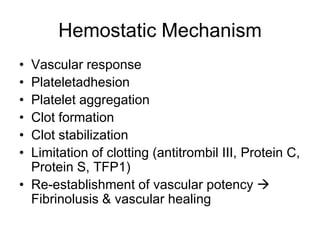
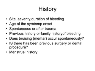
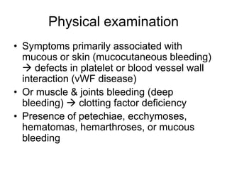
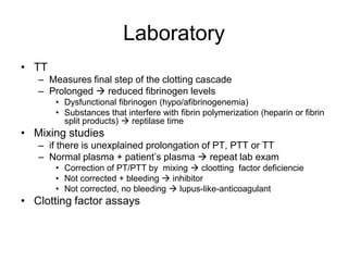
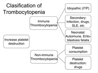
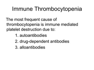
Hemostasis Disease
   In Children
     dr. Bertha
Hemostatic Mechanism
• Vascular response
• Plateletadhesion
• Platelet aggregation
• Clot formation
• Clot stabilization
• Limitation of clotting (antitrombil III, Protein C,
  Protein S, TFP1)
• Re-establishment of vascular potency 
  Fibrinolusis & vascular healing
History
• Site, severity,duration of bleeding
• Age of the symtomp onset
• Spontaneous or after trauma
• Previous history or family historyof bleeding
• Does bruising (memar) occur spontaneously?
• IS there has been previous surgery or dental
  procedure?
• Menstrual history
Physical examination
• Symptoms primarily associated with
  mucous or skin (mucocutaneous bleeding)
   defects in platelet or blood vessel wall
  interaction (vWF disease)
• Or muscle & joints bleeding (deep
  bleeding)  clotting factor deficiency
• Presence of petechiae, ecchymoses,
  hematomas, hemarthroses, or mucous
  bleeding
Trombin time
                      vWF

-Platelet count
-Bleeding time
  -PT / aPTT

                  Specific work-up
Laboratory
• Bleeding time (BT)
   – Assesses platelet function & their interaction with vascular wall
   – Platelet < 100.000/µL  prolonged BT
   – Disproportionate BT  qualitative platelet defects or vWF
     disease

• aPTT
   – Measures the initiation of clotting (intrinsic pathway)
   – doesn’t measure factor VII, XIII or anticoagulant

• PT
   – Measures extrinsic pathway
   – Normal in defiencies offactor VIII, IX, XI or XIII
Laboratory
• TT
   – Measures final step of the clotting cascade
   – Prolonged  reduced fibrinogen levels
       • Dysfunctional fibrinogen (hypo/afibrinogenemia)
       • Substances that interfere with fibrin polymerization (heparin or fibrin
         split products)  reptilase time
• Mixing studies
   – if there is unexplained prolongation of PT, PTT or TT
   – Normal plasma + patient’s plasma  repeat lab exam
       • Correction of PT/PTT by mixing  clootting factor deficiencie
       • Not corrected + bleeding  inhibitor
       • Not corrected, no bleeding  lupus-like-anticoagulant
• Clotting factor assays
Hemophilia
• Hemophilia A  Factor VIII deficiencies
  (85%)
• Hemophilia B  Factor IX deficiencies (
  10-15%)
• Most common & serious congenital
  coagulation factor deficiencies
• Prevalence 1:5000 males
• No racial predilection
• Clinical finding  same
Hemophilia
• Classification
  – Severe deficiency  <1% factor activity
     • Spontaneous bleeding
  – Moderate deficiency  1-5% factor activity
     • Mild trauma to induce bleeding
  – Mild deficiency  >5% factor activity
     • May be asymptomatic, took years to diagnose
Hemophilia
• Clot formation is delayed & fragile
• When bleeding occurs in the closed space 
  tamponade
• Open wound  profuse bleeding
• Bleeding symptoms may be present in utero
• Neonates  intracranial bleeding
• Easy bruising, IM hematomas, hemarthroses
• Bleeding from minor trauma of the mouth 
  persist for days
Hemophilia
• Iliposoas bleeding  life threatening
  – Inability to extend the hip
  – Confirmed by UTZ or CT scan
  – Aggresive therapy
• Life threathening bleeding
  – CNS, Upper airways bleeding
  – External bleeding
  – GI bleeding
Hemophilia
• Prolonged PTT
• Factor assay:
  – Severe def.
  – Moderate def.
  – Mild def.
• Inhibitor assay
Treatment
•   Prevention of trauma
•   Phychosocial
•   Avoid aspirin & NSAID
•   Replacement therapy
    – Recombinant fact. VIII/IX
    – Cryoprecipitate/ cryosupernate
• Joint bleeding:
    – Ice pack
    – Elevate the limb
    – Immobilization of the limb
• Supportive therapy
• multidiciplinary
Chronic Complication
• Chronic joint destruction
• Risk of transfusions associated disease
• Development of inhibitor
Disseminated Intravascular
       Coagulation (DIC)
• Consumptive coagulopathy
• Consumption of clotting factors, platelets &
  anticoagulant protein
• Widespread intavascular deposition of
  fibrin  tissue ischemic & necrosis,
  generalized hemorrhagic state, hemolytic
  anemia
DIC
• Trigger factors:
  – Hypoxia
  – Acidosis
  – Tissue becrosis
  – Shock
  – Endothelial damage
  – Septic shock
  – Incompatible blood transfusion
  – Snake bite
DIC
• Manifestations:
  – Bleeding from surgical incision/venipuncture
    (pungsi vena)petechiae, ecchymoses
  – Organ damage
  – Anemia microangiopathic hemolytic anemia
DIC
• Labs:
  – Prolonged PT, PTT & TT
  – Thrombocytopenia
  – Hemolytic process on blood smear
  – FDP, d-dimer appear in blood
DIC
• Treatment:
  – Treat the cause
  – Restore normal homeostasis
    • Correct shock, acidosis, hypoxia
  – Blood component transfusions
    • Platelet concentrate, cryoprecipitate, FFP
  – Heparin infusions
    • For acute promyelocytic leukemia
    • Not indicated for septic shock, snack bite, massive
      head injury, incompatible transfusions
Platelet
The characteristic of platelets
• Size: 1-4 µm (younger platelets are larger)
• Mean platelet volume (MPV) : 8,9 ± 1.5 µm3
• Number : 150.000 – 400.000 / mm
• Distribution : 1/3 in the spleen, 2/3 in blood
  stream
• Life span : 7-10 days

Bleeding may occur because :
• Reduce in number (thrombsytopenia)
• Defective in function
Thrombocytopeni based on
           pletelet sized
Macrothrombocytes (MPV ↑)            Microthrombocytes (MPV ↓)
• ITP or condition with increased    • Wiskott –Aldrich syndrome
  platelet turnover (eg. DIC)
                                     •   TAR syndrome
•   Bernard-Soulier Syndrome
                                     •   Some storage pool diseases
•   May Heggin anomaly and other
    MYH-9-Related disease            •   Iron def. Anemia
•   Swiss Cheese platelet syndrome   Normal size (MPV normal)
                                     • Disease with hypocellular marrow
•   Montreal platelet syndrome         or infiltrated with malignant
                                       disease
•   Gray platelet syndorme

•   Various mucopolysaccharisoses
Clasification of Trombocytopenia

   Incrase platelet       Disorder of platelet
     destruction             distribution or
                                pooling

                 Thrombocyto
                    penia


   Decrased platelet    Pseudothrombocyto
      production              penia
Clasification of Trombocytopenia

                       Hypersplenism
                    (Portal hypertension,
                  Gaucher disease,cyanitic
 Disorder of      congenital, heart disease,
   platelets         neoplasm, infection)
distribution or
   pooling


                        Hypothermia
Clasification of                              Drugs:
                                       Chlorothiazide, ethanol
Trombocytopenia
                                           Constitusional:
                    Hyperplasia or          Rubella,…
                    suppresion of
                    megakaryocyte    Ineffective Thrombopoeisis:
                                                 ……..

                                         Disorder of control
 Decrase platelet                           mechanism:
   production                         Trombopoetin deficiency
                                        Acquired myelositic
                                             disorder:
                                              Drugs

                      Marrow                 Benign:
                     Infiltrative          Osteoporosis
                      Process
                                            Malignancy
Clasification of Trombocytopenia

                    Platelet inactivation
                        during bloof
                         collection

    Pseudo-
thrombocytopenia     Undercounting of
                    megathrombocytes

                   In vitro agglutination
                         of platelets
                           to EDTA
Clasification of                       Idiopathic (ITP)
Trombocytopenia
                                          Secondary:
                        Immune         Infection, drugs,
                    Thrombocytopenia       SLE, etc

                                           Neonatal:
                                       Autoimune, Erito-
Increase platelet                       blastosis fetalis
   destruction
                                           Platelet
                                         consumption
                      Non-immune
                    Thrombocytopenia      Platelet
                                         destruction:
                                           drugs
Immune Thrombocytopenia
The most frequent cause of
thrombocytopenia is immune mediated
platelet destruction due to:
     1. autoantibodies
     2. drug-dependent antibodies
     3. alloantibodies
Immune (Idiopathic)
     Thrombocytopenic Pupura
A syndrome characterized by
thrombocytopenia :
     1. Shortened platelet survival
     2. Presence of antiplatelet antibody in
        the plasma
     3. Increase megakaryocytes in the
             bone marrow
ITP
• The syndrome can be:
  – Acute
     • Platelet count return to normal within 6 month &
       relaps does not occur
     • Most in children
  – Chronic
     • Platelet count remain low beyond 6 month
     • More common in adult
  – Recurrent
     • Platelet count decrease after having returned to
       normal
Predisposing Factor
• 50-80% : infection (usually viral) prior to
  thrombocytopenia

• About 20% : a specific infection can be
  identified, eg. Rubella, measel, varicella,
  pertussis, mumps, infectious mononucleosis,
  CMV, parvovirus or bacterial

• Measel or smallpox vaccination
Clinical Manifestation
• Skin: Ecchymosess/purpira usually on the
  anterior surface of lower extremities and body
  prominences (ribs, scapula, shoulders, legs,
  pubic)
• Mucous membranes: subconjunctival, buccal
  mucosa, soft palate
• Menorrhagia
• Hematemesis & melena  infrequent
• Others: nose, gum, G.I, Kidnets (usually at the
  onset of the disease
Clinical manifestation
• Intracranial bleeding:
  – Usually preceded by:
     • Headache, dizziness, acute bleeding at other
       place
• Retinal hemorrage
• Middle ear  hearing impairment
• Deep muscle hematoma and hemarthrosis
     • Rare, seen after i.m injection or significant trauma
     • Characteristic of plasma coagulation
Laboratory Findings
• Low platelet count
   – Always <150.000 /mm3
   – Often <20.000 /mm3 in patients with severe generalized
     hemorrhagic manifestations
   – MPV ( N : 8.9 + 1.5 um3)
• Blood smear
   – Thormbocytopenia must be confirmed by peripheral
     blood examination to exclude the diagnosis
     pseudothrombocytopenia, the presence of
     megathrombocytes and other hematologic
     manifestation
   – Blood semar normal apart from thrombicytopenia
• Anemia present in proportion to amount of blood loss
Bone Marrow Aspiration
• Indication
  – Atypical presentation
  – Poor respone to therapy
  – To exclude other hematologic disorder sucg as
    leukemia
• Characteristic
  –   ↑ ,megakaryocytes, immature and asence of budding
  –   Nomlar erythroid and myeloid cells
  –   Occasionally eosinophilia
  –   Erythroid hyperplasia if significant blood loss
Intracranial Hemorrhage
• Incidence : 0,1 – 0,5 %
• Age: 13 month – 16 years
• Platelet count :
  – < 10.000 /mm3 in 73% cases
  – 10-20.000 /mm3 in 25% of cases
  – >20.000 /mm3 in 2% of cases
• Interval between diagnosis of ITP and ICH:
  – <4 wekks in 51% of cases
  – 4 weeks – 9 years in 49%of cases (mean 27 weeks)
Intracranial Hemorrhage
• Risk Factors in 45 % cases of ICH
  include:
   –   Head injury (29%)
   –   Aspirin treatment (5%)
   –   AV malformation (17%)
   –   Mucocutaneous hemorrhage (49%)
• Site of ICH:
   – Intra cerebral (77%)
   – Subdural hematoma (23%)
• 50 % had prior tretament with steroid
  and / or IVIG
• 54% survival, most without
  permanent damage
Supportive Treatment
• No treatment is required when platelet count
  >20.000 /mm3 , asymptomatic or has mild
  bruising but no evidence of mucous membrane
  bleeding
• Competitive sport should be avoided
• Depoprovera or any other long-acting
  progesteron in suspending menstruation for
  several month
• Aspirin, Nonsteroidal antiinflammatory agents
  and any other drug the interfere with platelet
  function should not be given
Farmacological Treatment
• Treatment choice : Steroid, IVIG, and anti–D
• Indication
  – Platelet count <20.000 /mm3 and significant
    mucous membrane bleeding
  – Platelet <10.000 /mm3 and minor purpura
Steroid Therapy
• Mechanisms:
   – Inhibits phagocytosis of antibody coated platelet in
     the spleen  prolongs platelet survival
   – Improves capillary resistance and thereby improve
     platelet economy
• Dose and Duration:
   – Dose : 2mg/kg/day (max. 60/mg/day) in divided dose.
     Tap off in 5-7 day interval and stopped at the end of
     21-28 days, regardless of the response
   – In severe cases methylprednisolone 30mg/kg/day
     (max 1 g/day) for 3 days
• Prolonged case of steroid in undesirable:
   – Worsen the thrombocytopenia and depress platelet
     [rpduction
   – Side effect : weight gain, caushingoid facies, fluid
     retention, acne, hyperglycemia, hypertension, mood
     swings, pseudotumor cerebri, cataracts, growth
     retradation , avascular necrosis
IVIG
• Mechanism of action
  – Reticuloendothelial Fc-receptor blockade
  – Activation of inhibitor pathways
  – Decrease autoantibody synthesis
• Indication
  – Neonatal Symptomatic Immune Thrombocytopenia 
    Infant less than 2 y.o are generally more refractory to
    steroid treatment
  – Alternative therapy to corticosteroid therapy
• Much more expensive and has significant side
  effects
Anti –D Therapy
• Plasma derived gamma immune globulin of anti
  –Rh antigen
• Mechanism action :
   – Blockade of Fc receptor of reticuloendothelial cell
• Platelet is increase after 48 hours, therefore the
  therapy is not appropriate for emergency
  treatment
• Patients who have not undergone splenectomy
  and Rh positive are more likely to respond to IV
  Anti-D
Splenectomy
• Indication
  – Severe acute ITP with acute life-threatening bleeding
    and not responsive to medical treatment
  – Chronic ITP with bleeding symptom or platelet count
    persistently below 30.000 /mm3 an not responsive to
    medical treatment for several years
  – In very active patient subject to frequent trauma, early
    splenectomy may be indicated

• Because the hazard of overwhelming
  postsplenectomy infection (OPSI) the procedure
  should be performed after clear indication
Splenectomy
• Indication for splenectomy are rare because of
  judicious use if steroid and IVIG
• It is rarely necessary to perform splenectomy
  before 2 years adter diagnosis
• Laparoscopic splenectomy is preferable to open
  splenectomy
• Up to 70% have complete and long-lasting
  recovery
• 40% wuth persistent thrombocytopenia after
  splenectomy have acsseory spleen
Treatment Algorithm

Yang skema itu.. Gak keliatan di foto.. Maaf
                ya kawan..
Live Threatening Hemorrahage
• Platelet transfusion
• Methylprednisolone 500 mg/m2 IV per day
  for 3 days
• IVIG 2 /kg for 12 hours infusion
• Emergency splenecomy
Prognosis
• Excellent, 50 % recover within 1 month % 70-80% within
  6 month

• Spontaneous remission after 1 year in uncommon, but
  may occur even after several years

• When demonstration underlying cause, the prognosis is
  related to the cause

• Age older than 10 years, insidious onset, female are
  associated with chronic ITP

• 50-60 % chronic ITP………….. without any other therapy
  and without splenectomy

Hemostastis Pada Anak

  • 1.
    Hemostasis Disease In Children dr. Bertha
  • 2.
    Hemostatic Mechanism • Vascularresponse • Plateletadhesion • Platelet aggregation • Clot formation • Clot stabilization • Limitation of clotting (antitrombil III, Protein C, Protein S, TFP1) • Re-establishment of vascular potency  Fibrinolusis & vascular healing
  • 3.
    History • Site, severity,durationof bleeding • Age of the symtomp onset • Spontaneous or after trauma • Previous history or family historyof bleeding • Does bruising (memar) occur spontaneously? • IS there has been previous surgery or dental procedure? • Menstrual history
  • 4.
    Physical examination • Symptomsprimarily associated with mucous or skin (mucocutaneous bleeding)  defects in platelet or blood vessel wall interaction (vWF disease) • Or muscle & joints bleeding (deep bleeding)  clotting factor deficiency • Presence of petechiae, ecchymoses, hematomas, hemarthroses, or mucous bleeding
  • 5.
    Trombin time vWF -Platelet count -Bleeding time -PT / aPTT Specific work-up
  • 6.
    Laboratory • Bleeding time(BT) – Assesses platelet function & their interaction with vascular wall – Platelet < 100.000/µL  prolonged BT – Disproportionate BT  qualitative platelet defects or vWF disease • aPTT – Measures the initiation of clotting (intrinsic pathway) – doesn’t measure factor VII, XIII or anticoagulant • PT – Measures extrinsic pathway – Normal in defiencies offactor VIII, IX, XI or XIII
  • 7.
    Laboratory • TT – Measures final step of the clotting cascade – Prolonged  reduced fibrinogen levels • Dysfunctional fibrinogen (hypo/afibrinogenemia) • Substances that interfere with fibrin polymerization (heparin or fibrin split products)  reptilase time • Mixing studies – if there is unexplained prolongation of PT, PTT or TT – Normal plasma + patient’s plasma  repeat lab exam • Correction of PT/PTT by mixing  clootting factor deficiencie • Not corrected + bleeding  inhibitor • Not corrected, no bleeding  lupus-like-anticoagulant • Clotting factor assays
  • 8.
    Hemophilia • Hemophilia A Factor VIII deficiencies (85%) • Hemophilia B  Factor IX deficiencies ( 10-15%) • Most common & serious congenital coagulation factor deficiencies • Prevalence 1:5000 males • No racial predilection • Clinical finding  same
  • 9.
    Hemophilia • Classification – Severe deficiency  <1% factor activity • Spontaneous bleeding – Moderate deficiency  1-5% factor activity • Mild trauma to induce bleeding – Mild deficiency  >5% factor activity • May be asymptomatic, took years to diagnose
  • 10.
    Hemophilia • Clot formationis delayed & fragile • When bleeding occurs in the closed space  tamponade • Open wound  profuse bleeding • Bleeding symptoms may be present in utero • Neonates  intracranial bleeding • Easy bruising, IM hematomas, hemarthroses • Bleeding from minor trauma of the mouth  persist for days
  • 12.
    Hemophilia • Iliposoas bleeding life threatening – Inability to extend the hip – Confirmed by UTZ or CT scan – Aggresive therapy • Life threathening bleeding – CNS, Upper airways bleeding – External bleeding – GI bleeding
  • 13.
    Hemophilia • Prolonged PTT •Factor assay: – Severe def. – Moderate def. – Mild def. • Inhibitor assay
  • 15.
    Treatment • Prevention of trauma • Phychosocial • Avoid aspirin & NSAID • Replacement therapy – Recombinant fact. VIII/IX – Cryoprecipitate/ cryosupernate • Joint bleeding: – Ice pack – Elevate the limb – Immobilization of the limb • Supportive therapy • multidiciplinary
  • 16.
    Chronic Complication • Chronicjoint destruction • Risk of transfusions associated disease • Development of inhibitor
  • 17.
    Disseminated Intravascular Coagulation (DIC) • Consumptive coagulopathy • Consumption of clotting factors, platelets & anticoagulant protein • Widespread intavascular deposition of fibrin  tissue ischemic & necrosis, generalized hemorrhagic state, hemolytic anemia
  • 18.
    DIC • Trigger factors: – Hypoxia – Acidosis – Tissue becrosis – Shock – Endothelial damage – Septic shock – Incompatible blood transfusion – Snake bite
  • 19.
    DIC • Manifestations: – Bleeding from surgical incision/venipuncture (pungsi vena)petechiae, ecchymoses – Organ damage – Anemia microangiopathic hemolytic anemia
  • 20.
    DIC • Labs: – Prolonged PT, PTT & TT – Thrombocytopenia – Hemolytic process on blood smear – FDP, d-dimer appear in blood
  • 21.
    DIC • Treatment: – Treat the cause – Restore normal homeostasis • Correct shock, acidosis, hypoxia – Blood component transfusions • Platelet concentrate, cryoprecipitate, FFP – Heparin infusions • For acute promyelocytic leukemia • Not indicated for septic shock, snack bite, massive head injury, incompatible transfusions
  • 22.
  • 23.
    The characteristic ofplatelets • Size: 1-4 µm (younger platelets are larger) • Mean platelet volume (MPV) : 8,9 ± 1.5 µm3 • Number : 150.000 – 400.000 / mm • Distribution : 1/3 in the spleen, 2/3 in blood stream • Life span : 7-10 days Bleeding may occur because : • Reduce in number (thrombsytopenia) • Defective in function
  • 24.
    Thrombocytopeni based on pletelet sized Macrothrombocytes (MPV ↑) Microthrombocytes (MPV ↓) • ITP or condition with increased • Wiskott –Aldrich syndrome platelet turnover (eg. DIC) • TAR syndrome • Bernard-Soulier Syndrome • Some storage pool diseases • May Heggin anomaly and other MYH-9-Related disease • Iron def. Anemia • Swiss Cheese platelet syndrome Normal size (MPV normal) • Disease with hypocellular marrow • Montreal platelet syndrome or infiltrated with malignant disease • Gray platelet syndorme • Various mucopolysaccharisoses
  • 25.
    Clasification of Trombocytopenia Incrase platelet Disorder of platelet destruction distribution or pooling Thrombocyto penia Decrased platelet Pseudothrombocyto production penia
  • 26.
    Clasification of Trombocytopenia Hypersplenism (Portal hypertension, Gaucher disease,cyanitic Disorder of congenital, heart disease, platelets neoplasm, infection) distribution or pooling Hypothermia
  • 27.
    Clasification of Drugs: Chlorothiazide, ethanol Trombocytopenia Constitusional: Hyperplasia or Rubella,… suppresion of megakaryocyte Ineffective Thrombopoeisis: …….. Disorder of control Decrase platelet mechanism: production Trombopoetin deficiency Acquired myelositic disorder: Drugs Marrow Benign: Infiltrative Osteoporosis Process Malignancy
  • 28.
    Clasification of Trombocytopenia Platelet inactivation during bloof collection Pseudo- thrombocytopenia Undercounting of megathrombocytes In vitro agglutination of platelets to EDTA
  • 29.
    Clasification of Idiopathic (ITP) Trombocytopenia Secondary: Immune Infection, drugs, Thrombocytopenia SLE, etc Neonatal: Autoimune, Erito- Increase platelet blastosis fetalis destruction Platelet consumption Non-immune Thrombocytopenia Platelet destruction: drugs
  • 30.
    Immune Thrombocytopenia The mostfrequent cause of thrombocytopenia is immune mediated platelet destruction due to: 1. autoantibodies 2. drug-dependent antibodies 3. alloantibodies
  • 31.
    Immune (Idiopathic) Thrombocytopenic Pupura A syndrome characterized by thrombocytopenia : 1. Shortened platelet survival 2. Presence of antiplatelet antibody in the plasma 3. Increase megakaryocytes in the bone marrow
  • 32.
    ITP • The syndromecan be: – Acute • Platelet count return to normal within 6 month & relaps does not occur • Most in children – Chronic • Platelet count remain low beyond 6 month • More common in adult – Recurrent • Platelet count decrease after having returned to normal
  • 33.
    Predisposing Factor • 50-80%: infection (usually viral) prior to thrombocytopenia • About 20% : a specific infection can be identified, eg. Rubella, measel, varicella, pertussis, mumps, infectious mononucleosis, CMV, parvovirus or bacterial • Measel or smallpox vaccination
  • 34.
    Clinical Manifestation • Skin:Ecchymosess/purpira usually on the anterior surface of lower extremities and body prominences (ribs, scapula, shoulders, legs, pubic) • Mucous membranes: subconjunctival, buccal mucosa, soft palate • Menorrhagia • Hematemesis & melena  infrequent • Others: nose, gum, G.I, Kidnets (usually at the onset of the disease
  • 35.
    Clinical manifestation • Intracranialbleeding: – Usually preceded by: • Headache, dizziness, acute bleeding at other place • Retinal hemorrage • Middle ear  hearing impairment • Deep muscle hematoma and hemarthrosis • Rare, seen after i.m injection or significant trauma • Characteristic of plasma coagulation
  • 36.
    Laboratory Findings • Lowplatelet count – Always <150.000 /mm3 – Often <20.000 /mm3 in patients with severe generalized hemorrhagic manifestations – MPV ( N : 8.9 + 1.5 um3) • Blood smear – Thormbocytopenia must be confirmed by peripheral blood examination to exclude the diagnosis pseudothrombocytopenia, the presence of megathrombocytes and other hematologic manifestation – Blood semar normal apart from thrombicytopenia • Anemia present in proportion to amount of blood loss
  • 37.
    Bone Marrow Aspiration •Indication – Atypical presentation – Poor respone to therapy – To exclude other hematologic disorder sucg as leukemia • Characteristic – ↑ ,megakaryocytes, immature and asence of budding – Nomlar erythroid and myeloid cells – Occasionally eosinophilia – Erythroid hyperplasia if significant blood loss
  • 38.
    Intracranial Hemorrhage • Incidence: 0,1 – 0,5 % • Age: 13 month – 16 years • Platelet count : – < 10.000 /mm3 in 73% cases – 10-20.000 /mm3 in 25% of cases – >20.000 /mm3 in 2% of cases • Interval between diagnosis of ITP and ICH: – <4 wekks in 51% of cases – 4 weeks – 9 years in 49%of cases (mean 27 weeks)
  • 39.
    Intracranial Hemorrhage • RiskFactors in 45 % cases of ICH include: – Head injury (29%) – Aspirin treatment (5%) – AV malformation (17%) – Mucocutaneous hemorrhage (49%) • Site of ICH: – Intra cerebral (77%) – Subdural hematoma (23%) • 50 % had prior tretament with steroid and / or IVIG • 54% survival, most without permanent damage
  • 40.
    Supportive Treatment • Notreatment is required when platelet count >20.000 /mm3 , asymptomatic or has mild bruising but no evidence of mucous membrane bleeding • Competitive sport should be avoided • Depoprovera or any other long-acting progesteron in suspending menstruation for several month • Aspirin, Nonsteroidal antiinflammatory agents and any other drug the interfere with platelet function should not be given
  • 41.
    Farmacological Treatment • Treatmentchoice : Steroid, IVIG, and anti–D • Indication – Platelet count <20.000 /mm3 and significant mucous membrane bleeding – Platelet <10.000 /mm3 and minor purpura
  • 42.
    Steroid Therapy • Mechanisms: – Inhibits phagocytosis of antibody coated platelet in the spleen  prolongs platelet survival – Improves capillary resistance and thereby improve platelet economy • Dose and Duration: – Dose : 2mg/kg/day (max. 60/mg/day) in divided dose. Tap off in 5-7 day interval and stopped at the end of 21-28 days, regardless of the response – In severe cases methylprednisolone 30mg/kg/day (max 1 g/day) for 3 days • Prolonged case of steroid in undesirable: – Worsen the thrombocytopenia and depress platelet [rpduction – Side effect : weight gain, caushingoid facies, fluid retention, acne, hyperglycemia, hypertension, mood swings, pseudotumor cerebri, cataracts, growth retradation , avascular necrosis
  • 43.
    IVIG • Mechanism ofaction – Reticuloendothelial Fc-receptor blockade – Activation of inhibitor pathways – Decrease autoantibody synthesis • Indication – Neonatal Symptomatic Immune Thrombocytopenia  Infant less than 2 y.o are generally more refractory to steroid treatment – Alternative therapy to corticosteroid therapy • Much more expensive and has significant side effects
  • 44.
    Anti –D Therapy •Plasma derived gamma immune globulin of anti –Rh antigen • Mechanism action : – Blockade of Fc receptor of reticuloendothelial cell • Platelet is increase after 48 hours, therefore the therapy is not appropriate for emergency treatment • Patients who have not undergone splenectomy and Rh positive are more likely to respond to IV Anti-D
  • 45.
    Splenectomy • Indication – Severe acute ITP with acute life-threatening bleeding and not responsive to medical treatment – Chronic ITP with bleeding symptom or platelet count persistently below 30.000 /mm3 an not responsive to medical treatment for several years – In very active patient subject to frequent trauma, early splenectomy may be indicated • Because the hazard of overwhelming postsplenectomy infection (OPSI) the procedure should be performed after clear indication
  • 46.
    Splenectomy • Indication forsplenectomy are rare because of judicious use if steroid and IVIG • It is rarely necessary to perform splenectomy before 2 years adter diagnosis • Laparoscopic splenectomy is preferable to open splenectomy • Up to 70% have complete and long-lasting recovery • 40% wuth persistent thrombocytopenia after splenectomy have acsseory spleen
  • 47.
    Treatment Algorithm Yang skemaitu.. Gak keliatan di foto.. Maaf ya kawan..
  • 48.
    Live Threatening Hemorrahage •Platelet transfusion • Methylprednisolone 500 mg/m2 IV per day for 3 days • IVIG 2 /kg for 12 hours infusion • Emergency splenecomy
  • 49.
    Prognosis • Excellent, 50% recover within 1 month % 70-80% within 6 month • Spontaneous remission after 1 year in uncommon, but may occur even after several years • When demonstration underlying cause, the prognosis is related to the cause • Age older than 10 years, insidious onset, female are associated with chronic ITP • 50-60 % chronic ITP………….. without any other therapy and without splenectomy