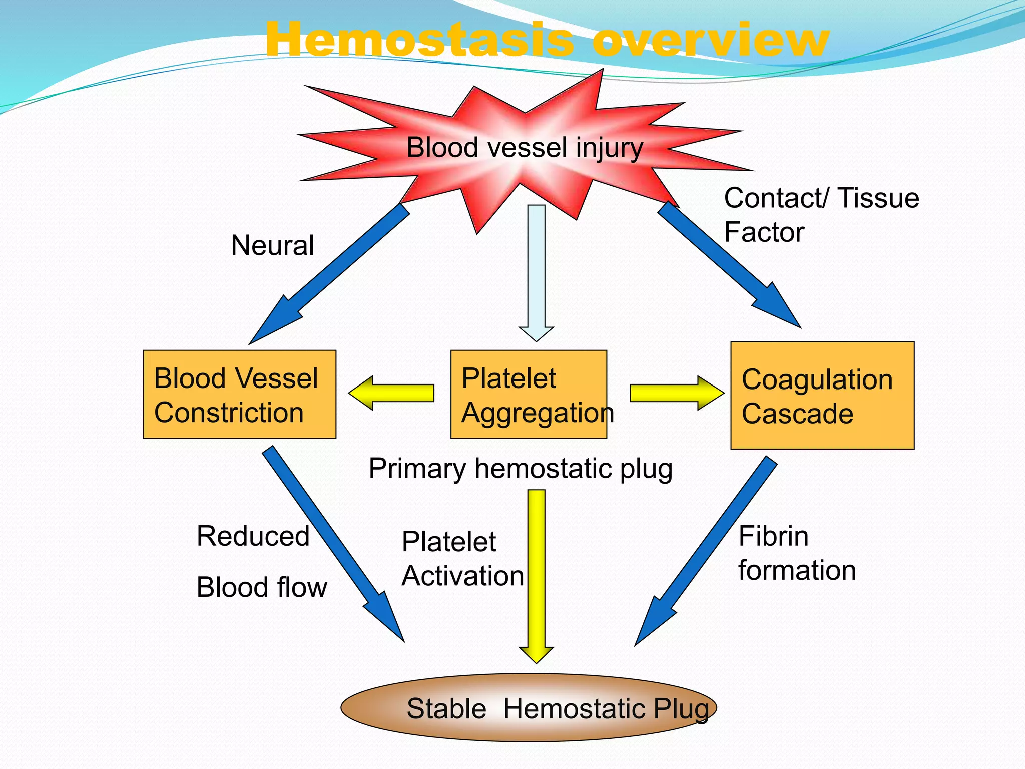
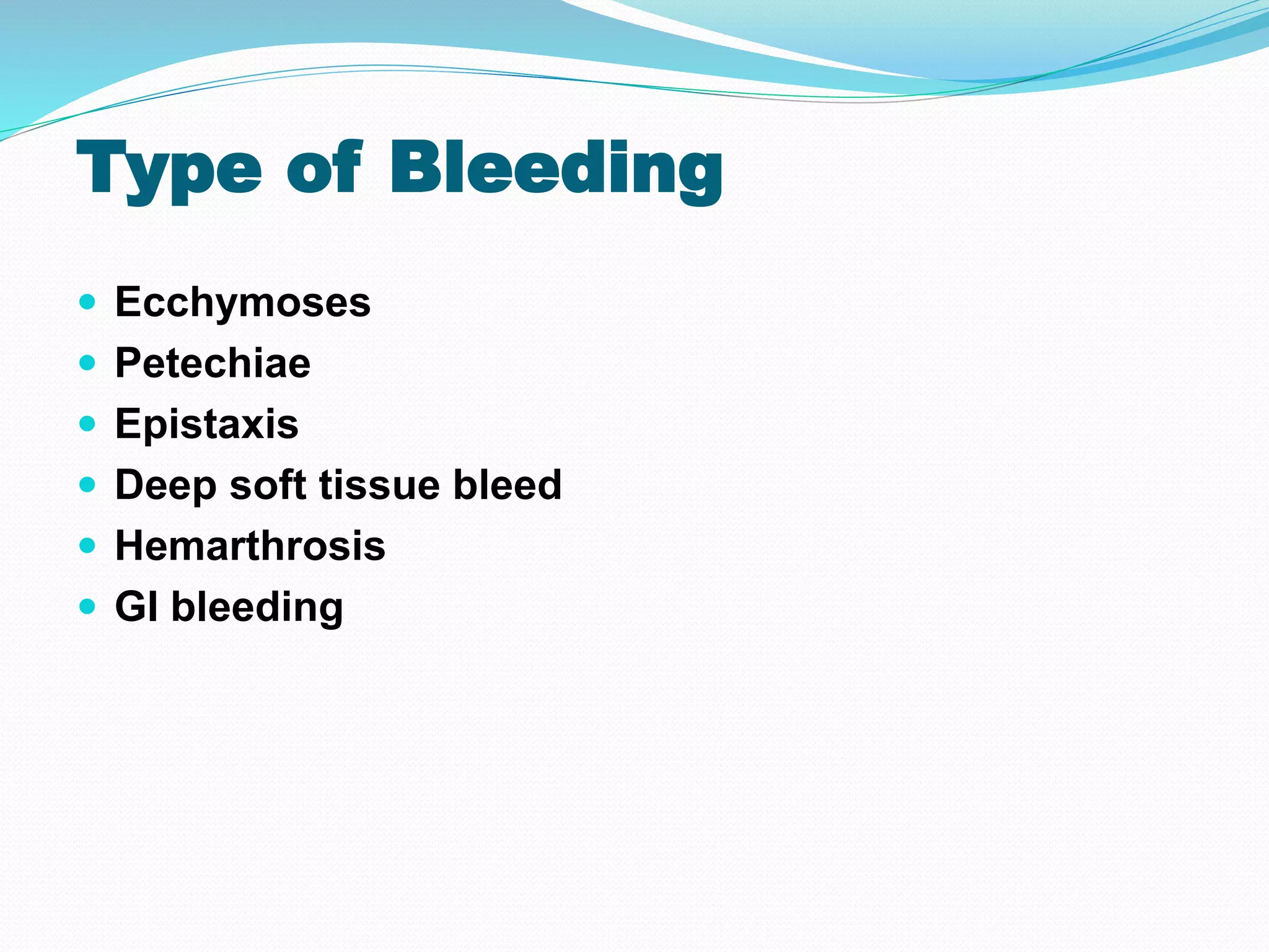
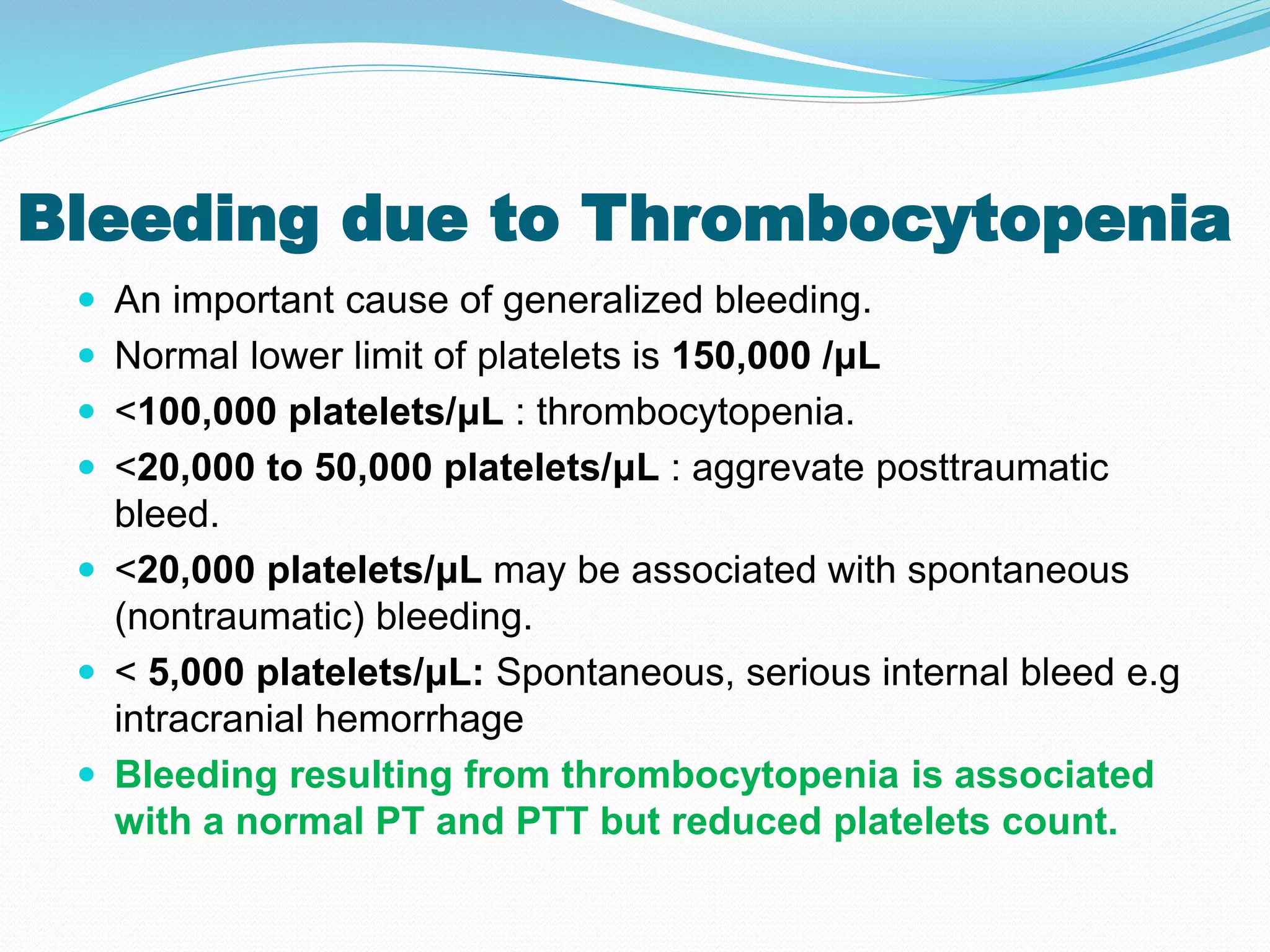
Dr. Umme Habiba
Hemostasis
 Normal hemostasis comprises a series of regulated
processes that maintain blood in a fluid ,clot-free
state in normal vessels while rapidly forming a
localized haemostatic plug at the site of vascular
injury
Hemostasis overview
Blood vessel injury
Platelet
Aggregation
Platelet
Activation
Blood Vessel
Constriction
Coagulation
Cascade
Stable Hemostatic Plug
Fibrin
formation
Reduced
Blood flow
Contact/ Tissue
Factor
Primary hemostatic plug
Neural
Vessel Wall Damage
Platelet Plug
Clot
1. Vessel Wall Abnormalities
2. Platelets Deficiency or
Dysfunction
3. Derangement of Clotting
Factors
ETIOLOGY OF BLEEDING
DISRODERS:
NORMAL COAGULATION
There are 3 stages in normal coagulation:
 Primary hemostasis platelets (adhesion &
aggregation).
 Secondary hemostasis the clotting factors.
 Tertiary hemostasis is the formation of fibrin polymers
(Fibrin cross linking)& subsequent resolution through
fibrinolysis.
Tests For Evaluation of Bleeding
Disorders
 Bleeding time: 2-9 minutes. Prolonged in thrombocytopenia/platelet
function defects
 Platelet counts:150-450X103/mm3
 Prothrombin time (PT): 10-15 sec. For the extrinsic (V, VII, X)&
common (prothrombin, fibrinogen) coagulation pathways
 Activated Partial thromboplastin time (aPTT): 30 – 50 sec. For the
intrinsic (V, VIII, IX, XI, VII) & common (prothrombin, fibrinogen)
coagulation pathways
 Thrombin time:9-13 sec. Time for thrombin to convert fibrinogen to
fibrin
 Other specific tests
INR
D-Dimers
FDPs
Evaluation of the patient
 History
Spontaneous bleeding, Post traumatic, Umblical
stump/post circumsicion, dental extractions.
Menorrahgia
 Physical Examination
Ecchymoses / Petechiae/ Epistaxis/ Deep soft tissue
bleed/ Hemarthroses/ GI bleed
 Laboratory Evaluation
CBC,PT, aPTT, Fibrinogen, Platelet functional Assays
Type of Bleeding
 Ecchymoses
 Petechiae
 Epistaxis
 Deep soft tissue bleed
 Hemarthrosis
 GI bleeding
Petechiae
Do not blanch with pressure
(D/D. angiomas)
Not palpable
(D/D. vasculitis)
(typical of platelet disorders)
Petechiae → Purpura → Ecchymoses
Hemarthrosis
 Most common problem in haemophilia
 Symptoms: prolonged bleeding, pain & disabled joints
 Repeated joint bleed → synovial inflammation &↑ vascularity
→thickening of the synovium.
Thigh muscle bleedings
Clinical Features of Bleeding Disorders
Platelet Coagulation
disorders factor disorders
Site of bleeding Skin Deep in soft tissues
Mucous membranes (joints, muscles)
(epistaxis, gum,
vaginal, GI tract)
Petechiae Yes No
Ecchymoses (“bruises”) Small, superficial Large (hematomas),
deep
Hemarthrosis / muscle bleeding Extremely rare Common
Bleeding after cuts & scratches Yes No
Bleeding after surgery or trauma Immediate, Delayed (1-2 days),
usually mild often severe
Laboratory Assessment
 Guided by history
 Screening tests
 PT
 aPTT
 platelet count
 fibrinogen
 thrombin time
Bleeding Disorders Caused by
Vessel Wall Abnormalities
 Disorders within this category are relatively common.
 But do not usually cause serious bleeding problems.
 Mostly present with small hemorrhages .
 Petechiae and purpura in the skin or mucous
membranes, particularly the gingivae.
 On occasion hemorrhages occur into joints, muscles,
and subperiosteal locations, or take the form of
menorrhagia, nosebleeds, gastrointestinal bleeding, or
hematuria.
 The platelet count and tests of coagulation (PT, PTT)
are usually normal.
VESSEL WALL ABNORMALITIES:
 Infections
 Drug Reactions
 Scurvy
 Ehlers-Danlos Syndrome
 Henoch-Schönlein Purpura
 Hereditary Hemorrhagic Telangiectasia
 Perivascular Amyloidosis
Bleeding due to Thrombocytopenia
 An important cause of generalized bleeding.
 Normal lower limit of platelets is 150,000 /μL
 <100,000 platelets/μL : thrombocytopenia.
 <20,000 to 50,000 platelets/μL : aggrevate posttraumatic
bleed.
 <20,000 platelets/μL may be associated with spontaneous
(nontraumatic) bleeding.
 < 5,000 platelets/μL: Spontaneous, serious internal bleed e.g
intracranial hemorrhage
 Bleeding resulting from thrombocytopenia is associated
with a normal PT and PTT but reduced platelets count.
PLATELET DISORDERS:
DEFICIENCY
Decreased Production
Increased Destruction
Abnormal Distribution
Dilution
DYSFUNCTION
Defects of Adhesion
Defects of Aggregation
Disorders of Platelet Secretion
Decreased Production of Platelets
1:Selective impairment of platelet
production
Drug-induced: alcohol, thiazides, cytotoxic drugs
Infections: measles, human immunodeficiency virus
(HIV)
Nutritional deficiencies: B12, folate deficiency
(megaloblastic)
Bone marrow failure: Aplastic anemia
Bone marrow replacement: Leukemia, disseminated
cancer, granulomatous disease
Ineffective hematopoiesis: Myelodysplastic syndromes
Decreased Platelet Survival:
 Immunologic destruction
Primary autoimmune: Chromic ITP, Acute ITP
Secondary autoimmune: SLE, B-cell lymphoid neoplasms
Alloimmunization: post-transfusion and neonatal
Drug-associated: quinidine, heparin, sulfa compounds
Infections: HIV, infectious mononucleosis (transient, mild), dengue fever
 Non immunologic destruction
Disseminated intravascular coagulation
Thrombotic microangiopathies
Giant Hemangioma
Sequestration
 There is abnormal redistribution of platelets.
Platelets get trapped in splenic circulation more than
systemic circulation. Conditions causing
Hypersplenism like CLD
Dilutional
 Pegnancy, Transfusions
Immune Thrombocytopenic Purpura (ITP)
 Autoimmune disorder
 Destruction of platelets
 Anti-platelet antibodies
 Types:
 Acute
 Chronic
EPIDEMIOLOGY:
• ACUTE
• Lasts < 6 months
• Mostly seen in children
• Follows a viral infection
• Self limiting
 CHRONIC
• Lasts > 6 months
• Mainly seen in adults
• ♂ :♀ ratio = 1:3
• Is known to be associated with HIV, SLE, CLL, NHL
• Spontaneous remission uncommon
PATHOGENESIS:
Sensitized
Platelet
Anti platelet
Autoantibodies
Fc receptor
Fc portion
of antibody
PATHOGENESIS:
 In approximately 60% of the patients, the cause is not idiopathic but
autoimmune
 Antibodies of IgG type bind to the platelet membrane glycoproteins
IIb-IIIa or Ib-IX.
 The sensitized platelets become susceptible to opsonization and
phagocytosis by the splenic macrophages.
 The life span of platelets is reduced to a few hours (normal 5-7 days).
Clinical Features
 Chronic ITP occurs most commonly in adult women
younger than 40 years of age.
 The female-to-male ratio is 3 : 1.
 It is often insidious in onset.
 Clinical presentation depends on severity:
Asymptomatic/Petechiae/Ecchymoses/Gum
Bleeding/Epistaxis/Menorrhagia/Subarachnoid
hemorrhage/Intracerebral hemorrhage
DIAGNOSIS: (mainly a diagnosis of exclusion)
 History: Viral infection
 Clinical Features: Petechiae, ecchymoses
 Complete Blood Counts: Isolated thrombocytopenia
 Peripheral Film: Mega thrombocytes (giant Platelets)
 Bone Marrow: Megakaryocytosis with left shift
 The PT and PTT are normal.
 Platelet auto antibodies: Not widely available, non specific
Bone Marrow
 The marrow reveals a modestly increased number of
megakaryocytes.
 Some are apparently immature, with large, non-
lobulated, single nuclei.
 The importance of bone marrow examination is to
rule out thrombocytopenia resulting from bone
marrow failure or other disorders.
 The peripheral blood often reveals abnormally large
platelets (megathrombocytes), which are a sign of
accelerated thrombopoiesis.
MORPHOLOGY
Normal ITP
MANAGEMENT:
Steroids: Prednisolone 0.5 -1 mg per kg per day
IV Immunoglobulins: 400mg per kg per day for 5 days
Surgery: Splenectomy
Immunosuppression: Cyclosporine, Azathioprine
Platelet transfusion: CONTRAINDICATED except in case of life
threatening bleed
Drug-Induced Thrombocytopenia
Direct effects on platelets .
Secondary to immunologically mediated platelet
destruction.
The drugs most commonly implicated : Quinine,
Quinidine, and Vancomycin. All of which bind platelet
glycoproteins & create antigenic determinants that
are recognized by antibodies.
Drugs may induce true autoantibodies through
unknown mechanisms.
Heparin-induced thrombocytopenia
(HIT)
 Thrombocytopenia occurs in about 5% of persons receiving
heparin.
 Type I HIT: occurs rapidly after the onset of therapy and is of
little clinical importance.
 Sometimes resolved despite the continuation of therapy.
 It most likely results from a direct platelet-aggregating effect of
heparin.
 Type II HIT: is less common but of much greater clinical
significance.
 It occurs 5-14 days after therapy begins (or sooner if the
person has been sensitized to heparin)
 Type 2 HIT often leads to life threatening venous and
arterial thrombosis.
 This severe form of HIT is caused by antibodies that
recognize complexes of heparin and platelet factor 4,
which is a normal component of platelet granules.
 Binding of antibody to these complexes activates
platelets and promotes thrombosis,even in the setting of
thrombocytopenia.
Pathophysiology of HIT
Complications of HIT:
 Unless therapy is immediately discontinued and an
alternative non-heparin anticoagulant instituted.
 Clots within large arteries: vascular insufficiency and limb
loss.
 Emboli from DVT: fatal pulmonary thromboembolism.
 The risk of severe HIT is lowered, but not completely
eliminated, by the use of LMWH (low-molecular-weight
heparin).
 Unfortunately, once severe HIT develops, even LMWH
exacerbate the thrombotic tendency and must be
avoided.
HIV-Associated Thrombocytopenia
 Thrombocytopenia is one of the most common
hematologic manifestations of HIV infection.
 Both impaired platelet production and increased
destruction contribute.
Thrombotic Thrombocytopenic Purpura (TTP)
& Hemolytic-Uremic Syndrome (HUS)
 Thrombotic microangiopathies encompass a
spectrum of clinical syndromes that includes TTP
and HUS.
 They are caused by insults that lead to excessive
activation of platelets, which deposit as thrombi in
small blood vessels.
TTP
 Defined as the pentad of :
 Fever,
 Thrombocytopenia,
 Microangiopathic hemolytic anemia,
 Transient neurologic deficits,
 Renal failure.
HUS
 Associated with : Microangiopathic hemolytic anemia
 Thrombocytopenia But is distinguished by :
 Absence of neurologic symptoms.
 Prominence of acute renal failure.
 Its frequent occurrence in children.
 Many adult patients with “TTP” lack one or more of the
five criteria.
 Some patients with “HUS” have fever and neurologic
dysfunction.
Symptoms
 Intravascular thrombi cause a microangiopathic hemolytic
anemia and widespread organ dysfunction,
 Attendant consumption of platelets leads to
thrombocytopenia.
 In TTP and HUS (unlike DIC ), activation of the
coagulation cascade is not of primary importance.
 PT and PTT, are usually normal.
Pathogenesis
 TTP is usually associated with a deficiency in a plasma
enzyme called ADAMTS13, also designated “vWF
metalloprotease.”
 ADAMTS13 normally degrades very high-molecular-
weight multimers of von Willebrand factor (vWF).
 These multimers accumulate in plasma and tend to
promote platelet activation and aggregation.
 Superimposition of endothelial cell injury (caused by
some other condition) may further promote the formation
of platelet microaggregates, thus initiating or
exacerbating clinically evident TTP.
Thrombotic Microangiopathies: Causes
and Associations
Thrombotic Microangiopathies:
Causes and Associations
Platelet Functions Disorders
 Qualitative defects of platelet function can be
 Inherited
 Acquired.
Inherited disorders of platelet function
Classified into three pathogenically distinct groups:
 (1) defects of adhesion.
 (2) defects of aggregation.
 (3) disorders of platelet secretion (release
reaction).
Functions of Platelet
Bernard-Soulier syndrome
 Defective adhesion of platelets to subendothelial matrix.
 Caused by an inherited deficiency of the platelet
membrane glycoprotein complex Ib-IX.
 This glycoprotein is a receptor for vWF .
 Essential for normal platelet adhesion to the
subendothelial extracellular matrix .
 Affected patients have a variable, often severe, bleeding
tendency.
Glanzmann Thrombasthenia
 Platelet Aggregation Defect.
 Transmitted as an autosomal recessive trait.
 Thrombasthenic platelets fail to aggregate in
response to adenosine diphosphate (ADP), collagen,
epinephrine, or thrombin.
 Deficiency or dysfunction of glycoprotein IIb- IIIa.
 An integrin that participates in “bridge formation”
between platelets by binding fibrinogen.
 The associated bleeding tendency is often severe.
Disorders of Platelet Secretion
 Characterized by the defective release of certain
mediators of platelet activation such as
thromboxanes and granule-bound ADP.
 (ἀ granules): Grey Platelet Syndrome
 (ẟ granules): Wiskott-Aldrich syndrome, Storage
Pool Defect, Chediak-Higashii
Acquired Defects of Platelet function
 1:Caused by ingestion of aspirin and other nonsteroidal
anti-inflammatory drugs.
 Aspirin is a potent, irreversible inhibitor of the enzyme
cyclooxygenase.
 Required for the synthesis of thromboxane A2 and
prostaglandins .
 These mediators play important roles in platelet
aggregation and subsequent release reactions.
 The antiplatelet effects of aspirin form the basis for its use
in the prophylaxis of coronary thrombosis .
 2:Uremia
 The pathogenesis of platelet dysfunction in uremia is
complex and involves defects in adhesion, granule
secretion, and aggregation.
SUMMARY
BLEEDING DISORDERS
(aka, Hemorrhagic “DIATHESES”)
 Blood vessel wall abnormalities √
 Reduced platelets √
 Decreased platelet function √
 Abnormal clotting factors √
 DIC (Disseminated INTRA-vascular Coagulation), also has ↓
plats.
THROMBOCYTOPENIAS
 Like RBCs:
 DE-creased production
 IN-creased destruction
 Sequestration (Hypersplenism)
 Dilutional
 Normal value 150K-300K
ITP
 ADULTS AND ELDERLY
 ACUTE OR CHRONIC
 AUTO-IMMUNE
 ANTI-PLATELET ANTIBODIES PRESENT
 INCREASED MARROW MEGAKARYOCYTES
 PERIPHERAL SMEAR GIANT PLATELETS.
 Rx: STEROIDS
DRUGS
 Quinine
 Quinidine
 Sulfonamide antibiotics
 HEPARIN: Type II HIT
HIV
 BOTH DE-creased production &
 IN-creased destruction factors are present
Thrombotic Microangiopathies
 BOTH are very SERIOUS CONDITIONS with a HIGH
mortality:
 TTP (THROMBOTIC THROMBOCYTOPENIC
PURPURA)
 H.U.S. (HEMOLYTIC UREMIC SYNDROME)
 These can also be called “consumptive”
coagulopathies, just like a DIC
Worksheet for students
 Tabulate differences between bleeding by platelet
disorders and clotting factor deficiencies.
 Diagrammatically explain pathophysiology of ITP.
 Classify Inherited Platelet Function Defects with one
example for each.
Bleeding disorders Pathology Dr. UMME HABIBA

Bleeding disorders Pathology Dr. UMME HABIBA

  • 1.
  • 2.
    Hemostasis  Normal hemostasiscomprises a series of regulated processes that maintain blood in a fluid ,clot-free state in normal vessels while rapidly forming a localized haemostatic plug at the site of vascular injury
  • 3.
    Hemostasis overview Blood vesselinjury Platelet Aggregation Platelet Activation Blood Vessel Constriction Coagulation Cascade Stable Hemostatic Plug Fibrin formation Reduced Blood flow Contact/ Tissue Factor Primary hemostatic plug Neural
  • 4.
    Vessel Wall Damage PlateletPlug Clot 1. Vessel Wall Abnormalities 2. Platelets Deficiency or Dysfunction 3. Derangement of Clotting Factors ETIOLOGY OF BLEEDING DISRODERS:
  • 5.
    NORMAL COAGULATION There are3 stages in normal coagulation:  Primary hemostasis platelets (adhesion & aggregation).  Secondary hemostasis the clotting factors.  Tertiary hemostasis is the formation of fibrin polymers (Fibrin cross linking)& subsequent resolution through fibrinolysis.
  • 6.
    Tests For Evaluationof Bleeding Disorders  Bleeding time: 2-9 minutes. Prolonged in thrombocytopenia/platelet function defects  Platelet counts:150-450X103/mm3  Prothrombin time (PT): 10-15 sec. For the extrinsic (V, VII, X)& common (prothrombin, fibrinogen) coagulation pathways  Activated Partial thromboplastin time (aPTT): 30 – 50 sec. For the intrinsic (V, VIII, IX, XI, VII) & common (prothrombin, fibrinogen) coagulation pathways  Thrombin time:9-13 sec. Time for thrombin to convert fibrinogen to fibrin  Other specific tests INR D-Dimers FDPs
  • 7.
    Evaluation of thepatient  History Spontaneous bleeding, Post traumatic, Umblical stump/post circumsicion, dental extractions. Menorrahgia  Physical Examination Ecchymoses / Petechiae/ Epistaxis/ Deep soft tissue bleed/ Hemarthroses/ GI bleed  Laboratory Evaluation CBC,PT, aPTT, Fibrinogen, Platelet functional Assays
  • 8.
    Type of Bleeding Ecchymoses  Petechiae  Epistaxis  Deep soft tissue bleed  Hemarthrosis  GI bleeding
  • 9.
    Petechiae Do not blanchwith pressure (D/D. angiomas) Not palpable (D/D. vasculitis) (typical of platelet disorders) Petechiae → Purpura → Ecchymoses
  • 10.
    Hemarthrosis  Most commonproblem in haemophilia  Symptoms: prolonged bleeding, pain & disabled joints  Repeated joint bleed → synovial inflammation &↑ vascularity →thickening of the synovium.
  • 11.
  • 12.
    Clinical Features ofBleeding Disorders Platelet Coagulation disorders factor disorders Site of bleeding Skin Deep in soft tissues Mucous membranes (joints, muscles) (epistaxis, gum, vaginal, GI tract) Petechiae Yes No Ecchymoses (“bruises”) Small, superficial Large (hematomas), deep Hemarthrosis / muscle bleeding Extremely rare Common Bleeding after cuts & scratches Yes No Bleeding after surgery or trauma Immediate, Delayed (1-2 days), usually mild often severe
  • 13.
    Laboratory Assessment  Guidedby history  Screening tests  PT  aPTT  platelet count  fibrinogen  thrombin time
  • 14.
    Bleeding Disorders Causedby Vessel Wall Abnormalities  Disorders within this category are relatively common.  But do not usually cause serious bleeding problems.  Mostly present with small hemorrhages .  Petechiae and purpura in the skin or mucous membranes, particularly the gingivae.  On occasion hemorrhages occur into joints, muscles, and subperiosteal locations, or take the form of menorrhagia, nosebleeds, gastrointestinal bleeding, or hematuria.  The platelet count and tests of coagulation (PT, PTT) are usually normal.
  • 15.
    VESSEL WALL ABNORMALITIES: Infections  Drug Reactions  Scurvy  Ehlers-Danlos Syndrome  Henoch-Schönlein Purpura  Hereditary Hemorrhagic Telangiectasia  Perivascular Amyloidosis
  • 16.
    Bleeding due toThrombocytopenia  An important cause of generalized bleeding.  Normal lower limit of platelets is 150,000 /μL  <100,000 platelets/μL : thrombocytopenia.  <20,000 to 50,000 platelets/μL : aggrevate posttraumatic bleed.  <20,000 platelets/μL may be associated with spontaneous (nontraumatic) bleeding.  < 5,000 platelets/μL: Spontaneous, serious internal bleed e.g intracranial hemorrhage  Bleeding resulting from thrombocytopenia is associated with a normal PT and PTT but reduced platelets count.
  • 17.
    PLATELET DISORDERS: DEFICIENCY Decreased Production IncreasedDestruction Abnormal Distribution Dilution DYSFUNCTION Defects of Adhesion Defects of Aggregation Disorders of Platelet Secretion
  • 18.
    Decreased Production ofPlatelets 1:Selective impairment of platelet production Drug-induced: alcohol, thiazides, cytotoxic drugs Infections: measles, human immunodeficiency virus (HIV) Nutritional deficiencies: B12, folate deficiency (megaloblastic) Bone marrow failure: Aplastic anemia Bone marrow replacement: Leukemia, disseminated cancer, granulomatous disease Ineffective hematopoiesis: Myelodysplastic syndromes
  • 19.
    Decreased Platelet Survival: Immunologic destruction Primary autoimmune: Chromic ITP, Acute ITP Secondary autoimmune: SLE, B-cell lymphoid neoplasms Alloimmunization: post-transfusion and neonatal Drug-associated: quinidine, heparin, sulfa compounds Infections: HIV, infectious mononucleosis (transient, mild), dengue fever  Non immunologic destruction Disseminated intravascular coagulation Thrombotic microangiopathies Giant Hemangioma
  • 20.
    Sequestration  There isabnormal redistribution of platelets. Platelets get trapped in splenic circulation more than systemic circulation. Conditions causing Hypersplenism like CLD Dilutional  Pegnancy, Transfusions
  • 21.
    Immune Thrombocytopenic Purpura(ITP)  Autoimmune disorder  Destruction of platelets  Anti-platelet antibodies  Types:  Acute  Chronic
  • 22.
    EPIDEMIOLOGY: • ACUTE • Lasts< 6 months • Mostly seen in children • Follows a viral infection • Self limiting
  • 23.
     CHRONIC • Lasts> 6 months • Mainly seen in adults • ♂ :♀ ratio = 1:3 • Is known to be associated with HIV, SLE, CLL, NHL • Spontaneous remission uncommon
  • 24.
  • 25.
    PATHOGENESIS:  In approximately60% of the patients, the cause is not idiopathic but autoimmune  Antibodies of IgG type bind to the platelet membrane glycoproteins IIb-IIIa or Ib-IX.  The sensitized platelets become susceptible to opsonization and phagocytosis by the splenic macrophages.  The life span of platelets is reduced to a few hours (normal 5-7 days).
  • 27.
    Clinical Features  ChronicITP occurs most commonly in adult women younger than 40 years of age.  The female-to-male ratio is 3 : 1.  It is often insidious in onset.  Clinical presentation depends on severity: Asymptomatic/Petechiae/Ecchymoses/Gum Bleeding/Epistaxis/Menorrhagia/Subarachnoid hemorrhage/Intracerebral hemorrhage
  • 28.
    DIAGNOSIS: (mainly adiagnosis of exclusion)  History: Viral infection  Clinical Features: Petechiae, ecchymoses  Complete Blood Counts: Isolated thrombocytopenia  Peripheral Film: Mega thrombocytes (giant Platelets)  Bone Marrow: Megakaryocytosis with left shift  The PT and PTT are normal.  Platelet auto antibodies: Not widely available, non specific
  • 29.
    Bone Marrow  Themarrow reveals a modestly increased number of megakaryocytes.  Some are apparently immature, with large, non- lobulated, single nuclei.  The importance of bone marrow examination is to rule out thrombocytopenia resulting from bone marrow failure or other disorders.  The peripheral blood often reveals abnormally large platelets (megathrombocytes), which are a sign of accelerated thrombopoiesis.
  • 30.
  • 31.
    MANAGEMENT: Steroids: Prednisolone 0.5-1 mg per kg per day IV Immunoglobulins: 400mg per kg per day for 5 days Surgery: Splenectomy Immunosuppression: Cyclosporine, Azathioprine Platelet transfusion: CONTRAINDICATED except in case of life threatening bleed
  • 32.
    Drug-Induced Thrombocytopenia Direct effectson platelets . Secondary to immunologically mediated platelet destruction. The drugs most commonly implicated : Quinine, Quinidine, and Vancomycin. All of which bind platelet glycoproteins & create antigenic determinants that are recognized by antibodies. Drugs may induce true autoantibodies through unknown mechanisms.
  • 34.
    Heparin-induced thrombocytopenia (HIT)  Thrombocytopeniaoccurs in about 5% of persons receiving heparin.  Type I HIT: occurs rapidly after the onset of therapy and is of little clinical importance.  Sometimes resolved despite the continuation of therapy.  It most likely results from a direct platelet-aggregating effect of heparin.  Type II HIT: is less common but of much greater clinical significance.  It occurs 5-14 days after therapy begins (or sooner if the person has been sensitized to heparin)
  • 35.
     Type 2HIT often leads to life threatening venous and arterial thrombosis.  This severe form of HIT is caused by antibodies that recognize complexes of heparin and platelet factor 4, which is a normal component of platelet granules.  Binding of antibody to these complexes activates platelets and promotes thrombosis,even in the setting of thrombocytopenia.
  • 36.
  • 37.
    Complications of HIT: Unless therapy is immediately discontinued and an alternative non-heparin anticoagulant instituted.  Clots within large arteries: vascular insufficiency and limb loss.  Emboli from DVT: fatal pulmonary thromboembolism.  The risk of severe HIT is lowered, but not completely eliminated, by the use of LMWH (low-molecular-weight heparin).  Unfortunately, once severe HIT develops, even LMWH exacerbate the thrombotic tendency and must be avoided.
  • 38.
    HIV-Associated Thrombocytopenia  Thrombocytopeniais one of the most common hematologic manifestations of HIV infection.  Both impaired platelet production and increased destruction contribute.
  • 39.
    Thrombotic Thrombocytopenic Purpura(TTP) & Hemolytic-Uremic Syndrome (HUS)  Thrombotic microangiopathies encompass a spectrum of clinical syndromes that includes TTP and HUS.  They are caused by insults that lead to excessive activation of platelets, which deposit as thrombi in small blood vessels.
  • 40.
    TTP  Defined asthe pentad of :  Fever,  Thrombocytopenia,  Microangiopathic hemolytic anemia,  Transient neurologic deficits,  Renal failure.
  • 41.
    HUS  Associated with: Microangiopathic hemolytic anemia  Thrombocytopenia But is distinguished by :  Absence of neurologic symptoms.  Prominence of acute renal failure.  Its frequent occurrence in children.  Many adult patients with “TTP” lack one or more of the five criteria.  Some patients with “HUS” have fever and neurologic dysfunction.
  • 42.
    Symptoms  Intravascular thrombicause a microangiopathic hemolytic anemia and widespread organ dysfunction,  Attendant consumption of platelets leads to thrombocytopenia.  In TTP and HUS (unlike DIC ), activation of the coagulation cascade is not of primary importance.  PT and PTT, are usually normal.
  • 43.
    Pathogenesis  TTP isusually associated with a deficiency in a plasma enzyme called ADAMTS13, also designated “vWF metalloprotease.”  ADAMTS13 normally degrades very high-molecular- weight multimers of von Willebrand factor (vWF).  These multimers accumulate in plasma and tend to promote platelet activation and aggregation.  Superimposition of endothelial cell injury (caused by some other condition) may further promote the formation of platelet microaggregates, thus initiating or exacerbating clinically evident TTP.
  • 45.
  • 46.
  • 47.
    Platelet Functions Disorders Qualitative defects of platelet function can be  Inherited  Acquired.
  • 48.
    Inherited disorders ofplatelet function Classified into three pathogenically distinct groups:  (1) defects of adhesion.  (2) defects of aggregation.  (3) disorders of platelet secretion (release reaction).
  • 49.
  • 50.
    Bernard-Soulier syndrome  Defectiveadhesion of platelets to subendothelial matrix.  Caused by an inherited deficiency of the platelet membrane glycoprotein complex Ib-IX.  This glycoprotein is a receptor for vWF .  Essential for normal platelet adhesion to the subendothelial extracellular matrix .  Affected patients have a variable, often severe, bleeding tendency.
  • 51.
    Glanzmann Thrombasthenia  PlateletAggregation Defect.  Transmitted as an autosomal recessive trait.  Thrombasthenic platelets fail to aggregate in response to adenosine diphosphate (ADP), collagen, epinephrine, or thrombin.  Deficiency or dysfunction of glycoprotein IIb- IIIa.  An integrin that participates in “bridge formation” between platelets by binding fibrinogen.  The associated bleeding tendency is often severe.
  • 52.
    Disorders of PlateletSecretion  Characterized by the defective release of certain mediators of platelet activation such as thromboxanes and granule-bound ADP.  (ἀ granules): Grey Platelet Syndrome  (ẟ granules): Wiskott-Aldrich syndrome, Storage Pool Defect, Chediak-Higashii
  • 53.
    Acquired Defects ofPlatelet function  1:Caused by ingestion of aspirin and other nonsteroidal anti-inflammatory drugs.  Aspirin is a potent, irreversible inhibitor of the enzyme cyclooxygenase.  Required for the synthesis of thromboxane A2 and prostaglandins .  These mediators play important roles in platelet aggregation and subsequent release reactions.  The antiplatelet effects of aspirin form the basis for its use in the prophylaxis of coronary thrombosis .  2:Uremia  The pathogenesis of platelet dysfunction in uremia is complex and involves defects in adhesion, granule secretion, and aggregation.
  • 54.
  • 55.
    BLEEDING DISORDERS (aka, Hemorrhagic“DIATHESES”)  Blood vessel wall abnormalities √  Reduced platelets √  Decreased platelet function √  Abnormal clotting factors √  DIC (Disseminated INTRA-vascular Coagulation), also has ↓ plats.
  • 56.
    THROMBOCYTOPENIAS  Like RBCs: DE-creased production  IN-creased destruction  Sequestration (Hypersplenism)  Dilutional  Normal value 150K-300K
  • 57.
    ITP  ADULTS ANDELDERLY  ACUTE OR CHRONIC  AUTO-IMMUNE  ANTI-PLATELET ANTIBODIES PRESENT  INCREASED MARROW MEGAKARYOCYTES  PERIPHERAL SMEAR GIANT PLATELETS.  Rx: STEROIDS
  • 58.
    DRUGS  Quinine  Quinidine Sulfonamide antibiotics  HEPARIN: Type II HIT
  • 59.
    HIV  BOTH DE-creasedproduction &  IN-creased destruction factors are present
  • 60.
    Thrombotic Microangiopathies  BOTHare very SERIOUS CONDITIONS with a HIGH mortality:  TTP (THROMBOTIC THROMBOCYTOPENIC PURPURA)  H.U.S. (HEMOLYTIC UREMIC SYNDROME)  These can also be called “consumptive” coagulopathies, just like a DIC
  • 61.
    Worksheet for students Tabulate differences between bleeding by platelet disorders and clotting factor deficiencies.  Diagrammatically explain pathophysiology of ITP.  Classify Inherited Platelet Function Defects with one example for each.