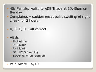
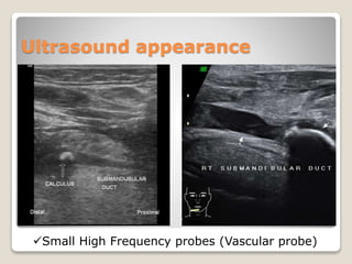
ENT Case Presentation
Runal Shah
PGY-2
Masters in Emergency Medicine
KDAH
 45/ Female, walks to A&E Triage at 10.45pm on
Sunday
 Complaints – sudden onset pain, swelling of right
cheek for 2 hours.
 A, B, C, D – all correct
 Vitals
◦ T- Afebrile
◦ P- 84/min
◦ R- 16/min
◦ BP- 120/70 mmHg
◦ SpO2- 97% on room air
 Pain Score – 5/10
 So, what is the problem we are dealing with ??
 What should be done ?
 Consider resources and act wisely !
 Triage category ???
 Interventions –
◦ IV access – scalp vein
◦ Inj Dynapar 75mg in 20ml NS iv
 Investigate –
◦ CBC
◦ Imaging – CT scan / USG soft parts ??
 S – Pain and swelling of the right cheek, for
approx 2 hours, following dinner, No fever,
similar complaints in the past
 A – no allergies
 M – no meds on routine basis
 L – dinner at 8.30pm
 E – none
 Secondary survey –
 HEENT –
 Inspection –
◦ vague swelling of right cheek, comparable increase in
size as to left cheek
◦ asymmetry of face !
◦ Oral mucosa healthy, no purulent discharge.
 Palpation –
◦ mild tenderness over right cheek
◦ on per-oral palpation no stones palpable
 Rest of the systems – unremarkable
 Reassessment
 Pain score 1/10 – discomfort only
 Swelling was same
 Discussed with pt regarding need of imaging for
the same to rule out sialolithiasis
 USG soft parts –
◦ right parotid gland duct stone of 1.7mm !
◦ No obvious inflammatory changes within right parotid
gland
 CBC
◦ 13.2 / 40 / 7800 / 2,90,000
 Disposition
 Explained the condition to the patient,
discharged with ENT follow up in OPD.
 On discharge Rx
◦ Tab Voveran-SR 75mg 1—0—0 x 3 days
◦ Tab Pantocid 40mg 1—0—0 x 3 days, 30min before
breakfast
◦ Warm compresses over right cheek
 Concrements (stones) formed in the salivary
gland parenchyma or duct due to increased
viscosity or stasis.
 1% population
 M > F
 Age group : 30-50
 Submandibular gland (80-90%) >> Sublingual >
Parotid
Case Discussion – Sialolithiasis
 Differentials :
1) Infections –
 Bacterial
(Staphylococcus,
Strep Viridans,
Pneumococcus, H
influenza)
 Viral – Mumps
2) Inflammation
3) Granulamtous
4) Neoplastic
 Commonly forms around an organic nidus.
 Clinical presentation
 Pain, swelling, tenderness overlying the gland,
Purulent discharge from the duct
 Difficult to differentiate between parotitis and
sialolithiasis clinically as they may co-exist or
may be causative of each other !!
 Features favoring Sialolithiasis :
◦ Typically Unilateral
◦ Pain, swelling aggravated post meals
 Management in ED
 Diagnosis is clinical
 Per-oral palpation with gland massage may
reveal stone or purulent discharge
 Imaging –
◦ X-ray
◦ USG
◦ CT
◦ MRI
◦ Sialography
 Pain relief by NSAIDs
Ultrasound appearance
Small High Frequency probes (Vascular probe)
Disposition
 If palpable stone is removed in ED – f/up with
ENT in 3-4 days
 If not, ENT f/up within 24 hours
 Discharge treatment –
1) Hydration
2) Moist heat
3) Massage
4) Analgesics
5) Sialogogues (Lemon
drops)
6) Antibiotics
Thank You…
 Ref :
◦ Rosen 8/e
◦ Tintinalli 7/e
◦ http://radiopaedia.org/articles/sialolithiasis

ENT case

  • 1.
    ENT Case Presentation RunalShah PGY-2 Masters in Emergency Medicine KDAH
  • 2.
     45/ Female,walks to A&E Triage at 10.45pm on Sunday  Complaints – sudden onset pain, swelling of right cheek for 2 hours.  A, B, C, D – all correct  Vitals ◦ T- Afebrile ◦ P- 84/min ◦ R- 16/min ◦ BP- 120/70 mmHg ◦ SpO2- 97% on room air  Pain Score – 5/10
  • 3.
     So, whatis the problem we are dealing with ??  What should be done ?  Consider resources and act wisely !  Triage category ???
  • 4.
     Interventions – ◦IV access – scalp vein ◦ Inj Dynapar 75mg in 20ml NS iv  Investigate – ◦ CBC ◦ Imaging – CT scan / USG soft parts ??
  • 5.
     S –Pain and swelling of the right cheek, for approx 2 hours, following dinner, No fever, similar complaints in the past  A – no allergies  M – no meds on routine basis  L – dinner at 8.30pm  E – none
  • 6.
     Secondary survey–  HEENT –  Inspection – ◦ vague swelling of right cheek, comparable increase in size as to left cheek ◦ asymmetry of face ! ◦ Oral mucosa healthy, no purulent discharge.  Palpation – ◦ mild tenderness over right cheek ◦ on per-oral palpation no stones palpable  Rest of the systems – unremarkable
  • 7.
     Reassessment  Painscore 1/10 – discomfort only  Swelling was same  Discussed with pt regarding need of imaging for the same to rule out sialolithiasis  USG soft parts – ◦ right parotid gland duct stone of 1.7mm ! ◦ No obvious inflammatory changes within right parotid gland  CBC ◦ 13.2 / 40 / 7800 / 2,90,000
  • 8.
     Disposition  Explainedthe condition to the patient, discharged with ENT follow up in OPD.  On discharge Rx ◦ Tab Voveran-SR 75mg 1—0—0 x 3 days ◦ Tab Pantocid 40mg 1—0—0 x 3 days, 30min before breakfast ◦ Warm compresses over right cheek
  • 9.
     Concrements (stones)formed in the salivary gland parenchyma or duct due to increased viscosity or stasis.  1% population  M > F  Age group : 30-50  Submandibular gland (80-90%) >> Sublingual > Parotid Case Discussion – Sialolithiasis
  • 10.
     Differentials : 1)Infections –  Bacterial (Staphylococcus, Strep Viridans, Pneumococcus, H influenza)  Viral – Mumps 2) Inflammation 3) Granulamtous 4) Neoplastic  Commonly forms around an organic nidus.
  • 11.
     Clinical presentation Pain, swelling, tenderness overlying the gland, Purulent discharge from the duct  Difficult to differentiate between parotitis and sialolithiasis clinically as they may co-exist or may be causative of each other !!  Features favoring Sialolithiasis : ◦ Typically Unilateral ◦ Pain, swelling aggravated post meals
  • 12.
     Management inED  Diagnosis is clinical  Per-oral palpation with gland massage may reveal stone or purulent discharge  Imaging – ◦ X-ray ◦ USG ◦ CT ◦ MRI ◦ Sialography  Pain relief by NSAIDs
  • 13.
    Ultrasound appearance Small HighFrequency probes (Vascular probe)
  • 14.
    Disposition  If palpablestone is removed in ED – f/up with ENT in 3-4 days  If not, ENT f/up within 24 hours  Discharge treatment – 1) Hydration 2) Moist heat 3) Massage 4) Analgesics 5) Sialogogues (Lemon drops) 6) Antibiotics
  • 15.
    Thank You…  Ref: ◦ Rosen 8/e ◦ Tintinalli 7/e ◦ http://radiopaedia.org/articles/sialolithiasis