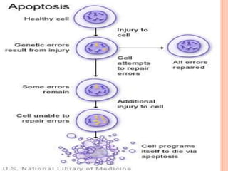
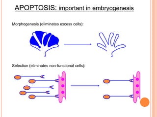
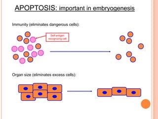
APOPTOSIS
MOHD SAQUIB KHAN
M.Sc 3rd SEMESTER
DEPARTMENT OF BIOCHEMISTRY &
MOLECULAR BIOLOGY
PONDICHERRY UNIVERSITY
INTRODUCTION
 Apoptosis is the process of programmed cell
death.
 Biochemical events lead to characteristic cell
changes (morphology) and death. These changes
include blebbing, cell shrinkage, nuclear
fragmentation, chromatin condensation, and
chromosomal DNA fragmentation.
 Between 50 and 70 billion cells die each day due to
apoptosis in the average human adult. For an
average child between the ages of 8 and 14,
approximately 20 billion to 30 billion cells die a day.
HISTORY
 German scientist Carl Vogt was first to describe the
principle of apoptosis in 1842.
 In 1972 Kerr first introduced the term apoptosis in a
publication.
 Kerr received the Paul Ehrlich and Ludwig
Darmstaedter Prize on March 14, 2000, for his
description of apoptosis.
 The 2002 Nobel Prize in Medicine was awarded to
Sydney Brenner, Horvitz and John E. Sulston for
their work identifying genes that control apoptosis.
APOPTOSIS: PATHWAYS
Death
Ligands
Effector
Caspase 3
Death
Receptors
Initiator
Caspase 8
Cell
death
DNA
damage
& p53
Mitochondria/
Cytochrome C
Initiator
Caspase 9
“Extrinsic Pathway”
“Intrinsic Pathway”
EXTRINSIC PATHWAY
 The extrinsic signaling pathway leading to apoptosis
involves transmembrane death receptors that are
members of the tumor necrosis factor (TNF) receptor
gene superfamily. Members of this receptor family bind to
extrinsic ligands and transduce intracellular signals that
ultimately result in the destruction of the cell.
 The most well characterized ligands of these receptors to
date are FasL, TNF-alpha, Apo3L, and Apo2L.
Corresponding receptors are FasR, TNFR1, DR3, and
DR4/DR5, respectively.
 The signal transduction of the extrinsic pathway involves
several caspases which are proteases with specific
cellular targets. Once activated, the caspases affect
several cellular functions as part of a process that results
in the death of the cells.
THE INTRINSIC PATHWAY
 The intrinsic signaling pathway for programmed cell death
involves non-receptor–mediated intracellular signals, inducing
activities in the mitochondria that initiate apoptosis.
 Stimuli for the intrinsic pathway include viral infections or
damage to the cell by toxins, free radicals, or radiation.
Damage to the cellular DNA can also induce the activation of
the intrinsic pathway for programmed cell death.
 Pro-apoptotic proteins activate caspases that mediate the
destruction of the cell through many pathways. These proteins
also translocate into the cellular nucleus, inducing DNA
fragmentation, a hallmark of apoptosis.
 The regulation of pro-apoptotic events in the mitochondria
occurs through activity of members of the Bcl-2 family of
proteins and the tumor suppressor protein p53.
 Members of the Bcl-2 family of proteins may be pro- or
anti-apoptotic.
 The anti-apoptotic proteins are Bcl-2, Bcl-x, Bcl-xL, Bcl-XS,
Bcl-w, and BAG.
 Pro-apoptotic proteins include Bcl-10, Bax, Bak, Bid, Bad,
Bim, Bik, and Blk.
CASPASES
 Caspases or cysteine-aspartic proteases or cysteine-
dependent aspartate-directed proteases are a family of
cysteine proteases that play essential roles in apoptosis
(programmed cell death), necrosis, and inflammation.
 Single chain of pro-enzymes.
 Contains an N-terminal domain, a small subunit and a
large subunit (similar to a ribosome).
 Apoptotic stimulus Activation Substrate Cleavage
Enzyme.
3 TYPES OF CASPASES
 Inflammatory Caspases: 1, 4, and 5
 Initiator Caspases: 2, 8, 9, and 10
 Long N-terminal domain
 Interact with effector caspases
 Effector Caspases: 3, 6, and 7
 Little to no N-terminal domain
 Initiate cell death
IMPORTANCE OF
APOPTOSIS
o Important in normal physiology / development
– Development: Immune systems maturation,
Morphogenesis, Neural development
– Adult: Immune privilege, DNA damage and wound
repair.
o Excess apoptosis
– Neurodegenerative diseases
o Deficient apoptosis
– Cancer
– Autoimmunity
APOPTOSIS: important in embryogenesis
Morphogenesis (eliminates excess cells):
Selection (eliminates non-functional cells):
APOPTOSIS: important in embryogenesis
Immunity (eliminates dangerous cells):
Self antigen
recognizing cell
Organ size (eliminates excess cells):
apoptosis-131115234500-phpapp02 (1).pdf

apoptosis-131115234500-phpapp02 (1).pdf

  • 1.
    APOPTOSIS MOHD SAQUIB KHAN M.Sc3rd SEMESTER DEPARTMENT OF BIOCHEMISTRY & MOLECULAR BIOLOGY PONDICHERRY UNIVERSITY
  • 2.
    INTRODUCTION  Apoptosis isthe process of programmed cell death.  Biochemical events lead to characteristic cell changes (morphology) and death. These changes include blebbing, cell shrinkage, nuclear fragmentation, chromatin condensation, and chromosomal DNA fragmentation.  Between 50 and 70 billion cells die each day due to apoptosis in the average human adult. For an average child between the ages of 8 and 14, approximately 20 billion to 30 billion cells die a day.
  • 3.
    HISTORY  German scientistCarl Vogt was first to describe the principle of apoptosis in 1842.  In 1972 Kerr first introduced the term apoptosis in a publication.  Kerr received the Paul Ehrlich and Ludwig Darmstaedter Prize on March 14, 2000, for his description of apoptosis.  The 2002 Nobel Prize in Medicine was awarded to Sydney Brenner, Horvitz and John E. Sulston for their work identifying genes that control apoptosis.
  • 5.
    APOPTOSIS: PATHWAYS Death Ligands Effector Caspase 3 Death Receptors Initiator Caspase8 Cell death DNA damage & p53 Mitochondria/ Cytochrome C Initiator Caspase 9 “Extrinsic Pathway” “Intrinsic Pathway”
  • 6.
  • 7.
     The extrinsicsignaling pathway leading to apoptosis involves transmembrane death receptors that are members of the tumor necrosis factor (TNF) receptor gene superfamily. Members of this receptor family bind to extrinsic ligands and transduce intracellular signals that ultimately result in the destruction of the cell.  The most well characterized ligands of these receptors to date are FasL, TNF-alpha, Apo3L, and Apo2L. Corresponding receptors are FasR, TNFR1, DR3, and DR4/DR5, respectively.  The signal transduction of the extrinsic pathway involves several caspases which are proteases with specific cellular targets. Once activated, the caspases affect several cellular functions as part of a process that results in the death of the cells.
  • 8.
  • 9.
     The intrinsicsignaling pathway for programmed cell death involves non-receptor–mediated intracellular signals, inducing activities in the mitochondria that initiate apoptosis.  Stimuli for the intrinsic pathway include viral infections or damage to the cell by toxins, free radicals, or radiation. Damage to the cellular DNA can also induce the activation of the intrinsic pathway for programmed cell death.  Pro-apoptotic proteins activate caspases that mediate the destruction of the cell through many pathways. These proteins also translocate into the cellular nucleus, inducing DNA fragmentation, a hallmark of apoptosis.
  • 10.
     The regulationof pro-apoptotic events in the mitochondria occurs through activity of members of the Bcl-2 family of proteins and the tumor suppressor protein p53.  Members of the Bcl-2 family of proteins may be pro- or anti-apoptotic.  The anti-apoptotic proteins are Bcl-2, Bcl-x, Bcl-xL, Bcl-XS, Bcl-w, and BAG.  Pro-apoptotic proteins include Bcl-10, Bax, Bak, Bid, Bad, Bim, Bik, and Blk.
  • 12.
    CASPASES  Caspases orcysteine-aspartic proteases or cysteine- dependent aspartate-directed proteases are a family of cysteine proteases that play essential roles in apoptosis (programmed cell death), necrosis, and inflammation.  Single chain of pro-enzymes.  Contains an N-terminal domain, a small subunit and a large subunit (similar to a ribosome).  Apoptotic stimulus Activation Substrate Cleavage Enzyme.
  • 13.
    3 TYPES OFCASPASES  Inflammatory Caspases: 1, 4, and 5  Initiator Caspases: 2, 8, 9, and 10  Long N-terminal domain  Interact with effector caspases  Effector Caspases: 3, 6, and 7  Little to no N-terminal domain  Initiate cell death
  • 14.
    IMPORTANCE OF APOPTOSIS o Importantin normal physiology / development – Development: Immune systems maturation, Morphogenesis, Neural development – Adult: Immune privilege, DNA damage and wound repair. o Excess apoptosis – Neurodegenerative diseases o Deficient apoptosis – Cancer – Autoimmunity
  • 15.
    APOPTOSIS: important inembryogenesis Morphogenesis (eliminates excess cells): Selection (eliminates non-functional cells):
  • 16.
    APOPTOSIS: important inembryogenesis Immunity (eliminates dangerous cells): Self antigen recognizing cell Organ size (eliminates excess cells):