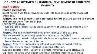
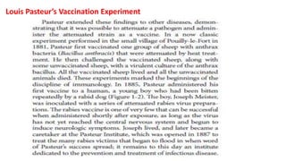
SlideShare a Scribd company logo
1 of 104
INTRODUCTION TO IMMUNE SYSTEM
Immunity – State of protection against infectious
pathogens.
Immune system – Defense system of host
protecting against pathogens
Immune response – The response created by host
against infectious pathogens.
Immunology – Study on body’s defense against
infection.
Brief History
EDWARD JENNER (1798)
Injecting the fluid from cowpox pustule into humans can protect against
small pox.
Reason: The fluid contains some protective factors that are carried to humans
and protect them from small pox.
LOUIS PASTEUR (1885).
• Old culture of bacteria caused less severity of Cholera in chicken after
injection.
Reason: The ageing had weakened the virulence of the bacteria.
This weakened (attenuated) strain was named as VACCINE.
• Previously exposed chicken with cholera causing bacteria were protected
from disease on second infection.
Reason: Protective immunity developed in already exposed chicken,
therefore, they become resistant to second infection.
EMIL VON BEHRING (1890) - Serum of animals immunized with attenuated
Diptheria/Tetanus virus could cure fresh animals injected with these
Q 1 - GIVE AN OVERVIEW ON HISTORICAL DEVELOPMENT OF PROTECTIVE
IMMUNITY
Louis Pasteur’s Vaccination Experiment
Herd Immunity
As a critical mass of people acquire protective immunity, either through
vaccination or infection, they can serve as a buffer for the rest. This principle,
called herd immunity, works by decreasing the number of individuals who can
harbor and spread an infectious agent, significantly decreasing the chances that
susceptible individuals will become infected. This presents an important altruistic
consideration: although many of us could survive infectious diseases for which
we receive a vaccine (such as the flu), this is not true for everyone. Some
individuals cannot receive the vaccine (e.g., the very young or immune
compromised), and vaccination is never 100% effective. In other words, the
susceptible, nonimmune individuals among us can benefit from the pervasive
immunity of their neighbors.
Immunity involves both cellular and humoral components
HUMORAL IMMUNITY Humors – Body fluid
Immunity mediated by BODY FLUID
Example:
Serum of animals immunized with attenuated
Diptheria virus provided resistance in fresh
animals infected with these pathogens. (Emil Von
Behring & Kitasato,1890).
Components:
Fraction of γ globulins (Immunoglobulins) - Serum
component carrying the immunity. Elvin Kabat, 1930.
CELL MEDIATED IMMUNITY
Immunity mediated by CELLS
Example:
WBCs collected from previously injected guinea
pigs with Mycobacterium tuberculosis could
transfer immunity to healthy guinea pigs. (Merrill
Chase, 1940).
Types: Lymphocytes, Monocytes, Neutrophils
Immunity involves both cellular and humoral components
Passive Immunity – The immunity that is
transferred from one individual to the other.
Examples –
1. Serum containing antibodies to snake venom
is passively transferred to individual with
snake bite.
2. New borne babies get protection by maternal
antibodies from circulation through passive
immunity.
• It provides short lived protection
Active Immunity – The immunity developed in an
individual against an antigen.
Example – Immune response developed against a
QUESTIONS
• What is active immunity?
• Serum of animals immunized with attenuated Tetanus virus could cure fresh
animals injected with these pathogens – Active/Passive immunity?
• Vaccine induced immunity – Active/Passive?
• Anti venom therapy – Humoral or cell mediated immunity?
• Which pathogen can induce better immune response – Non-attenuated
or attenuated?
• Humoral immunity is active or passive?
HAEMATOPOESIS
Process of blood cell formation
From HSC.
Self renewal
HAEMATOPOESIS DURING EMBRYONIC DEVELOPMENT
Cells of Innate and Adaptive Immune
Response
Primary lymphoid organs - Development
and differentiation of mature immune cells
from immature cells.
Examples – Bone marrow, Thymus
Secondary lymphoid organs - Interaction between
antigens and matured immune cells occurs and
immune response is generated.
Examples: Spleen, Lymph nodes, MALT (Peyer’s
patches, Tonsils, Nasopharynx & Bronchus)
ORGANS OF IMMUNE SYSTEM
Bone marrow
Thymus
Spleen
Lymphoid Organs – Organs of lymphatic system
(A vascular system associated with immunity)
Immune response – The defense response of host
to
fight against pathogens
Types: Innate and Adaptive response
Innate Immune Response
• It is primitive, includes built-in molecular
structures encoded in germline DNA and is
present at birth.
• Body’s first line of defense.
• The process includes rapid recognition,
phagocytosis and destruction of pathogens.
• Antigen non specific.
Components
Types of immune response
Adaptive Immune Response
• This response makes the host suitable (adapts)
to better recognize, eliminate and remember the
pathogens.
• Slow response, as it takes time to develop after
initial exposure due to clonal selection.
• Antigen specific and leads to memory response
(the body can recognize the pathogen in second
encounter).
Components
T lymphocytes, B lymphocytes, Cytokines,
Antibodies
Strength of Innate and Adaptive Immune Response
A VACCINE PRODUCES IMMUNE RESPONSE THAT IS AUGMENTED AFTER A BOOSTER DOSE
Difference between Innate and Adaptive
Immune Response
Antigen non-specific Antigen specific
Cells of Innate Immune System
Granulocytes.
• Neutrophils
• Basophils
• Mast cells
• Eosinophils
Myeloid antigen presenting cells.
• Monocytes
• Macrophages
• Dendritic cells
Erythroid cells.
Megakaryocytes.
Granulocytes
• They are at front line of pathogen attack and a part of innate
immune system.
• Multilobed nuclei, cytoplasm is filled with granules that are released
to outside on pathogen attack.
• Part of white blood cells.
1. Neutrophils
• Constitute 50%-70% of circulating leucocytes.
• They migrate to site of infection and cause inflammation.
• Their numbers increase with respect to various infections in
circulation – Leukocytosis.
• Neutrophils phagocytose bacteria effectively.
2. Basophils.
• Constitute < 1% of circulating leucocytes.
• Non phagocytic granulocytes with large granules containing
basophilic proteins. Ex. Histamine.
• They are critical to provide immune response against helminth
parasites.
Bilobed nuclei
Histamine: It increases blood vessel permeability and smooth muscle
activity
3. Mast cells
• Constitute < 1% of circulating leucocytes.
• They are found in skin, connective tissues, mucosal epithelial
tissues in many parts of body.
• They contain cytoplasmic granules secreting histamine.
• They play a role in fighting against allergic reactions.
Round nucleus
4. Eosinophils
• Constitute 1%-3% of circulating leucocytes.
• They migrate from blood to site of infections.
• Play a role in defense against different worms. These cells
are found clustering around worms whose membranes are found
damaged.
Filaria worm
Eosinophils
Myeloid antigen presenting cells
1. Monocytes
• Constitute 5%-10% of circulating leucocytes.
• They migrate into tissues to become macrophages/dendritic cells
• Inflammatory monocytes move to tissues in response to infection.
• Patrolling monocytes stay in tissues as reservoir in the absence of
infection.
• These include a group of phagocytic cells – Antigen Presenting Cells.
• Serve as cellular bridge between innate and adaptive immune system
2. Macrophages
• Monocytes migrate into tissues and differentiate to form
macrophages
• Macrophages respond to infection and become effective phagocytic
cells to clear infection from hosts.
OPSONIZATION: When a bacterium is coated with antibody, the rate
of phagocytosis by macrophages gets enhanced compared to uncoated
ones. Antibody is termed as Opsonin and the process is known as
Opsonization.
3. Dendritic cells
• Both myeloid and lymphoid origin.
• Possess long extensions on surface resembling dendrites of nerve
cells.
• Discovered by Ralph Steinman in mid 1970 (Nobel prize – 2011)
• Professional antigen presenting cells
• Follicular dendritic cells help in B cell maturation and diversification.
Erythroid cells
These cells are of erythroid origin – Erythrocytes (Red Blood Cells)
Possess hemoglobin and that carries oxygen to cells and tissues.
Immune functions? Secrete few free radicals playing role in innate
immunity – Research is in progress.
Megakaryocytes
Large myeloid cells and give rise to platelets.
Platelets also release proteolytic substances that kill pathogens
Cells of Adaptive Immune System
• Lymphocytes
T lymphocytes
B lymphocytes
• Natural Killer Cells
• Natural Killer T Cells
Lymphocytes
• Key players to induce adaptive immune response
• 20-40% in circulating lymphocytes and 99% in
lymph nodes.
Clusters of Differentiation (CDs)
Surface proteins expressed on immune cells used for identification.
T LYMPHOCYTES
• Maturation takes place in thymus.
• They express binding receptors - T cell receptors.
• T cell receptors can recognize antigens loaded on MHC molecules
• T cell clone – A group of T cells that is formed when a T cell bearing
antigen specific receptors divides and form daughter cells with
identical receptor specificity.
T cell types
CD4+ T cells (Th) CD8+ T cells (Tc)
Th1 cells
Regulate immune
response to
intracellular
pathogens
IFNγ
IL2
Th2 cells
Regulates immune
response to
extracellular
pathogens
IL4
IL5
IL10
IL13
Th 17
Regulates
Immune response
against fungi
IL17
Tregs
CD25
FoxP3
TGFβ
TFH
CXCR5
PD-1
Regulate B cell
development
Virus elimination
Tumor cell elimination
Granzymes
Perforin
Regulatory T cells
• Also known as ‘suppressor T cells’
• CD4+CD25+FoxP3+ cells producing TGFβ cytokine
• They downregulate the effector functions in immune system.
Two types:
1. Natural Tregs – Mature in thymus and control the immune
tolerance to self antigens.
2. Induced Tregs – Developed in an antigen dependent manner
in response to any infection.
B LYMPHOCYTES
• Mature in ‘Bursa of Fabricius’ of birds and ‘bone marrow’ in mammals.
• They express B cell receptors – membrane bound immunoglobulins.
• B cell clone - A group of B cells that is formed when a B cell bearing
antigen specific receptors divides and form daughter cells with
identical receptor specificity.
Plasma cells:
• Terminally differentiated B cell capable of secreting antibodies
• Two types
Short lived – Generated in response to an infection in
peripheral tissue.
Long lived – Few short lived cells move to bone marrow
and stay for long time to continuously secrete antibodies.
Natural killer cells
• Express a surface marker termed as NK1.1
• They are efficient killers for pathogens (virus, tumor cells)
by cytotoxic granules.
• NK cells also bear immunoglobulin receptors like B cells.
Natural killer T cells
• They share common features resembling T cells and NK cells.
• Like T cells, they also bear T cell receptors
• Like NK cells, they also bear antibody receptors.
• Identified as CD3+CD56+ cells
• They secrete a lot of cytotoxic granules as well as many cytokines.
PAMPs and PRRs
Pathogen Associated Molecular Patterns (PAMPs).
• PAMPs represent unique antigenic structures
present on the
surface of most pathogens; not present in
hosts.
• Effective phagocytosis occurs through
recognition of PAMPs
by phagocytes.
• Initiation of innate immune response occurs
through interaction between PAMPs and host
cells.
• Also called as MAMPs as they are expressed on
microbes
whether they are pathogenic or not.
Pattern Recognition Receptors (PRRs).
• Host cells have evolved several surface receptors/proteins
that quickly recognize PAMPs.
Damage Associated Molecular Patterns (DAMPs).
• Phagocytosis of dead, dying, and aging cells occurs by
macrophages
• Macrophages phagocytose dead cells through recognition
of few components leading to clearance from body:
referred as DAMPs.
• DAMPs are never expressed on live host cells/tissue
PRRs and corresponding PAMPs to promote
phagocytosis
Toll Like Receptors (TLRs)
Toll-like receptors (TLRs) are a set of PRRs that are well
characterized in terms of their structure, mode of
interaction with PAMPs to activate cells and induction of
innate immune response.
Toll Like Receptors: Discovery
The ‘Toll’ gene encoding a cell membrane protein was first found in
Drosophila.
There was a defect in embryonic development in Drosophila due to
mutation in Toll gene.
Defects in Toll gene also made the flies susceptible to a fungal infection
indicating that that Toll has a role in regulating innate responses in
invertebrates.
The cytoplasmic domain for Toll protein in Drosophila was similar to IL1R
domain in vertebrates.
Effort was made to find cytoplasmic domains for Toll and IL1R in humans
and finally TLRs were discovered (Janeway and Medzhitov, 1997)
Types of TLRs
There are 13 different TLRs identified in both mice and humans
functioning as PRRs.
Structurally, TLR1-10 are conserved between humans and mice.
TLR11-13 is mouse specific, not present in humans.
General structure of a TLR
Membrane
spanning
domain
WHY LEUCINE RICH? – LRR (leucine rich repeats) are conserved in many proteins
associated with innate immunity in plants, invertebrates and vertebrates..
Toll/IL1R domain
(Cytoplasmic domain)
Signal Transduction
A process of transmission for molecular signals from cell’s exterior
to interior to make an appropriate response.
General Steps
1. Receptor activation by extracellular/ intracellular signal.
2. Binding of adapter proteins to receptor domains
3. Recruitment of other proteins
4. Phosphorylation, Ubiquitination, Acetylation etc.
5. Activation.
6. Repeat of step 3,4,5 (If required).
7. Activation of transcription factor
8. Synthesis of desired proteins.
ADAPTER PROTEINS: Adaptor proteins are types of connecting molecules that
regulates signal transduction by engaging the surface receptors.
Signaling pathway through
TLR4
MyD88 DEPENDENT PATHWAY
Activation of cell membrane
bound TLR4 occurs through
ligands.
TRIF DEPENDENT SIGNALING
Activation of endosomal TLR4
through ligands.
Plasma membrane
Nuclear membrane
MyD88 – Myeloid Differentiation
TRIF- TIR domain containing adapter inducing interferon-β
Signaling pathway through
TLR4
Signaling through cell membrane bound
TLR4
• LPS activation causes binding of
MyD88/TRAP.
• IRAK ubiquitinates and activates TRAF6.
• TRAF6 ubiquitinates TAB and NEMO.
• TAK1 activation occurs.
Further signaling can be
directed in two ways)
1a. TAK1 phosphorylates IKK complex.
1b. IKK complex ubiquitinates and
phosphorylates Ikb of NFkB
causing entry of NFkB into nucleus that
causes transcription of innate response
genes. The detached Ikb is degraded.
Signaling pathway through
TLR4
2a. TAK1 also activates MAP kinase
pathway alternatively.
2b. AP-1 dimer gets activated which
causes transcription of IFNά and β genes
(Type 1 Interferons).
Signaling pathway through
TLR4
Signaling through endosomal TLR4
• Viral protein activation causes
binding of TRIF/TRAM.
Further signaling can be directed in
two ways.
1a. RIP1 ubiqutinates and activates
TRAF6.
1b. Steps for signaling through
membrane bound TLR4 should be
followed.
2a. TRIFF/TRAM binding causes
activation of TRAF3/TBK-1/IKK
complex
2b. The complex phosphorylates
IRF3/7 which enter nucleus to
transcribe cytokine genes
Signaling pathway through
TLR4
MHC Molecules
• TYPES AND STRUCTURE OF MHC MOLECULES.
• MHC-PEPTIDE INTERACTIONS.
MHC (Major Histocompatibility Complex)
A set of genes encoding for proteins found on the surfaces of cells
that help the immune system recognize foreign substances.
Brief History. These genes played a role in acceptance/rejection fate
of a tissue transplant between two individuals.
• First defined in mice. – H2 complex
• In humans – HLA (Human Leucocyte Antigen)
Antigen recognition by T cells through MHC – Zinkernagel and
Doherty, 1997
Types of MHC proteins
Two types: MHC I & MHC II
Class I MHC
• Expressed in all nucleated cells of hosts (including APCs).
• Present antigens (Intracellular antigens) available in cytosol: Viral
proteins.
• The antigens are presented to CD8+ cytotoxic T cells.
Class II MHC
• Expressed only on a subset of leucocytes – APCs.
• Present antigen fragments from pathogens engulfed by these cells
(extracellular antigens): Bacteria.
• Present the antigenic fragments to CD4+ helper T cells.
Structure of MHC I molecule
Two polypeptide chains
# α chain (45 kDa) is organized into
• α1, α2, α3 domains
• Transmembrane domain
• Cytoplasmic anchor
# β2-microglobulin (12 kDa)
• Appears as α3 domain
• No transmembrane region
• Non covalent bond with α chain
The peptide binding groove (PBG) is
present between α1 and α2 domains with
a single polypeptide chain
Homology in aa sequence between
α3 and β2-microglobulin
β2-microglobulin
Peptide binding
groove
Interacting domains in MHC I
• Membrane distal domains
α1, α2.
• Membrane proximal domains
α3, β2-microglobulin
β2-microglobulin
Peptide binding
groove
ROLE OF β2 -MICROGLOBULIN
• Enhances the stability of MHC I
• Helps MHC I for adequate expression
surface of APC
Structure of MHC II molecule
Two polypeptide chains
# α chain (33 kDa) is organized into
• α1 & α2 domains
• Transmembrane domain
• Cytoplasmic anchor
# β chain (28 kDa)
• β1 & β2 domains
• Transmembrane domain
• Cytoplasmic anchor
The PBG is formed by α1 and β1
involving 2 peptide chains
Homology in aa sequence between
α2 and β2 domains
Peptide binding
groove
Transmembrane
segment
Cytoplasmic
anchor
Membrane
distal
domain
Membrane
proximal
domain
Interacting domains in MHC II
• Membrane distal domains
α1, β1.
• Membrane proximal domains
α2, β2
Peptide binding
groove
Membrane
distal
domain
Transmembrane
segment
Cytoplasmic
anchor
Membrane
proximal
domain
How limited numbers of MHC molecules can recognize a wide
variety of antigenic peptides?
Many ‘allelic variants’ exist in MHC I and II molecules dividing them into
many classes.
These variants result due to polymorphism in the region where the
peptide binds.
PROSMISCUITY OF PEPTIDE BINDING: Many different peptides ‘match
up’ with the peptide binding grooves of MHC molecules.
Peptide-MHC interactions
Class I MHC - peptide Interaction
α1 domain
α2 domain
Peptide in PBG
• MHC I presents peptides derived from endogenous intracellular
proteins present in cytosol
• Both the ends of PBG is closed - Peptides of maximum 8-10 amino acids
can fit.
• Specific amino acids near N and C ends of peptides are needed for
binding to groove – Anchor residues.
Ribbon model of PBG of MHC I
PBG
Peptide
Domains
• Peptides of different lengths can fit into PBG displaying variations in
confirmation.
• Shorter peptides stay flat in the groove, whereas longer ones bulge
in the middle to better fit!!!!
Example
The main contacts between class I MHC and the a nona-peptide involve
residue 2 at the N-terminal end and residue 9 at the C-terminus of the peptide.
• Contact of peptides to MHC is made through H- bonds.
Class II MHC - peptide Interaction
PBG
• MHC II presents peptides derived from foreign proteins degraded
after phagocytosis/endocytosis of antigen by APCs.
• Both the ends of PBG are open.
• Peptide fragments of maximum 13-18 amino acids can best fit.
• No anchor residues.
• No bulging effect like MHC1!!!! Peptides maintain a roughly constant
elevation on the floor of the binding groove.
• Longer peptides (>13 residues) can fit into PBG, the ability to bind to
MHC is determined by centrally placed residues.
• Contact of peptide to PBG is through H- bonds
Antigen processing and presentation by MHC
Antigen processing - A process involving fragmentation of antigens into
peptides inside cells by enzymatic digestion.
Antigen presentation – The processed antigen must be presented to T cells
and T cell recognize these through T cell receptors.
Demonstrate that an antigen should be processed before presentation to T cell.
An Overview
Two pathways
• Endogenous (Virus) – MHC I mediated
• Exogenous (Bacteria) – MHCII mediated
PATHWAYS OF ANTIGEN PRESENTATION
ENDOGENOUS PATHWAY
• Degradation of proteins by proteasomes
• Transport of peptides from cytosol into rough
endoplasmic reticulum (MHC I is present in RER)
• Attachment of peptides to MHC I molecule
• Presentation of MHC loaded peptides to surface
# 1 – Degradation of proteins by proteasomes
Proteasomes – A cytosolic proteolytic system present in cells degrading
unwanted proteins.
Immunoproteasomes – Proteasomes
present in professional APCs with better
proteolytic activity.
• Proteins are targeted for
proteolysis after attaching to
Ubiquitin.
• Ub-Protein complex enters
into proteasome complex.
• The peptide bonds in protein
are cleaved in an ATP dependent
process.
Cell membrane
# 2 – Transport of peptides from cytosol to RER.
TAP – ‘Transporter associated with antigen processing’- a membrane
spanning protein on RER membrane required to transport peptides to RER
lumen from cytosol.
It is a heterodimer – TAP1 & TAP2 proteins
Two domains:
# ATP binding domain in cytosol,
# Domain in RER lumen.
Transmembrane segments
• Peptides in the cytosol are
translocated to RER lumen
by TAP requiring the hydrolysis
of ATP.
• TAP has is highly efficient to
send peptide of 8-10 aa.
• Bigger peptides can be
translocated into RER with less
efficiency.
• Bigger peptides are trimmed
to smaller lengths by ERAP
(Endoplasmic Reticulum
Amino Peptidase).
# 3 – Attachment of peptides to MHC I molecules
• α and β2-microglobulin of MHC I are synthesized on ribosomes
of RER.
• Molecular chaperones facilitate the folding and assembly of
polypeptides in MHC I molecule.
• Calnexin (Chaperone) and ERp57 associate with the free MHC I alpha
chain to promote its folding.
• Binding of β2-microglobulin to α chain causes release of Calnexin
leading to Calreticulin and Tapasin (Chaperones) attachment
• Tapasin brings TAP transporter in close proximity to MHC I, and the peptide is captured
by MHC I.
• The peptide may be trimmed by ERAP if bigger.
• Binding of peptide will cause release of two chaperones and ERp57 and the MHC-
peptide complex is released to cytosol.
EXOGENOUS PATHWAY
• Generation of peptides from internalized antigens in
endocytic vesicles.
• Transport of Class II MHC to endocytic vesicles
through cytoplasm.
• Assembly of peptides to MHC II molecule
# 1 - Generation of peptides from internalized antigens in
endocytic vesicles.
Internalization of antigen into APCs can be mediated by
• Phagocytosis
• Receptor mediated endocytosis.
The internalized antigen passes
through increasingly acidic
compartments of endosomes.
Early endosomes (pH 6.0-6.5)
Late endosomes (pH 4.5-5.0)
Peptides of 13-18 aa length are
generated after being degraded by
hydrolytic enzymes.
Late
endosomes
Receptor mediated
endocytosis
Late
endosomes
# 2 – Transport of Class II MHC to endocytic vesicles
INVARIANT CHAIN
The αβ chains of MHC II inside RER are attached to an invariant chain
(IC) which has following role:
• It binds to PBG and prevents the
binding of endogenously derived
peptides.
• It helps in proper folding of α
and β chain
• It helps MHC II to exit RER and
routing them to late endosomes
containing peptides.
Late endosomes containing peptides associate with MHC II carrying
transport vesicles and MIIC late endosomes are formed (pH- 4.5-5.0)
Late
endosomes
In RER
# 3 - Assembly of peptides to class II molecules
• The invariant chain is degraded due to acidic conditions in endosomes. However a
small fragment ‘CLIP’ remains attached to MHC II in late endosomes.
• The exchange between CLIP and antigenic peptides is made by few HLA-DM (non
classical MHC II).
• HLA-DO acts as a negative regulator. It prevents binding of self-peptides to MHC
by not removing CLIP – HLA-DO MAINTAINS SELF TOLERANCE.
Exposed on surface of cell
Membrane of APC
RER
Late endosomes
Brush Up…….
Vesicles
PRESENTATION OF NON-PEPTIDE ANTIGENS
How is non-protein antigens recognized by T cells??
Example:
T cells can be activated by mycolic acid (a lipid antigen) derived
from Mycobacterium tuberculosis
CD1 molecules present non-protein antigens to T cells
Structure of CD1 molecule
Lipid
binding
pockets
Lipid
binding
tunnel
α
domains
Transmembrane
domain
Cytoplasmic tail
• Structurally similar to MHC I
protein.
• Composed of 3 alpha domains
and one β2 microglobulin.
• Expressed on all cell types.
• Lipid binding pockets
• Lipid binding tunnel
The bound antigen slips from
pocket like a foot sliding into a
shoe and binds by hydrophobic
interaction.
• Types of antigens
Lipoproteins or Glycolipids.
Lipid
binding
pockets
Lipid
binding
tunnel
α
domains
Transmembrane
domain
Cytoplasmic tail
Structure of CD1 molecule
Mechanism of lipid antigen presentation
Mechanism of presentation matches with MHC II mediated antigen
presentation.
• CD1 molecule is synthesized in RER.
• It moves from RER to cytosol to endosomes.
and bind to processed lipid antigens that are exogenously
derived.
• Finally, it presents the antigen to T cells
Self MHC Restriction of T cells
T cells does not recognize antigen
due to tolerance
T cells recognizes antigen
due to tolerance
T cells does not recognize antigen
Self MHC
Restriction
Self MHC Restriction of T cells is due to different MHC haplotypes
Mouse MHC Haplotypes
MHC gene is encoded with several alleles. Mouse has H2 MHC encoded by 6
alleles (K, IA, IB, S and D). There is a variation in MHC alleles even in same type
mf animal (Mouse) but in different strains of mice.
This set of linked alleles – HAPLOTYPE (A set of genetic variant inherited together to offspring)
CBA – H-2k
DBA/2 – H2d
C57BL/10 – H2b
Can T cells in a mouse of one MHC haplotype recognize
an antigen presented by MHC molecule in another
mouse strain with a different haplotype?
Answer - NO
Self MHC Restriction of T cells
H2k or H2b H2k or H2b
Ag primed T
cells
Ag pulsed macrophages
H2b H2k
H2b + -
H2k - +
T cells can only recognize
antigen presented by APC
if the MHC haplotype of
APC matches with T cells
SELF MHC RESTRICTION
T CELLS
T cell proliferation
Rosenthal and Shevach Experiment
SELF MHC T
CELL RESTRICTION
Self MHC Class II restriction
Self MHC Class I restriction
CD4+ Th cells and APC should have similar MHC II haplotype
background
CD8+ Th cells and APC should have similar MHC I haplotype
background
ANTIGENICITY AND IMMUNOGENICITY
Antigen - Any foreign substance for a host.
Immunogen – Any antigen capable of inducing immune response.
Hapten – Any antigen capable to eliciting immune response when
bound to a carrier molecule.
Antigenicity
The ability of a substance to combine with T cell receptors or
B cell receptors (immunoglobulins).
Factors affecting antigencity
• Bigger the size - better antigenicity
• More complex - better antigenicity
Immunogenicity
Capacity of a substance to induce immune response (both T and B
cell response)
Example – Immunogenicity of vaccines.
Vaccines against diseases induce protective immunity against the
concerned pathogens due to their immunogenicity
ALL IMMUNOGENS ARE ANTIGENS BUT THE REVERSE IS NOT TRUE
T CELL AND B CELL EPITOPES
EPITOPES
• Specific determinants or sites on the whole antigen to which the
immune cells interact.
• Carbohydrate residues, proteins, amino acid sequence, lipid
residues present on the antigens.
Types of protein epitopes
LINEAR EPITOPE: An epitope, composed of a single segment of a
polypeptide chain
CONFORMATIONAL EPITOPE: An epitope containing a complex
folded protein.
T cell epitopes
• Epitopes on antigens recognized by T cells.
• For protein antigens, these are internal peptides, therefore not
easily accessible to T cells.
• Proteins undergo enzymatic digestion, recognized by T cell
receptor after attaching to MHC molecule.
• Hydrophobic in nature
T cell
epitope
Janeway’s Immunobiology
B cell epitopes
• Epitopes on antigens recognized by B cells.
• These are present on antigen surface for easy
accessibility to B cell receptors (immunoglobulins).
• Hydrophilic in nature.
• The internal amino acids present in a protein can be B
cell epitopes after the protein is denatured.
B cell
epitope
B cell epitope types
Sequential B cell
epitopes
Non-sequential B cell
epitopes
Sequential B cell epitopes
The aminoacid sequence (epitopes)
are present as sequential contiguous
residues on a polypeptide chain.
Sperm whale myoglobin
The B cell epitope containing few contiguous
amino acids
Sequential
B cell epitopes
Non sequential B cell epitopes
• The amino acid residues (epitopes) are far apart in the linear
sequence on the polypeptide chains
• The residues are brought together by protein folding forming
tertiary structures.
Hen egg white lysozyme
Non sequential
B cell epitopes
Structure of antibodies
MOLECULAR WEIGHT – 25kDa
MOLECULAR WEIGHT –
50-60 kDa
Complementarity
Determining
Region (CDR)
Light chains – Kappa (k) and Lambda (λ)
Only in constant regions
Isotype determines the antibody class
HINGE
J CHAIN
DIMER
MONOMER
PENTAMER
Antibody Classes
β-pleated sheet of light chain
of immunoglobulin
Pepsin digestion – One F(ab)2 + one Fc
Papain digestion – Two Fab fragments + one Fc
General functions of antibodies
Generation of antigen specific T cells
T CELL MATURATION IN THYMUS
LINEAGE COMMITMENT IN THYMUS
MIGRATION TO TISSUES
DEVELOPMENT OF T CELL PRECURSORS IN BONE MARROW
T CELL ACTIVATION IN RESPONSE TO ANTIGEN STIMULATION
T CELL PROLIFERATION
GENERATION OF ANTIGEN SPECIFIC T CELLS
EARLY THYMOCYTE DEVELOPMENT
POSITIVE AND NEGATIVE SELECTION
LINEAGE COMMITMENT
T cells precursors migrate to
thymic cortex.
The ‘notch’ receptors/ligand
interaction in thymic epithelium
initiates maturation signaling.
DN thymocytes
• Double negative thymocytes
• Lack CD4 and CD8 on surface
• Pass through 4 different stages
and differ in C kit, CD44 and CD25
expression..
Early Thymocyte Development
DN1 ( c kit++, CD44+, CD25-)
DN2 ( c kit++, CD44+, CD25+)
DN3 ( c kit+, CD44-, CD25+)
DN4 ( c kitlow, CD44-, CD25-)
T cell
commitment
TCRα gene arrangement
TCRβ gene arrangement
β- selection
Double positive
(CD4+CD8+) cells bearing TCRαβ receptors
TCR genes (γ, δ and β) arrangement.
Differentiation of DN T cells
γδ T cell lineage
β- selection
A process by which DN cells successfully
rearrange β genes and expand.
• Expression of Pre-Tα chain near TCRβ.
• Pre-Tα forms a complex with TCRβ
and CD3 termed as Pre-TCR complex.
• A pre-TCR signaling will be initiated
causing maturation of DN3 and DN4
T cells.
CD3 CD3
Pre-TCR complex
DN3 T cells
• TCR α receptor is expressed after
successful arrangement of TCR α
gene.
• TCR α associates with TCRβ and pre
T- α is not expressed anymore.
• TCR/CD3 complex is formed
• CD4 and CD8 proteins are expressed
on surface - DP thymocytes.
CD3 CD3
TCR/CD3 complex
DN4 T cells
Thymic selection.
DP T cells bearing functional and mature
TCRαβ receptors undergo selection
processes to survive/dye.
DEATH BY NEGLECT
DP thymocytes with no affinity for self MHC/
Self peptide complex expressed on cTEC
get eliminated (90-96%)
NEGATIVE SELECTION
DP thymocytes with very high affinity to
self MHC/self peptide complex get eliminated
(2-5%)
POSITIVE SELECTION
DP thymocytes with low affinity for
self-MHC/self peptide complex survive (2-5%)
Formation of SP cells

More Related Content

Similar to Ppt for Immunology_MSc 2nd Sem.pptx

Bio202 20 22-immune_system
Bio202 20 22-immune_systemBio202 20 22-immune_system
Bio202 20 22-immune_systemswasticcc
 
Introductory Immunology Presentation
Introductory Immunology PresentationIntroductory Immunology Presentation
Introductory Immunology PresentationAndrea Henle
 
Immunomodulators screening methods
Immunomodulators screening methodsImmunomodulators screening methods
Immunomodulators screening methodsVIGNESHROSS
 
Basic immunology and hypersensitive disorders beba
Basic immunology and hypersensitive disorders bebaBasic immunology and hypersensitive disorders beba
Basic immunology and hypersensitive disorders bebaBISRATGETACHEWMD
 
Immunity, antigen and antibody- Introduction
Immunity, antigen and antibody- IntroductionImmunity, antigen and antibody- Introduction
Immunity, antigen and antibody- IntroductionTara Bhatta
 
Unit 2 immune systems(1)
Unit 2  immune systems(1)Unit 2  immune systems(1)
Unit 2 immune systems(1)ntando mthethwa
 
Overview of the immune system.pptx
Overview of the immune system.pptxOverview of the immune system.pptx
Overview of the immune system.pptxRachaelAdeleke
 
Fish and Shrimp immune response power point.ppt
Fish and Shrimp immune response power point.pptFish and Shrimp immune response power point.ppt
Fish and Shrimp immune response power point.pptAnh Nguyen
 
Immunology (Innate and adaptive immune systems) (ANTIGENS (Ag))
Immunology (Innate and adaptive immune systems) (ANTIGENS (Ag)) Immunology (Innate and adaptive immune systems) (ANTIGENS (Ag))
Immunology (Innate and adaptive immune systems) (ANTIGENS (Ag)) Amany Elsayed
 
Unit 2 immune systems
Unit 2  immune systemsUnit 2  immune systems
Unit 2 immune systemsObey Maduna
 
The immune response1
The immune response1The immune response1
The immune response1Gamal Dawood
 
Chapter 574585
Chapter 574585Chapter 574585
Chapter 574585jullz03
 

Similar to Ppt for Immunology_MSc 2nd Sem.pptx (20)

Bio202 20 22-immune_system
Bio202 20 22-immune_systemBio202 20 22-immune_system
Bio202 20 22-immune_system
 
Introductory Immunology Presentation
Introductory Immunology PresentationIntroductory Immunology Presentation
Introductory Immunology Presentation
 
Immunomodulators screening methods
Immunomodulators screening methodsImmunomodulators screening methods
Immunomodulators screening methods
 
Immunity of chicken
Immunity of chickenImmunity of chicken
Immunity of chicken
 
Basic immunology and hypersensitive disorders beba
Basic immunology and hypersensitive disorders bebaBasic immunology and hypersensitive disorders beba
Basic immunology and hypersensitive disorders beba
 
Immunity, antigen and antibody- Introduction
Immunity, antigen and antibody- IntroductionImmunity, antigen and antibody- Introduction
Immunity, antigen and antibody- Introduction
 
The immune system
The immune systemThe immune system
The immune system
 
Unit 2 immune systems(1)
Unit 2  immune systems(1)Unit 2  immune systems(1)
Unit 2 immune systems(1)
 
Unit 2 immune systems
Unit 2  immune systemsUnit 2  immune systems
Unit 2 immune systems
 
Immune System slides
Immune System slidesImmune System slides
Immune System slides
 
Overview of the immune system.pptx
Overview of the immune system.pptxOverview of the immune system.pptx
Overview of the immune system.pptx
 
Fish and Shrimp immune response power point.ppt
Fish and Shrimp immune response power point.pptFish and Shrimp immune response power point.ppt
Fish and Shrimp immune response power point.ppt
 
Immunology (Innate and adaptive immune systems) (ANTIGENS (Ag))
Immunology (Innate and adaptive immune systems) (ANTIGENS (Ag)) Immunology (Innate and adaptive immune systems) (ANTIGENS (Ag))
Immunology (Innate and adaptive immune systems) (ANTIGENS (Ag))
 
Immune System
Immune SystemImmune System
Immune System
 
Immunology
ImmunologyImmunology
Immunology
 
Unit 2 immune systems
Unit 2  immune systemsUnit 2  immune systems
Unit 2 immune systems
 
The immune response1
The immune response1The immune response1
The immune response1
 
Chapter 574585
Chapter 574585Chapter 574585
Chapter 574585
 
Basic immunology
Basic immunologyBasic immunology
Basic immunology
 
Immunity of bovine
Immunity of bovineImmunity of bovine
Immunity of bovine
 

More from DevPanda5

PSII and PSI.ppt
PSII and PSI.pptPSII and PSI.ppt
PSII and PSI.pptDevPanda5
 
d47nZcToS53i5I7I758.pptx
d47nZcToS53i5I7I758.pptxd47nZcToS53i5I7I758.pptx
d47nZcToS53i5I7I758.pptxDevPanda5
 
Roll 27.pptx
Roll 27.pptxRoll 27.pptx
Roll 27.pptxDevPanda5
 
210705180005.pptx
210705180005.pptx210705180005.pptx
210705180005.pptxDevPanda5
 
210705180010 sangita das.pptx
210705180010 sangita das.pptx210705180010 sangita das.pptx
210705180010 sangita das.pptxDevPanda5
 
prvnppt-141230053156-conversion-gate02.pdf
prvnppt-141230053156-conversion-gate02.pdfprvnppt-141230053156-conversion-gate02.pdf
prvnppt-141230053156-conversion-gate02.pdfDevPanda5
 
mod_i-12_lead_ekgs.ppt
mod_i-12_lead_ekgs.pptmod_i-12_lead_ekgs.ppt
mod_i-12_lead_ekgs.pptDevPanda5
 
Roll 27.pptx
Roll 27.pptxRoll 27.pptx
Roll 27.pptxDevPanda5
 
220705180062 cyanotoxin.pptx
220705180062 cyanotoxin.pptx220705180062 cyanotoxin.pptx
220705180062 cyanotoxin.pptxDevPanda5
 
220705180043Media Formulation.pptx
220705180043Media Formulation.pptx220705180043Media Formulation.pptx
220705180043Media Formulation.pptxDevPanda5
 
210705180008.pptx
210705180008.pptx210705180008.pptx
210705180008.pptxDevPanda5
 
220705180049 Genetic diversity of viruses.pptx
220705180049 Genetic diversity of viruses.pptx220705180049 Genetic diversity of viruses.pptx
220705180049 Genetic diversity of viruses.pptxDevPanda5
 
42 AKASH PRADIP DASH.pptx
42 AKASH PRADIP DASH.pptx42 AKASH PRADIP DASH.pptx
42 AKASH PRADIP DASH.pptxDevPanda5
 
220705180076 Entner - Doudoroff Pathway.pptx
220705180076 Entner - Doudoroff Pathway.pptx220705180076 Entner - Doudoroff Pathway.pptx
220705180076 Entner - Doudoroff Pathway.pptxDevPanda5
 
fungal diseases of fish(220705180075) 1.pptx
fungal diseases of fish(220705180075) 1.pptxfungal diseases of fish(220705180075) 1.pptx
fungal diseases of fish(220705180075) 1.pptxDevPanda5
 
220705180068 MEASUREMENT AND GROWTH MONITORING IN CULTURE.pptx
220705180068 MEASUREMENT AND GROWTH MONITORING IN CULTURE.pptx220705180068 MEASUREMENT AND GROWTH MONITORING IN CULTURE.pptx
220705180068 MEASUREMENT AND GROWTH MONITORING IN CULTURE.pptxDevPanda5
 
ABN 523 lecture.ppt
ABN 523 lecture.pptABN 523 lecture.ppt
ABN 523 lecture.pptDevPanda5
 
220705180077protozoan disease in fish.pptx
220705180077protozoan disease in fish.pptx220705180077protozoan disease in fish.pptx
220705180077protozoan disease in fish.pptxDevPanda5
 
MODULE-VII-22.6.22.pptx
MODULE-VII-22.6.22.pptxMODULE-VII-22.6.22.pptx
MODULE-VII-22.6.22.pptxDevPanda5
 
Module-V- Fish n shellfish -23.5.22.pptx
Module-V- Fish n shellfish -23.5.22.pptxModule-V- Fish n shellfish -23.5.22.pptx
Module-V- Fish n shellfish -23.5.22.pptxDevPanda5
 

More from DevPanda5 (20)

PSII and PSI.ppt
PSII and PSI.pptPSII and PSI.ppt
PSII and PSI.ppt
 
d47nZcToS53i5I7I758.pptx
d47nZcToS53i5I7I758.pptxd47nZcToS53i5I7I758.pptx
d47nZcToS53i5I7I758.pptx
 
Roll 27.pptx
Roll 27.pptxRoll 27.pptx
Roll 27.pptx
 
210705180005.pptx
210705180005.pptx210705180005.pptx
210705180005.pptx
 
210705180010 sangita das.pptx
210705180010 sangita das.pptx210705180010 sangita das.pptx
210705180010 sangita das.pptx
 
prvnppt-141230053156-conversion-gate02.pdf
prvnppt-141230053156-conversion-gate02.pdfprvnppt-141230053156-conversion-gate02.pdf
prvnppt-141230053156-conversion-gate02.pdf
 
mod_i-12_lead_ekgs.ppt
mod_i-12_lead_ekgs.pptmod_i-12_lead_ekgs.ppt
mod_i-12_lead_ekgs.ppt
 
Roll 27.pptx
Roll 27.pptxRoll 27.pptx
Roll 27.pptx
 
220705180062 cyanotoxin.pptx
220705180062 cyanotoxin.pptx220705180062 cyanotoxin.pptx
220705180062 cyanotoxin.pptx
 
220705180043Media Formulation.pptx
220705180043Media Formulation.pptx220705180043Media Formulation.pptx
220705180043Media Formulation.pptx
 
210705180008.pptx
210705180008.pptx210705180008.pptx
210705180008.pptx
 
220705180049 Genetic diversity of viruses.pptx
220705180049 Genetic diversity of viruses.pptx220705180049 Genetic diversity of viruses.pptx
220705180049 Genetic diversity of viruses.pptx
 
42 AKASH PRADIP DASH.pptx
42 AKASH PRADIP DASH.pptx42 AKASH PRADIP DASH.pptx
42 AKASH PRADIP DASH.pptx
 
220705180076 Entner - Doudoroff Pathway.pptx
220705180076 Entner - Doudoroff Pathway.pptx220705180076 Entner - Doudoroff Pathway.pptx
220705180076 Entner - Doudoroff Pathway.pptx
 
fungal diseases of fish(220705180075) 1.pptx
fungal diseases of fish(220705180075) 1.pptxfungal diseases of fish(220705180075) 1.pptx
fungal diseases of fish(220705180075) 1.pptx
 
220705180068 MEASUREMENT AND GROWTH MONITORING IN CULTURE.pptx
220705180068 MEASUREMENT AND GROWTH MONITORING IN CULTURE.pptx220705180068 MEASUREMENT AND GROWTH MONITORING IN CULTURE.pptx
220705180068 MEASUREMENT AND GROWTH MONITORING IN CULTURE.pptx
 
ABN 523 lecture.ppt
ABN 523 lecture.pptABN 523 lecture.ppt
ABN 523 lecture.ppt
 
220705180077protozoan disease in fish.pptx
220705180077protozoan disease in fish.pptx220705180077protozoan disease in fish.pptx
220705180077protozoan disease in fish.pptx
 
MODULE-VII-22.6.22.pptx
MODULE-VII-22.6.22.pptxMODULE-VII-22.6.22.pptx
MODULE-VII-22.6.22.pptx
 
Module-V- Fish n shellfish -23.5.22.pptx
Module-V- Fish n shellfish -23.5.22.pptxModule-V- Fish n shellfish -23.5.22.pptx
Module-V- Fish n shellfish -23.5.22.pptx
 

Recently uploaded

vip Krishna Nagar Call Girls 9999965857 Call or WhatsApp Now Book
vip Krishna Nagar Call Girls 9999965857 Call or WhatsApp Now Bookvip Krishna Nagar Call Girls 9999965857 Call or WhatsApp Now Book
vip Krishna Nagar Call Girls 9999965857 Call or WhatsApp Now Bookmanojkuma9823
 
Beautiful Sapna Call Girls CP 9711199012 ☎ Call /Whatsapps
Beautiful Sapna Call Girls CP 9711199012 ☎ Call /WhatsappsBeautiful Sapna Call Girls CP 9711199012 ☎ Call /Whatsapps
Beautiful Sapna Call Girls CP 9711199012 ☎ Call /Whatsappssapnasaifi408
 
(办理学位证)多伦多大学毕业证成绩单原版一比一
(办理学位证)多伦多大学毕业证成绩单原版一比一(办理学位证)多伦多大学毕业证成绩单原版一比一
(办理学位证)多伦多大学毕业证成绩单原版一比一C SSS
 
(MEGHA) Hinjewadi Call Girls Just Call 7001035870 [ Cash on Delivery ] Pune E...
(MEGHA) Hinjewadi Call Girls Just Call 7001035870 [ Cash on Delivery ] Pune E...(MEGHA) Hinjewadi Call Girls Just Call 7001035870 [ Cash on Delivery ] Pune E...
(MEGHA) Hinjewadi Call Girls Just Call 7001035870 [ Cash on Delivery ] Pune E...ranjana rawat
 
Call Girls Delhi {Rs-10000 Laxmi Nagar] 9711199012 Whats Up Number
Call Girls Delhi {Rs-10000 Laxmi Nagar] 9711199012 Whats Up NumberCall Girls Delhi {Rs-10000 Laxmi Nagar] 9711199012 Whats Up Number
Call Girls Delhi {Rs-10000 Laxmi Nagar] 9711199012 Whats Up NumberMs Riya
 
Call Girls Delhi {Rohini} 9711199012 high profile service
Call Girls Delhi {Rohini} 9711199012 high profile serviceCall Girls Delhi {Rohini} 9711199012 high profile service
Call Girls Delhi {Rohini} 9711199012 high profile servicerehmti665
 
Hifi Defence Colony Call Girls Service WhatsApp -> 9999965857 Available 24x7 ...
Hifi Defence Colony Call Girls Service WhatsApp -> 9999965857 Available 24x7 ...Hifi Defence Colony Call Girls Service WhatsApp -> 9999965857 Available 24x7 ...
Hifi Defence Colony Call Girls Service WhatsApp -> 9999965857 Available 24x7 ...srsj9000
 
(办理学位证)加州州立大学北岭分校毕业证成绩单原版一比一
(办理学位证)加州州立大学北岭分校毕业证成绩单原版一比一(办理学位证)加州州立大学北岭分校毕业证成绩单原版一比一
(办理学位证)加州州立大学北岭分校毕业证成绩单原版一比一Fi sss
 
Vip Udupi Call Girls 7001305949 WhatsApp Number 24x7 Best Services
Vip Udupi Call Girls 7001305949 WhatsApp Number 24x7 Best ServicesVip Udupi Call Girls 7001305949 WhatsApp Number 24x7 Best Services
Vip Udupi Call Girls 7001305949 WhatsApp Number 24x7 Best Servicesnajka9823
 
(ANIKA) Wanwadi Call Girls Just Call 7001035870 [ Cash on Delivery ] Pune Esc...
(ANIKA) Wanwadi Call Girls Just Call 7001035870 [ Cash on Delivery ] Pune Esc...(ANIKA) Wanwadi Call Girls Just Call 7001035870 [ Cash on Delivery ] Pune Esc...
(ANIKA) Wanwadi Call Girls Just Call 7001035870 [ Cash on Delivery ] Pune Esc...ranjana rawat
 
如何办理萨省大学毕业证(UofS毕业证)成绩单留信学历认证原版一比一
如何办理萨省大学毕业证(UofS毕业证)成绩单留信学历认证原版一比一如何办理萨省大学毕业证(UofS毕业证)成绩单留信学历认证原版一比一
如何办理萨省大学毕业证(UofS毕业证)成绩单留信学历认证原版一比一ga6c6bdl
 
Gaya Call Girls #9907093804 Contact Number Escorts Service Gaya
Gaya Call Girls #9907093804 Contact Number Escorts Service GayaGaya Call Girls #9907093804 Contact Number Escorts Service Gaya
Gaya Call Girls #9907093804 Contact Number Escorts Service Gayasrsj9000
 
Russian Call Girls Kolkata Chhaya 🤌 8250192130 🚀 Vip Call Girls Kolkata
Russian Call Girls Kolkata Chhaya 🤌  8250192130 🚀 Vip Call Girls KolkataRussian Call Girls Kolkata Chhaya 🤌  8250192130 🚀 Vip Call Girls Kolkata
Russian Call Girls Kolkata Chhaya 🤌 8250192130 🚀 Vip Call Girls Kolkataanamikaraghav4
 
Slim Call Girls Service Badshah Nagar * 9548273370 Naughty Call Girls Service...
Slim Call Girls Service Badshah Nagar * 9548273370 Naughty Call Girls Service...Slim Call Girls Service Badshah Nagar * 9548273370 Naughty Call Girls Service...
Slim Call Girls Service Badshah Nagar * 9548273370 Naughty Call Girls Service...nagunakhan
 
VIP Call Girl Saharanpur Aashi 8250192130 Independent Escort Service Saharanpur
VIP Call Girl Saharanpur Aashi 8250192130 Independent Escort Service SaharanpurVIP Call Girl Saharanpur Aashi 8250192130 Independent Escort Service Saharanpur
VIP Call Girl Saharanpur Aashi 8250192130 Independent Escort Service SaharanpurSuhani Kapoor
 
定制加拿大滑铁卢大学毕业证(Waterloo毕业证书)成绩单(文凭)原版一比一
定制加拿大滑铁卢大学毕业证(Waterloo毕业证书)成绩单(文凭)原版一比一定制加拿大滑铁卢大学毕业证(Waterloo毕业证书)成绩单(文凭)原版一比一
定制加拿大滑铁卢大学毕业证(Waterloo毕业证书)成绩单(文凭)原版一比一zul5vf0pq
 
Call Girls Service Kolkata Aishwarya 🤌 8250192130 🚀 Vip Call Girls Kolkata
Call Girls Service Kolkata Aishwarya 🤌  8250192130 🚀 Vip Call Girls KolkataCall Girls Service Kolkata Aishwarya 🤌  8250192130 🚀 Vip Call Girls Kolkata
Call Girls Service Kolkata Aishwarya 🤌 8250192130 🚀 Vip Call Girls Kolkataanamikaraghav4
 
NO1 Qualified Best Black Magic Specialist Near Me Spiritual Healer Powerful L...
NO1 Qualified Best Black Magic Specialist Near Me Spiritual Healer Powerful L...NO1 Qualified Best Black Magic Specialist Near Me Spiritual Healer Powerful L...
NO1 Qualified Best Black Magic Specialist Near Me Spiritual Healer Powerful L...Amil baba
 
定制(USF学位证)旧金山大学毕业证成绩单原版一比一
定制(USF学位证)旧金山大学毕业证成绩单原版一比一定制(USF学位证)旧金山大学毕业证成绩单原版一比一
定制(USF学位证)旧金山大学毕业证成绩单原版一比一ss ss
 

Recently uploaded (20)

vip Krishna Nagar Call Girls 9999965857 Call or WhatsApp Now Book
vip Krishna Nagar Call Girls 9999965857 Call or WhatsApp Now Bookvip Krishna Nagar Call Girls 9999965857 Call or WhatsApp Now Book
vip Krishna Nagar Call Girls 9999965857 Call or WhatsApp Now Book
 
Beautiful Sapna Call Girls CP 9711199012 ☎ Call /Whatsapps
Beautiful Sapna Call Girls CP 9711199012 ☎ Call /WhatsappsBeautiful Sapna Call Girls CP 9711199012 ☎ Call /Whatsapps
Beautiful Sapna Call Girls CP 9711199012 ☎ Call /Whatsapps
 
(办理学位证)多伦多大学毕业证成绩单原版一比一
(办理学位证)多伦多大学毕业证成绩单原版一比一(办理学位证)多伦多大学毕业证成绩单原版一比一
(办理学位证)多伦多大学毕业证成绩单原版一比一
 
(MEGHA) Hinjewadi Call Girls Just Call 7001035870 [ Cash on Delivery ] Pune E...
(MEGHA) Hinjewadi Call Girls Just Call 7001035870 [ Cash on Delivery ] Pune E...(MEGHA) Hinjewadi Call Girls Just Call 7001035870 [ Cash on Delivery ] Pune E...
(MEGHA) Hinjewadi Call Girls Just Call 7001035870 [ Cash on Delivery ] Pune E...
 
Low rate Call girls in Delhi Justdial | 9953330565
Low rate Call girls in Delhi Justdial | 9953330565Low rate Call girls in Delhi Justdial | 9953330565
Low rate Call girls in Delhi Justdial | 9953330565
 
Call Girls Delhi {Rs-10000 Laxmi Nagar] 9711199012 Whats Up Number
Call Girls Delhi {Rs-10000 Laxmi Nagar] 9711199012 Whats Up NumberCall Girls Delhi {Rs-10000 Laxmi Nagar] 9711199012 Whats Up Number
Call Girls Delhi {Rs-10000 Laxmi Nagar] 9711199012 Whats Up Number
 
Call Girls Delhi {Rohini} 9711199012 high profile service
Call Girls Delhi {Rohini} 9711199012 high profile serviceCall Girls Delhi {Rohini} 9711199012 high profile service
Call Girls Delhi {Rohini} 9711199012 high profile service
 
Hifi Defence Colony Call Girls Service WhatsApp -> 9999965857 Available 24x7 ...
Hifi Defence Colony Call Girls Service WhatsApp -> 9999965857 Available 24x7 ...Hifi Defence Colony Call Girls Service WhatsApp -> 9999965857 Available 24x7 ...
Hifi Defence Colony Call Girls Service WhatsApp -> 9999965857 Available 24x7 ...
 
(办理学位证)加州州立大学北岭分校毕业证成绩单原版一比一
(办理学位证)加州州立大学北岭分校毕业证成绩单原版一比一(办理学位证)加州州立大学北岭分校毕业证成绩单原版一比一
(办理学位证)加州州立大学北岭分校毕业证成绩单原版一比一
 
Vip Udupi Call Girls 7001305949 WhatsApp Number 24x7 Best Services
Vip Udupi Call Girls 7001305949 WhatsApp Number 24x7 Best ServicesVip Udupi Call Girls 7001305949 WhatsApp Number 24x7 Best Services
Vip Udupi Call Girls 7001305949 WhatsApp Number 24x7 Best Services
 
(ANIKA) Wanwadi Call Girls Just Call 7001035870 [ Cash on Delivery ] Pune Esc...
(ANIKA) Wanwadi Call Girls Just Call 7001035870 [ Cash on Delivery ] Pune Esc...(ANIKA) Wanwadi Call Girls Just Call 7001035870 [ Cash on Delivery ] Pune Esc...
(ANIKA) Wanwadi Call Girls Just Call 7001035870 [ Cash on Delivery ] Pune Esc...
 
如何办理萨省大学毕业证(UofS毕业证)成绩单留信学历认证原版一比一
如何办理萨省大学毕业证(UofS毕业证)成绩单留信学历认证原版一比一如何办理萨省大学毕业证(UofS毕业证)成绩单留信学历认证原版一比一
如何办理萨省大学毕业证(UofS毕业证)成绩单留信学历认证原版一比一
 
Gaya Call Girls #9907093804 Contact Number Escorts Service Gaya
Gaya Call Girls #9907093804 Contact Number Escorts Service GayaGaya Call Girls #9907093804 Contact Number Escorts Service Gaya
Gaya Call Girls #9907093804 Contact Number Escorts Service Gaya
 
Russian Call Girls Kolkata Chhaya 🤌 8250192130 🚀 Vip Call Girls Kolkata
Russian Call Girls Kolkata Chhaya 🤌  8250192130 🚀 Vip Call Girls KolkataRussian Call Girls Kolkata Chhaya 🤌  8250192130 🚀 Vip Call Girls Kolkata
Russian Call Girls Kolkata Chhaya 🤌 8250192130 🚀 Vip Call Girls Kolkata
 
Slim Call Girls Service Badshah Nagar * 9548273370 Naughty Call Girls Service...
Slim Call Girls Service Badshah Nagar * 9548273370 Naughty Call Girls Service...Slim Call Girls Service Badshah Nagar * 9548273370 Naughty Call Girls Service...
Slim Call Girls Service Badshah Nagar * 9548273370 Naughty Call Girls Service...
 
VIP Call Girl Saharanpur Aashi 8250192130 Independent Escort Service Saharanpur
VIP Call Girl Saharanpur Aashi 8250192130 Independent Escort Service SaharanpurVIP Call Girl Saharanpur Aashi 8250192130 Independent Escort Service Saharanpur
VIP Call Girl Saharanpur Aashi 8250192130 Independent Escort Service Saharanpur
 
定制加拿大滑铁卢大学毕业证(Waterloo毕业证书)成绩单(文凭)原版一比一
定制加拿大滑铁卢大学毕业证(Waterloo毕业证书)成绩单(文凭)原版一比一定制加拿大滑铁卢大学毕业证(Waterloo毕业证书)成绩单(文凭)原版一比一
定制加拿大滑铁卢大学毕业证(Waterloo毕业证书)成绩单(文凭)原版一比一
 
Call Girls Service Kolkata Aishwarya 🤌 8250192130 🚀 Vip Call Girls Kolkata
Call Girls Service Kolkata Aishwarya 🤌  8250192130 🚀 Vip Call Girls KolkataCall Girls Service Kolkata Aishwarya 🤌  8250192130 🚀 Vip Call Girls Kolkata
Call Girls Service Kolkata Aishwarya 🤌 8250192130 🚀 Vip Call Girls Kolkata
 
NO1 Qualified Best Black Magic Specialist Near Me Spiritual Healer Powerful L...
NO1 Qualified Best Black Magic Specialist Near Me Spiritual Healer Powerful L...NO1 Qualified Best Black Magic Specialist Near Me Spiritual Healer Powerful L...
NO1 Qualified Best Black Magic Specialist Near Me Spiritual Healer Powerful L...
 
定制(USF学位证)旧金山大学毕业证成绩单原版一比一
定制(USF学位证)旧金山大学毕业证成绩单原版一比一定制(USF学位证)旧金山大学毕业证成绩单原版一比一
定制(USF学位证)旧金山大学毕业证成绩单原版一比一
 

Ppt for Immunology_MSc 2nd Sem.pptx

  • 1. INTRODUCTION TO IMMUNE SYSTEM Immunity – State of protection against infectious pathogens. Immune system – Defense system of host protecting against pathogens Immune response – The response created by host against infectious pathogens. Immunology – Study on body’s defense against infection.
  • 2. Brief History EDWARD JENNER (1798) Injecting the fluid from cowpox pustule into humans can protect against small pox. Reason: The fluid contains some protective factors that are carried to humans and protect them from small pox. LOUIS PASTEUR (1885). • Old culture of bacteria caused less severity of Cholera in chicken after injection. Reason: The ageing had weakened the virulence of the bacteria. This weakened (attenuated) strain was named as VACCINE. • Previously exposed chicken with cholera causing bacteria were protected from disease on second infection. Reason: Protective immunity developed in already exposed chicken, therefore, they become resistant to second infection. EMIL VON BEHRING (1890) - Serum of animals immunized with attenuated Diptheria/Tetanus virus could cure fresh animals injected with these Q 1 - GIVE AN OVERVIEW ON HISTORICAL DEVELOPMENT OF PROTECTIVE IMMUNITY
  • 4. Herd Immunity As a critical mass of people acquire protective immunity, either through vaccination or infection, they can serve as a buffer for the rest. This principle, called herd immunity, works by decreasing the number of individuals who can harbor and spread an infectious agent, significantly decreasing the chances that susceptible individuals will become infected. This presents an important altruistic consideration: although many of us could survive infectious diseases for which we receive a vaccine (such as the flu), this is not true for everyone. Some individuals cannot receive the vaccine (e.g., the very young or immune compromised), and vaccination is never 100% effective. In other words, the susceptible, nonimmune individuals among us can benefit from the pervasive immunity of their neighbors.
  • 5. Immunity involves both cellular and humoral components HUMORAL IMMUNITY Humors – Body fluid Immunity mediated by BODY FLUID Example: Serum of animals immunized with attenuated Diptheria virus provided resistance in fresh animals infected with these pathogens. (Emil Von Behring & Kitasato,1890). Components: Fraction of γ globulins (Immunoglobulins) - Serum component carrying the immunity. Elvin Kabat, 1930.
  • 6. CELL MEDIATED IMMUNITY Immunity mediated by CELLS Example: WBCs collected from previously injected guinea pigs with Mycobacterium tuberculosis could transfer immunity to healthy guinea pigs. (Merrill Chase, 1940). Types: Lymphocytes, Monocytes, Neutrophils Immunity involves both cellular and humoral components
  • 7. Passive Immunity – The immunity that is transferred from one individual to the other. Examples – 1. Serum containing antibodies to snake venom is passively transferred to individual with snake bite. 2. New borne babies get protection by maternal antibodies from circulation through passive immunity. • It provides short lived protection Active Immunity – The immunity developed in an individual against an antigen. Example – Immune response developed against a
  • 8. QUESTIONS • What is active immunity? • Serum of animals immunized with attenuated Tetanus virus could cure fresh animals injected with these pathogens – Active/Passive immunity? • Vaccine induced immunity – Active/Passive? • Anti venom therapy – Humoral or cell mediated immunity? • Which pathogen can induce better immune response – Non-attenuated or attenuated? • Humoral immunity is active or passive?
  • 9. HAEMATOPOESIS Process of blood cell formation From HSC. Self renewal
  • 11. Cells of Innate and Adaptive Immune Response Primary lymphoid organs - Development and differentiation of mature immune cells from immature cells. Examples – Bone marrow, Thymus Secondary lymphoid organs - Interaction between antigens and matured immune cells occurs and immune response is generated. Examples: Spleen, Lymph nodes, MALT (Peyer’s patches, Tonsils, Nasopharynx & Bronchus) ORGANS OF IMMUNE SYSTEM Bone marrow Thymus Spleen Lymphoid Organs – Organs of lymphatic system (A vascular system associated with immunity)
  • 12.
  • 13. Immune response – The defense response of host to fight against pathogens Types: Innate and Adaptive response Innate Immune Response • It is primitive, includes built-in molecular structures encoded in germline DNA and is present at birth. • Body’s first line of defense. • The process includes rapid recognition, phagocytosis and destruction of pathogens. • Antigen non specific. Components Types of immune response
  • 14. Adaptive Immune Response • This response makes the host suitable (adapts) to better recognize, eliminate and remember the pathogens. • Slow response, as it takes time to develop after initial exposure due to clonal selection. • Antigen specific and leads to memory response (the body can recognize the pathogen in second encounter). Components T lymphocytes, B lymphocytes, Cytokines, Antibodies
  • 15. Strength of Innate and Adaptive Immune Response A VACCINE PRODUCES IMMUNE RESPONSE THAT IS AUGMENTED AFTER A BOOSTER DOSE
  • 16. Difference between Innate and Adaptive Immune Response Antigen non-specific Antigen specific
  • 17. Cells of Innate Immune System Granulocytes. • Neutrophils • Basophils • Mast cells • Eosinophils Myeloid antigen presenting cells. • Monocytes • Macrophages • Dendritic cells Erythroid cells. Megakaryocytes.
  • 18. Granulocytes • They are at front line of pathogen attack and a part of innate immune system. • Multilobed nuclei, cytoplasm is filled with granules that are released to outside on pathogen attack. • Part of white blood cells. 1. Neutrophils • Constitute 50%-70% of circulating leucocytes. • They migrate to site of infection and cause inflammation. • Their numbers increase with respect to various infections in circulation – Leukocytosis. • Neutrophils phagocytose bacteria effectively.
  • 19. 2. Basophils. • Constitute < 1% of circulating leucocytes. • Non phagocytic granulocytes with large granules containing basophilic proteins. Ex. Histamine. • They are critical to provide immune response against helminth parasites. Bilobed nuclei Histamine: It increases blood vessel permeability and smooth muscle activity
  • 20. 3. Mast cells • Constitute < 1% of circulating leucocytes. • They are found in skin, connective tissues, mucosal epithelial tissues in many parts of body. • They contain cytoplasmic granules secreting histamine. • They play a role in fighting against allergic reactions. Round nucleus
  • 21. 4. Eosinophils • Constitute 1%-3% of circulating leucocytes. • They migrate from blood to site of infections. • Play a role in defense against different worms. These cells are found clustering around worms whose membranes are found damaged. Filaria worm Eosinophils
  • 22. Myeloid antigen presenting cells 1. Monocytes • Constitute 5%-10% of circulating leucocytes. • They migrate into tissues to become macrophages/dendritic cells • Inflammatory monocytes move to tissues in response to infection. • Patrolling monocytes stay in tissues as reservoir in the absence of infection. • These include a group of phagocytic cells – Antigen Presenting Cells. • Serve as cellular bridge between innate and adaptive immune system
  • 23. 2. Macrophages • Monocytes migrate into tissues and differentiate to form macrophages • Macrophages respond to infection and become effective phagocytic cells to clear infection from hosts. OPSONIZATION: When a bacterium is coated with antibody, the rate of phagocytosis by macrophages gets enhanced compared to uncoated ones. Antibody is termed as Opsonin and the process is known as Opsonization.
  • 24. 3. Dendritic cells • Both myeloid and lymphoid origin. • Possess long extensions on surface resembling dendrites of nerve cells. • Discovered by Ralph Steinman in mid 1970 (Nobel prize – 2011) • Professional antigen presenting cells • Follicular dendritic cells help in B cell maturation and diversification.
  • 25. Erythroid cells These cells are of erythroid origin – Erythrocytes (Red Blood Cells) Possess hemoglobin and that carries oxygen to cells and tissues. Immune functions? Secrete few free radicals playing role in innate immunity – Research is in progress. Megakaryocytes Large myeloid cells and give rise to platelets. Platelets also release proteolytic substances that kill pathogens
  • 26. Cells of Adaptive Immune System • Lymphocytes T lymphocytes B lymphocytes • Natural Killer Cells • Natural Killer T Cells
  • 27. Lymphocytes • Key players to induce adaptive immune response • 20-40% in circulating lymphocytes and 99% in lymph nodes. Clusters of Differentiation (CDs) Surface proteins expressed on immune cells used for identification. T LYMPHOCYTES • Maturation takes place in thymus. • They express binding receptors - T cell receptors. • T cell receptors can recognize antigens loaded on MHC molecules • T cell clone – A group of T cells that is formed when a T cell bearing antigen specific receptors divides and form daughter cells with identical receptor specificity.
  • 28. T cell types CD4+ T cells (Th) CD8+ T cells (Tc) Th1 cells Regulate immune response to intracellular pathogens IFNγ IL2 Th2 cells Regulates immune response to extracellular pathogens IL4 IL5 IL10 IL13 Th 17 Regulates Immune response against fungi IL17 Tregs CD25 FoxP3 TGFβ TFH CXCR5 PD-1 Regulate B cell development Virus elimination Tumor cell elimination Granzymes Perforin
  • 29. Regulatory T cells • Also known as ‘suppressor T cells’ • CD4+CD25+FoxP3+ cells producing TGFβ cytokine • They downregulate the effector functions in immune system. Two types: 1. Natural Tregs – Mature in thymus and control the immune tolerance to self antigens. 2. Induced Tregs – Developed in an antigen dependent manner in response to any infection.
  • 30. B LYMPHOCYTES • Mature in ‘Bursa of Fabricius’ of birds and ‘bone marrow’ in mammals. • They express B cell receptors – membrane bound immunoglobulins. • B cell clone - A group of B cells that is formed when a B cell bearing antigen specific receptors divides and form daughter cells with identical receptor specificity. Plasma cells: • Terminally differentiated B cell capable of secreting antibodies • Two types Short lived – Generated in response to an infection in peripheral tissue. Long lived – Few short lived cells move to bone marrow and stay for long time to continuously secrete antibodies.
  • 31. Natural killer cells • Express a surface marker termed as NK1.1 • They are efficient killers for pathogens (virus, tumor cells) by cytotoxic granules. • NK cells also bear immunoglobulin receptors like B cells. Natural killer T cells • They share common features resembling T cells and NK cells. • Like T cells, they also bear T cell receptors • Like NK cells, they also bear antibody receptors. • Identified as CD3+CD56+ cells • They secrete a lot of cytotoxic granules as well as many cytokines.
  • 32. PAMPs and PRRs Pathogen Associated Molecular Patterns (PAMPs). • PAMPs represent unique antigenic structures present on the surface of most pathogens; not present in hosts. • Effective phagocytosis occurs through recognition of PAMPs by phagocytes. • Initiation of innate immune response occurs through interaction between PAMPs and host cells. • Also called as MAMPs as they are expressed on microbes whether they are pathogenic or not. Pattern Recognition Receptors (PRRs). • Host cells have evolved several surface receptors/proteins that quickly recognize PAMPs.
  • 33. Damage Associated Molecular Patterns (DAMPs). • Phagocytosis of dead, dying, and aging cells occurs by macrophages • Macrophages phagocytose dead cells through recognition of few components leading to clearance from body: referred as DAMPs. • DAMPs are never expressed on live host cells/tissue
  • 34. PRRs and corresponding PAMPs to promote phagocytosis
  • 35. Toll Like Receptors (TLRs) Toll-like receptors (TLRs) are a set of PRRs that are well characterized in terms of their structure, mode of interaction with PAMPs to activate cells and induction of innate immune response.
  • 36. Toll Like Receptors: Discovery The ‘Toll’ gene encoding a cell membrane protein was first found in Drosophila. There was a defect in embryonic development in Drosophila due to mutation in Toll gene. Defects in Toll gene also made the flies susceptible to a fungal infection indicating that that Toll has a role in regulating innate responses in invertebrates. The cytoplasmic domain for Toll protein in Drosophila was similar to IL1R domain in vertebrates. Effort was made to find cytoplasmic domains for Toll and IL1R in humans and finally TLRs were discovered (Janeway and Medzhitov, 1997)
  • 37. Types of TLRs There are 13 different TLRs identified in both mice and humans functioning as PRRs. Structurally, TLR1-10 are conserved between humans and mice. TLR11-13 is mouse specific, not present in humans.
  • 38. General structure of a TLR Membrane spanning domain WHY LEUCINE RICH? – LRR (leucine rich repeats) are conserved in many proteins associated with innate immunity in plants, invertebrates and vertebrates.. Toll/IL1R domain (Cytoplasmic domain)
  • 39. Signal Transduction A process of transmission for molecular signals from cell’s exterior to interior to make an appropriate response. General Steps 1. Receptor activation by extracellular/ intracellular signal. 2. Binding of adapter proteins to receptor domains 3. Recruitment of other proteins 4. Phosphorylation, Ubiquitination, Acetylation etc. 5. Activation. 6. Repeat of step 3,4,5 (If required). 7. Activation of transcription factor 8. Synthesis of desired proteins. ADAPTER PROTEINS: Adaptor proteins are types of connecting molecules that regulates signal transduction by engaging the surface receptors. Signaling pathway through TLR4
  • 40. MyD88 DEPENDENT PATHWAY Activation of cell membrane bound TLR4 occurs through ligands. TRIF DEPENDENT SIGNALING Activation of endosomal TLR4 through ligands. Plasma membrane Nuclear membrane MyD88 – Myeloid Differentiation TRIF- TIR domain containing adapter inducing interferon-β Signaling pathway through TLR4
  • 41. Signaling through cell membrane bound TLR4 • LPS activation causes binding of MyD88/TRAP. • IRAK ubiquitinates and activates TRAF6. • TRAF6 ubiquitinates TAB and NEMO. • TAK1 activation occurs. Further signaling can be directed in two ways) 1a. TAK1 phosphorylates IKK complex. 1b. IKK complex ubiquitinates and phosphorylates Ikb of NFkB causing entry of NFkB into nucleus that causes transcription of innate response genes. The detached Ikb is degraded. Signaling pathway through TLR4
  • 42. 2a. TAK1 also activates MAP kinase pathway alternatively. 2b. AP-1 dimer gets activated which causes transcription of IFNά and β genes (Type 1 Interferons). Signaling pathway through TLR4
  • 43. Signaling through endosomal TLR4 • Viral protein activation causes binding of TRIF/TRAM. Further signaling can be directed in two ways. 1a. RIP1 ubiqutinates and activates TRAF6. 1b. Steps for signaling through membrane bound TLR4 should be followed. 2a. TRIFF/TRAM binding causes activation of TRAF3/TBK-1/IKK complex 2b. The complex phosphorylates IRF3/7 which enter nucleus to transcribe cytokine genes Signaling pathway through TLR4
  • 44. MHC Molecules • TYPES AND STRUCTURE OF MHC MOLECULES. • MHC-PEPTIDE INTERACTIONS.
  • 45. MHC (Major Histocompatibility Complex) A set of genes encoding for proteins found on the surfaces of cells that help the immune system recognize foreign substances. Brief History. These genes played a role in acceptance/rejection fate of a tissue transplant between two individuals. • First defined in mice. – H2 complex • In humans – HLA (Human Leucocyte Antigen) Antigen recognition by T cells through MHC – Zinkernagel and Doherty, 1997
  • 46. Types of MHC proteins Two types: MHC I & MHC II Class I MHC • Expressed in all nucleated cells of hosts (including APCs). • Present antigens (Intracellular antigens) available in cytosol: Viral proteins. • The antigens are presented to CD8+ cytotoxic T cells. Class II MHC • Expressed only on a subset of leucocytes – APCs. • Present antigen fragments from pathogens engulfed by these cells (extracellular antigens): Bacteria. • Present the antigenic fragments to CD4+ helper T cells.
  • 47. Structure of MHC I molecule Two polypeptide chains # α chain (45 kDa) is organized into • α1, α2, α3 domains • Transmembrane domain • Cytoplasmic anchor # β2-microglobulin (12 kDa) • Appears as α3 domain • No transmembrane region • Non covalent bond with α chain The peptide binding groove (PBG) is present between α1 and α2 domains with a single polypeptide chain Homology in aa sequence between α3 and β2-microglobulin β2-microglobulin Peptide binding groove
  • 48. Interacting domains in MHC I • Membrane distal domains α1, α2. • Membrane proximal domains α3, β2-microglobulin β2-microglobulin Peptide binding groove ROLE OF β2 -MICROGLOBULIN • Enhances the stability of MHC I • Helps MHC I for adequate expression surface of APC
  • 49. Structure of MHC II molecule Two polypeptide chains # α chain (33 kDa) is organized into • α1 & α2 domains • Transmembrane domain • Cytoplasmic anchor # β chain (28 kDa) • β1 & β2 domains • Transmembrane domain • Cytoplasmic anchor The PBG is formed by α1 and β1 involving 2 peptide chains Homology in aa sequence between α2 and β2 domains Peptide binding groove Transmembrane segment Cytoplasmic anchor Membrane distal domain Membrane proximal domain
  • 50. Interacting domains in MHC II • Membrane distal domains α1, β1. • Membrane proximal domains α2, β2 Peptide binding groove Membrane distal domain Transmembrane segment Cytoplasmic anchor Membrane proximal domain
  • 51.
  • 52. How limited numbers of MHC molecules can recognize a wide variety of antigenic peptides? Many ‘allelic variants’ exist in MHC I and II molecules dividing them into many classes. These variants result due to polymorphism in the region where the peptide binds. PROSMISCUITY OF PEPTIDE BINDING: Many different peptides ‘match up’ with the peptide binding grooves of MHC molecules.
  • 53. Peptide-MHC interactions Class I MHC - peptide Interaction α1 domain α2 domain Peptide in PBG • MHC I presents peptides derived from endogenous intracellular proteins present in cytosol • Both the ends of PBG is closed - Peptides of maximum 8-10 amino acids can fit. • Specific amino acids near N and C ends of peptides are needed for binding to groove – Anchor residues. Ribbon model of PBG of MHC I PBG Peptide Domains
  • 54. • Peptides of different lengths can fit into PBG displaying variations in confirmation. • Shorter peptides stay flat in the groove, whereas longer ones bulge in the middle to better fit!!!! Example The main contacts between class I MHC and the a nona-peptide involve residue 2 at the N-terminal end and residue 9 at the C-terminus of the peptide. • Contact of peptides to MHC is made through H- bonds.
  • 55. Class II MHC - peptide Interaction PBG • MHC II presents peptides derived from foreign proteins degraded after phagocytosis/endocytosis of antigen by APCs. • Both the ends of PBG are open. • Peptide fragments of maximum 13-18 amino acids can best fit. • No anchor residues. • No bulging effect like MHC1!!!! Peptides maintain a roughly constant elevation on the floor of the binding groove. • Longer peptides (>13 residues) can fit into PBG, the ability to bind to MHC is determined by centrally placed residues. • Contact of peptide to PBG is through H- bonds
  • 56. Antigen processing and presentation by MHC Antigen processing - A process involving fragmentation of antigens into peptides inside cells by enzymatic digestion. Antigen presentation – The processed antigen must be presented to T cells and T cell recognize these through T cell receptors.
  • 57. Demonstrate that an antigen should be processed before presentation to T cell.
  • 58. An Overview Two pathways • Endogenous (Virus) – MHC I mediated • Exogenous (Bacteria) – MHCII mediated PATHWAYS OF ANTIGEN PRESENTATION
  • 59. ENDOGENOUS PATHWAY • Degradation of proteins by proteasomes • Transport of peptides from cytosol into rough endoplasmic reticulum (MHC I is present in RER) • Attachment of peptides to MHC I molecule • Presentation of MHC loaded peptides to surface
  • 60. # 1 – Degradation of proteins by proteasomes Proteasomes – A cytosolic proteolytic system present in cells degrading unwanted proteins. Immunoproteasomes – Proteasomes present in professional APCs with better proteolytic activity. • Proteins are targeted for proteolysis after attaching to Ubiquitin. • Ub-Protein complex enters into proteasome complex. • The peptide bonds in protein are cleaved in an ATP dependent process. Cell membrane
  • 61. # 2 – Transport of peptides from cytosol to RER. TAP – ‘Transporter associated with antigen processing’- a membrane spanning protein on RER membrane required to transport peptides to RER lumen from cytosol. It is a heterodimer – TAP1 & TAP2 proteins Two domains: # ATP binding domain in cytosol, # Domain in RER lumen. Transmembrane segments
  • 62. • Peptides in the cytosol are translocated to RER lumen by TAP requiring the hydrolysis of ATP. • TAP has is highly efficient to send peptide of 8-10 aa. • Bigger peptides can be translocated into RER with less efficiency. • Bigger peptides are trimmed to smaller lengths by ERAP (Endoplasmic Reticulum Amino Peptidase).
  • 63. # 3 – Attachment of peptides to MHC I molecules • α and β2-microglobulin of MHC I are synthesized on ribosomes of RER. • Molecular chaperones facilitate the folding and assembly of polypeptides in MHC I molecule.
  • 64. • Calnexin (Chaperone) and ERp57 associate with the free MHC I alpha chain to promote its folding. • Binding of β2-microglobulin to α chain causes release of Calnexin leading to Calreticulin and Tapasin (Chaperones) attachment
  • 65. • Tapasin brings TAP transporter in close proximity to MHC I, and the peptide is captured by MHC I. • The peptide may be trimmed by ERAP if bigger. • Binding of peptide will cause release of two chaperones and ERp57 and the MHC- peptide complex is released to cytosol.
  • 66. EXOGENOUS PATHWAY • Generation of peptides from internalized antigens in endocytic vesicles. • Transport of Class II MHC to endocytic vesicles through cytoplasm. • Assembly of peptides to MHC II molecule
  • 67. # 1 - Generation of peptides from internalized antigens in endocytic vesicles. Internalization of antigen into APCs can be mediated by • Phagocytosis • Receptor mediated endocytosis. The internalized antigen passes through increasingly acidic compartments of endosomes. Early endosomes (pH 6.0-6.5) Late endosomes (pH 4.5-5.0) Peptides of 13-18 aa length are generated after being degraded by hydrolytic enzymes. Late endosomes Receptor mediated endocytosis Late endosomes
  • 68. # 2 – Transport of Class II MHC to endocytic vesicles INVARIANT CHAIN The αβ chains of MHC II inside RER are attached to an invariant chain (IC) which has following role: • It binds to PBG and prevents the binding of endogenously derived peptides. • It helps in proper folding of α and β chain • It helps MHC II to exit RER and routing them to late endosomes containing peptides. Late endosomes containing peptides associate with MHC II carrying transport vesicles and MIIC late endosomes are formed (pH- 4.5-5.0) Late endosomes In RER
  • 69. # 3 - Assembly of peptides to class II molecules • The invariant chain is degraded due to acidic conditions in endosomes. However a small fragment ‘CLIP’ remains attached to MHC II in late endosomes. • The exchange between CLIP and antigenic peptides is made by few HLA-DM (non classical MHC II). • HLA-DO acts as a negative regulator. It prevents binding of self-peptides to MHC by not removing CLIP – HLA-DO MAINTAINS SELF TOLERANCE. Exposed on surface of cell Membrane of APC RER Late endosomes
  • 71. PRESENTATION OF NON-PEPTIDE ANTIGENS How is non-protein antigens recognized by T cells?? Example: T cells can be activated by mycolic acid (a lipid antigen) derived from Mycobacterium tuberculosis CD1 molecules present non-protein antigens to T cells
  • 72. Structure of CD1 molecule Lipid binding pockets Lipid binding tunnel α domains Transmembrane domain Cytoplasmic tail • Structurally similar to MHC I protein. • Composed of 3 alpha domains and one β2 microglobulin. • Expressed on all cell types.
  • 73. • Lipid binding pockets • Lipid binding tunnel The bound antigen slips from pocket like a foot sliding into a shoe and binds by hydrophobic interaction. • Types of antigens Lipoproteins or Glycolipids. Lipid binding pockets Lipid binding tunnel α domains Transmembrane domain Cytoplasmic tail Structure of CD1 molecule
  • 74. Mechanism of lipid antigen presentation Mechanism of presentation matches with MHC II mediated antigen presentation. • CD1 molecule is synthesized in RER. • It moves from RER to cytosol to endosomes. and bind to processed lipid antigens that are exogenously derived. • Finally, it presents the antigen to T cells
  • 75. Self MHC Restriction of T cells T cells does not recognize antigen due to tolerance T cells recognizes antigen due to tolerance T cells does not recognize antigen T cells does not recognize antigen Self MHC Restriction
  • 76. Self MHC Restriction of T cells is due to different MHC haplotypes Mouse MHC Haplotypes MHC gene is encoded with several alleles. Mouse has H2 MHC encoded by 6 alleles (K, IA, IB, S and D). There is a variation in MHC alleles even in same type mf animal (Mouse) but in different strains of mice. This set of linked alleles – HAPLOTYPE (A set of genetic variant inherited together to offspring) CBA – H-2k DBA/2 – H2d C57BL/10 – H2b
  • 77. Can T cells in a mouse of one MHC haplotype recognize an antigen presented by MHC molecule in another mouse strain with a different haplotype? Answer - NO Self MHC Restriction of T cells
  • 78. H2k or H2b H2k or H2b Ag primed T cells Ag pulsed macrophages H2b H2k H2b + - H2k - + T cells can only recognize antigen presented by APC if the MHC haplotype of APC matches with T cells SELF MHC RESTRICTION T CELLS T cell proliferation Rosenthal and Shevach Experiment
  • 79. SELF MHC T CELL RESTRICTION Self MHC Class II restriction Self MHC Class I restriction CD4+ Th cells and APC should have similar MHC II haplotype background CD8+ Th cells and APC should have similar MHC I haplotype background
  • 80.
  • 81. ANTIGENICITY AND IMMUNOGENICITY Antigen - Any foreign substance for a host. Immunogen – Any antigen capable of inducing immune response. Hapten – Any antigen capable to eliciting immune response when bound to a carrier molecule. Antigenicity The ability of a substance to combine with T cell receptors or B cell receptors (immunoglobulins). Factors affecting antigencity • Bigger the size - better antigenicity • More complex - better antigenicity
  • 82. Immunogenicity Capacity of a substance to induce immune response (both T and B cell response) Example – Immunogenicity of vaccines. Vaccines against diseases induce protective immunity against the concerned pathogens due to their immunogenicity ALL IMMUNOGENS ARE ANTIGENS BUT THE REVERSE IS NOT TRUE
  • 83. T CELL AND B CELL EPITOPES EPITOPES • Specific determinants or sites on the whole antigen to which the immune cells interact. • Carbohydrate residues, proteins, amino acid sequence, lipid residues present on the antigens. Types of protein epitopes LINEAR EPITOPE: An epitope, composed of a single segment of a polypeptide chain CONFORMATIONAL EPITOPE: An epitope containing a complex folded protein.
  • 84. T cell epitopes • Epitopes on antigens recognized by T cells. • For protein antigens, these are internal peptides, therefore not easily accessible to T cells. • Proteins undergo enzymatic digestion, recognized by T cell receptor after attaching to MHC molecule. • Hydrophobic in nature T cell epitope Janeway’s Immunobiology
  • 85. B cell epitopes • Epitopes on antigens recognized by B cells. • These are present on antigen surface for easy accessibility to B cell receptors (immunoglobulins). • Hydrophilic in nature. • The internal amino acids present in a protein can be B cell epitopes after the protein is denatured. B cell epitope
  • 86. B cell epitope types Sequential B cell epitopes Non-sequential B cell epitopes Sequential B cell epitopes The aminoacid sequence (epitopes) are present as sequential contiguous residues on a polypeptide chain. Sperm whale myoglobin The B cell epitope containing few contiguous amino acids Sequential B cell epitopes
  • 87. Non sequential B cell epitopes • The amino acid residues (epitopes) are far apart in the linear sequence on the polypeptide chains • The residues are brought together by protein folding forming tertiary structures. Hen egg white lysozyme Non sequential B cell epitopes
  • 88. Structure of antibodies MOLECULAR WEIGHT – 25kDa MOLECULAR WEIGHT – 50-60 kDa
  • 90.
  • 91.
  • 92. Light chains – Kappa (k) and Lambda (λ) Only in constant regions Only in constant regions Isotype determines the antibody class
  • 93.
  • 95. β-pleated sheet of light chain of immunoglobulin
  • 96. Pepsin digestion – One F(ab)2 + one Fc Papain digestion – Two Fab fragments + one Fc
  • 97. General functions of antibodies
  • 98. Generation of antigen specific T cells T CELL MATURATION IN THYMUS LINEAGE COMMITMENT IN THYMUS MIGRATION TO TISSUES DEVELOPMENT OF T CELL PRECURSORS IN BONE MARROW T CELL ACTIVATION IN RESPONSE TO ANTIGEN STIMULATION T CELL PROLIFERATION GENERATION OF ANTIGEN SPECIFIC T CELLS
  • 99. EARLY THYMOCYTE DEVELOPMENT POSITIVE AND NEGATIVE SELECTION LINEAGE COMMITMENT
  • 100. T cells precursors migrate to thymic cortex. The ‘notch’ receptors/ligand interaction in thymic epithelium initiates maturation signaling. DN thymocytes • Double negative thymocytes • Lack CD4 and CD8 on surface • Pass through 4 different stages and differ in C kit, CD44 and CD25 expression.. Early Thymocyte Development
  • 101. DN1 ( c kit++, CD44+, CD25-) DN2 ( c kit++, CD44+, CD25+) DN3 ( c kit+, CD44-, CD25+) DN4 ( c kitlow, CD44-, CD25-) T cell commitment TCRα gene arrangement TCRβ gene arrangement β- selection Double positive (CD4+CD8+) cells bearing TCRαβ receptors TCR genes (γ, δ and β) arrangement. Differentiation of DN T cells γδ T cell lineage
  • 102. β- selection A process by which DN cells successfully rearrange β genes and expand. • Expression of Pre-Tα chain near TCRβ. • Pre-Tα forms a complex with TCRβ and CD3 termed as Pre-TCR complex. • A pre-TCR signaling will be initiated causing maturation of DN3 and DN4 T cells. CD3 CD3 Pre-TCR complex DN3 T cells
  • 103. • TCR α receptor is expressed after successful arrangement of TCR α gene. • TCR α associates with TCRβ and pre T- α is not expressed anymore. • TCR/CD3 complex is formed • CD4 and CD8 proteins are expressed on surface - DP thymocytes. CD3 CD3 TCR/CD3 complex DN4 T cells
  • 104. Thymic selection. DP T cells bearing functional and mature TCRαβ receptors undergo selection processes to survive/dye. DEATH BY NEGLECT DP thymocytes with no affinity for self MHC/ Self peptide complex expressed on cTEC get eliminated (90-96%) NEGATIVE SELECTION DP thymocytes with very high affinity to self MHC/self peptide complex get eliminated (2-5%) POSITIVE SELECTION DP thymocytes with low affinity for self-MHC/self peptide complex survive (2-5%) Formation of SP cells

Editor's Notes

  1. Question: Why there are less numbers of TLRs in humans as compared to mice, although humans are evolutionary better than mice? Answer: There is about 90-93% genetic similarity between mice and humans. However, the evolution trees for both are different. Therefore, there may or may not be better molecules in humans as compared to mice.
  2. Questions: How leucine rich repeats are associated with innate immunity. Answers: There is no direct involvement of LRR with innate immunity, however the hydrophobic interaction between LRRs and PAMPS initiates a signaling that finally induces an innate response.