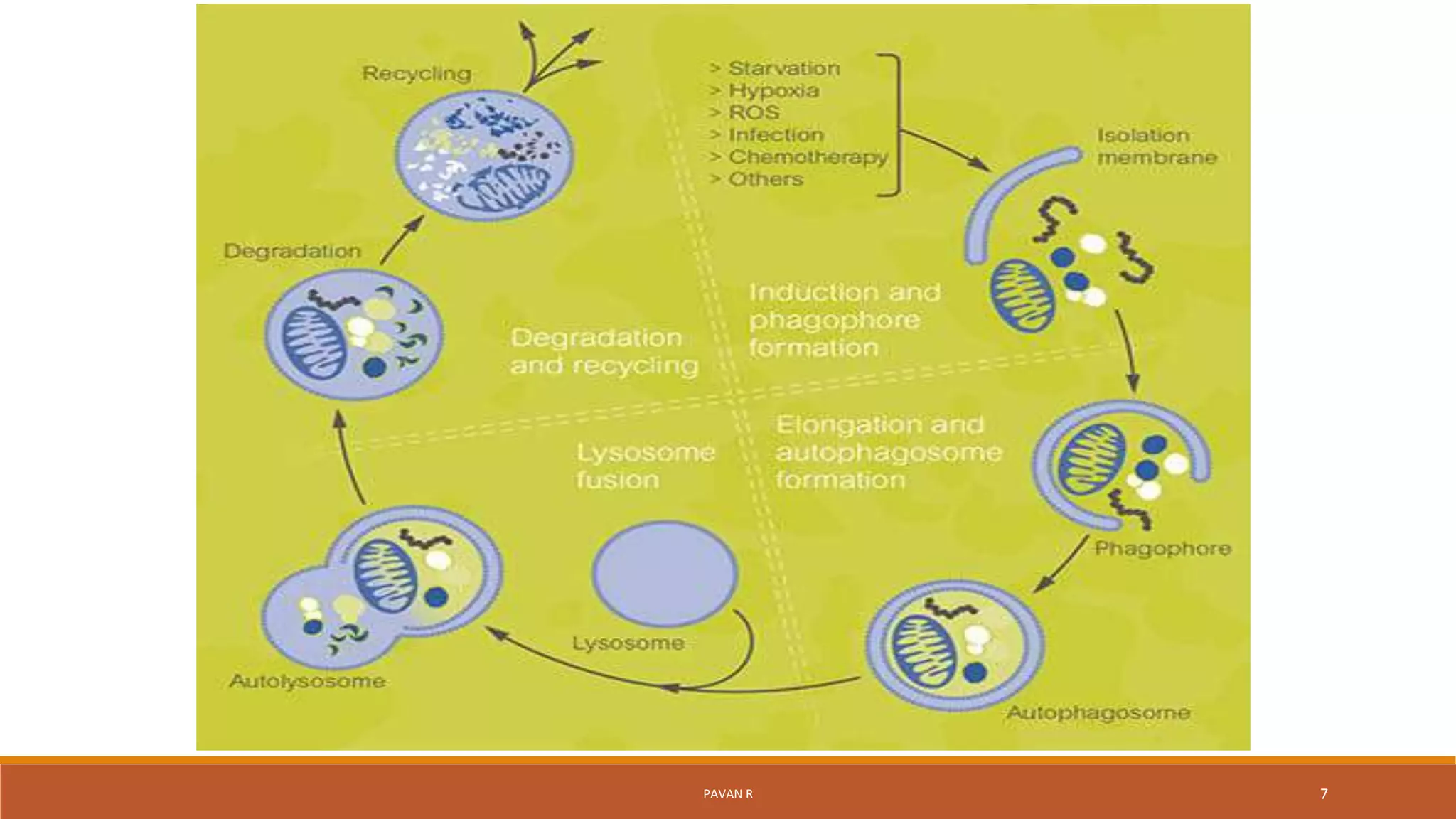
AUTOPHAGY
Submitted to Presented by
ASST PROF. VIMAL JOHN SAMUEL PAVAN R
DEPT of PHARMACOLOGY 1st SEM M.PHARM
(PHARMACOLOGY)
PAVAN R 1
CONTENTS
 Introduction
 History
Mechanism of Autophagy
 Types of Autophagy
 Reference
PAVAN R 2
Introduction
Definition:
Auto means self, phage means eating
Autophagy is a normal physiological process in the body that deals
with the destruction of cell in the body. It maintains homeostasis or normal
functioning by protein degradation turnover of the destroyed cell organelles
for new cell formation
During cellular stress the process of autophagy is upscaled and increased
PAVAN R 3
Introduction
Autophagy is a self-degradative process that is important for balancing sources
of energy at critical times in development and in response to nutrient stress.
The liver is rich in lysosomes and possesses high levels of metabolic-stress-
induced autophagy.
Autophagy regulated by concentrations of hormones and amino acids.
PAVAN R 4
History
 First time observed by Keith R. Porter and his student Thomas Ashford at the
Rockefeller Institute in 1962 soon after the discovery of lysosomes in rat liver.
 They called this autolysis after Christian de Duve and Alex B. Novikoff.
 A new era of autophagy research began in 1990s when several groups of
scientists independently discovered autophagy-related genes using the budding
yeast.
 6426 article were published about it from 2007 until 2012
PAVAN R 5
Mechanism
Different stages of autophagic process
1) Induction of autophagy and phagophore formation
2) Autophagophore elongation and formation
3) Fusion, degradation and recycling
PAVAN R 6
PAVAN R 7
PAVAN R 8
Induction and phagophore formation
The molecular mechanism of autophagy involves several conserved Atg
(autophagy-related) proteins. Various stimuli, such as nutrient starvation, lead
to the formation of the phagophore, a step that involves two protein
complexes:
• A complex that contains the class III PI3K/Vps34, Atg6/Beclin1, Atg14 and
Vps15/p150.73.
• A complex that includes the serine/threonine kinase Atg1/ULK1, an
essential positive regulator of autophagosome formation
PAVAN R 9
Autophagosome elongation and formation
 The elongation of the phagophore results in the formation of an
autophagosome, which is typically a double-membraned organelle.
 This step is a simple sequestration, and no degradation occurs.
 LC3B-II is found on both the inner and the outer surfaces of the
autophagosome.
 During autophagy, the synthesis and processing of LC3 is increased and it is
used as a marker to monitor levels of autophagy in cells.
PAVAN R 10
Fusion, degradation and recycling
 Completely formed autophagosome are fused with the lysosomes in the cell.
 Autophagosome-lysosome fusion is mediated by the same machinery that is
involved in homotypic vacuole membrane fusion.
 The degradation of the vesicular cargo is dependent on a series of
lysosomal/vacuolar acid hydrolases.
 The resulting small molecules from the degradation, particularly amino acids,
are transported back to the cytosol for protein synthesis and maintenance of
cellular functions
Types of Autophagy
1)Micro-autophagy
2)Macro-autophagy
3) Chaperone-mediated autophagy
PAVAN R 11
PAVAN R 12
MicroAutophagy
Microautophagy involves the direct engulfment of cytoplasmic cargo into the
lysosome through invagination of the lysosomal membrane.
Microautophagy is important in the maintenance of organellar size
PAVAN R 13
Microautophagy
1)Non-selective microautophagy
a)Membrane invagination and autophagic tubes formation
b)Vesicle formation
c)Vesicle expansion and scission
d)Vesicle degradation and recycling
2)Selective microautophagy
a) micropexophagy,
b)piecemeal microautophagy
c)micromitophagy
PAVAN R 14
Macroautophagy
Macroautophagy is a process in which cells form double-membrane vesicles,
called autophagosomes, around a portion of cytoplasm.
These autophagosomes ultimately fuse with lysosomes, resulting in degradation
of their contents.
PAVAN R 15
PAVAN R 16
Chaperone-mediated autophagy
Chaperone-mediated autophagy (CMA) refers to the chaperone-dependent
selection of soluble cytosolic proteins that are then targeted to lysosomes and
directly translocated across the lysosome membrane for degradation.
The unique features of this type of autophagy are the selectivity on the proteins
that are degraded by this pathway and the direct shuttling of these proteins
across the lysosomal membrane without the requirement for the formation of
additional vesicles
PAVAN R 17
Induce autophagy
Nutrition
Growth factors
Hormones
Intercellular calcium concentration
Hypoxia
Aggregation of misfolded proteins
PAVAN R 18
Some regulators
 Bcl2
 Reactive oxygen species (ROS)
 Calcium
 AMP-activated protein kinase
 BNIP3
 DRAM
 Calpain
 FADD
 IP3
PAVAN R 19
Reference
Glick D, Barth S, Macleod KF (2010). Autophagy: cellular and molecular mechanisms. J. Pathol.
221(1), 3-12
He C, Baba M, Cao Y, Klionsky DJ (2008). Self-interaction is critical for Atg9 transport and
function at the phagophore assembly site duting autophagy. Mol. Biol. Cell., 19(12), 5506-5516
He C & Klionsky DJ (2010). Analyzing autophagy in zebrafish. Autophagy, 6(5), 642-644
Kaur J & Debnath J (2015). Autophagy at the crossroads of catabolism and anabolism. Nat. Rev.
Mol. Cell Biol., 16(8), 461-472
Levine B & Kroemer G (2008). Autophagy in the pathogenesis of disease. Cell, 11(132), 27-42
Mizushima N (2007). Autophagy: process and function. Genes Dev., 21(22), 2861-2873.
PAVAN R 20
PAVAN R 21

Autophagy by pavan

  • 1.
    AUTOPHAGY Submitted to Presentedby ASST PROF. VIMAL JOHN SAMUEL PAVAN R DEPT of PHARMACOLOGY 1st SEM M.PHARM (PHARMACOLOGY) PAVAN R 1
  • 2.
    CONTENTS  Introduction  History Mechanismof Autophagy  Types of Autophagy  Reference PAVAN R 2
  • 3.
    Introduction Definition: Auto means self,phage means eating Autophagy is a normal physiological process in the body that deals with the destruction of cell in the body. It maintains homeostasis or normal functioning by protein degradation turnover of the destroyed cell organelles for new cell formation During cellular stress the process of autophagy is upscaled and increased PAVAN R 3
  • 4.
    Introduction Autophagy is aself-degradative process that is important for balancing sources of energy at critical times in development and in response to nutrient stress. The liver is rich in lysosomes and possesses high levels of metabolic-stress- induced autophagy. Autophagy regulated by concentrations of hormones and amino acids. PAVAN R 4
  • 5.
    History  First timeobserved by Keith R. Porter and his student Thomas Ashford at the Rockefeller Institute in 1962 soon after the discovery of lysosomes in rat liver.  They called this autolysis after Christian de Duve and Alex B. Novikoff.  A new era of autophagy research began in 1990s when several groups of scientists independently discovered autophagy-related genes using the budding yeast.  6426 article were published about it from 2007 until 2012 PAVAN R 5
  • 6.
    Mechanism Different stages ofautophagic process 1) Induction of autophagy and phagophore formation 2) Autophagophore elongation and formation 3) Fusion, degradation and recycling PAVAN R 6
  • 7.
  • 8.
    PAVAN R 8 Inductionand phagophore formation The molecular mechanism of autophagy involves several conserved Atg (autophagy-related) proteins. Various stimuli, such as nutrient starvation, lead to the formation of the phagophore, a step that involves two protein complexes: • A complex that contains the class III PI3K/Vps34, Atg6/Beclin1, Atg14 and Vps15/p150.73. • A complex that includes the serine/threonine kinase Atg1/ULK1, an essential positive regulator of autophagosome formation
  • 9.
    PAVAN R 9 Autophagosomeelongation and formation  The elongation of the phagophore results in the formation of an autophagosome, which is typically a double-membraned organelle.  This step is a simple sequestration, and no degradation occurs.  LC3B-II is found on both the inner and the outer surfaces of the autophagosome.  During autophagy, the synthesis and processing of LC3 is increased and it is used as a marker to monitor levels of autophagy in cells.
  • 10.
    PAVAN R 10 Fusion,degradation and recycling  Completely formed autophagosome are fused with the lysosomes in the cell.  Autophagosome-lysosome fusion is mediated by the same machinery that is involved in homotypic vacuole membrane fusion.  The degradation of the vesicular cargo is dependent on a series of lysosomal/vacuolar acid hydrolases.  The resulting small molecules from the degradation, particularly amino acids, are transported back to the cytosol for protein synthesis and maintenance of cellular functions
  • 11.
    Types of Autophagy 1)Micro-autophagy 2)Macro-autophagy 3)Chaperone-mediated autophagy PAVAN R 11
  • 12.
  • 13.
    MicroAutophagy Microautophagy involves thedirect engulfment of cytoplasmic cargo into the lysosome through invagination of the lysosomal membrane. Microautophagy is important in the maintenance of organellar size PAVAN R 13
  • 14.
    Microautophagy 1)Non-selective microautophagy a)Membrane invaginationand autophagic tubes formation b)Vesicle formation c)Vesicle expansion and scission d)Vesicle degradation and recycling 2)Selective microautophagy a) micropexophagy, b)piecemeal microautophagy c)micromitophagy PAVAN R 14
  • 15.
    Macroautophagy Macroautophagy is aprocess in which cells form double-membrane vesicles, called autophagosomes, around a portion of cytoplasm. These autophagosomes ultimately fuse with lysosomes, resulting in degradation of their contents. PAVAN R 15
  • 16.
  • 17.
    Chaperone-mediated autophagy Chaperone-mediated autophagy(CMA) refers to the chaperone-dependent selection of soluble cytosolic proteins that are then targeted to lysosomes and directly translocated across the lysosome membrane for degradation. The unique features of this type of autophagy are the selectivity on the proteins that are degraded by this pathway and the direct shuttling of these proteins across the lysosomal membrane without the requirement for the formation of additional vesicles PAVAN R 17
  • 18.
    Induce autophagy Nutrition Growth factors Hormones Intercellularcalcium concentration Hypoxia Aggregation of misfolded proteins PAVAN R 18
  • 19.
    Some regulators  Bcl2 Reactive oxygen species (ROS)  Calcium  AMP-activated protein kinase  BNIP3  DRAM  Calpain  FADD  IP3 PAVAN R 19
  • 20.
    Reference Glick D, BarthS, Macleod KF (2010). Autophagy: cellular and molecular mechanisms. J. Pathol. 221(1), 3-12 He C, Baba M, Cao Y, Klionsky DJ (2008). Self-interaction is critical for Atg9 transport and function at the phagophore assembly site duting autophagy. Mol. Biol. Cell., 19(12), 5506-5516 He C & Klionsky DJ (2010). Analyzing autophagy in zebrafish. Autophagy, 6(5), 642-644 Kaur J & Debnath J (2015). Autophagy at the crossroads of catabolism and anabolism. Nat. Rev. Mol. Cell Biol., 16(8), 461-472 Levine B & Kroemer G (2008). Autophagy in the pathogenesis of disease. Cell, 11(132), 27-42 Mizushima N (2007). Autophagy: process and function. Genes Dev., 21(22), 2861-2873. PAVAN R 20
  • 21.