Apoptosis is a tightly regulated and controlled process of programmed cell death. It is essential for normal development and maintenance of tissues as it removes unnecessary or damaged cells. During apoptosis, cells activate enzymes to degrade their own DNA and proteins. Apoptosis is initiated through either the extrinsic or intrinsic pathway which involve death ligands/receptors or mitochondrial signaling, respectively, ultimately activating caspase enzymes that kill the cell in a controlled manner. Apoptosis is important for development, the immune system, and removing pre-cancerous or infected cells, while deficiencies can lead to cancer or autoimmune disorders.
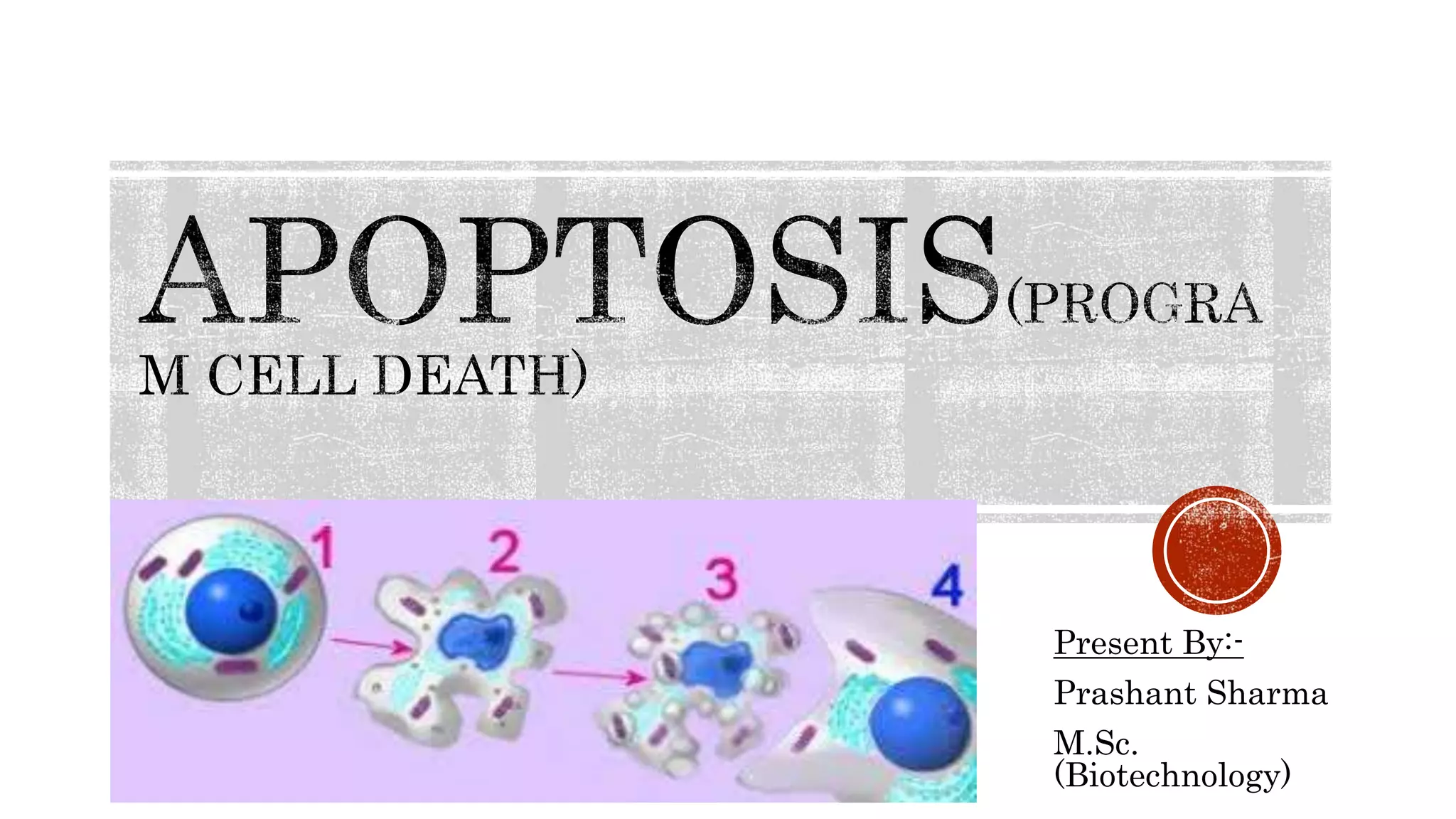
Apoptosis isthe process of programmed cell death
A pathway of cell death induced by a tightly regulated suicidal program, in which
the cells destined to die activate enzymes that degrade cells own nuclear DNA and
nuclear, cytoplasmic proteins.
Controlled by specific genes.
in which cells die due to injury
Between 50 and 70 billion cells die each day due to apoptosis in the average
human adult. For an average child between the ages of 8 and 14, approximately
20 billion to 30 billion cells die a day.
3.
In manyorganisms, programmed cell death is a normal part of development.
Apoptosis removes cells during development. It also eliminates pre-cancerous and
virus-infected cells, although “successful” cancer cells manage to escape apoptosis so
they can continue dividing.
Apoptosis maintains the balance of cells in the human body and is particularly
important in the immune system.
Apoptosis is needed for proper development
Examples:
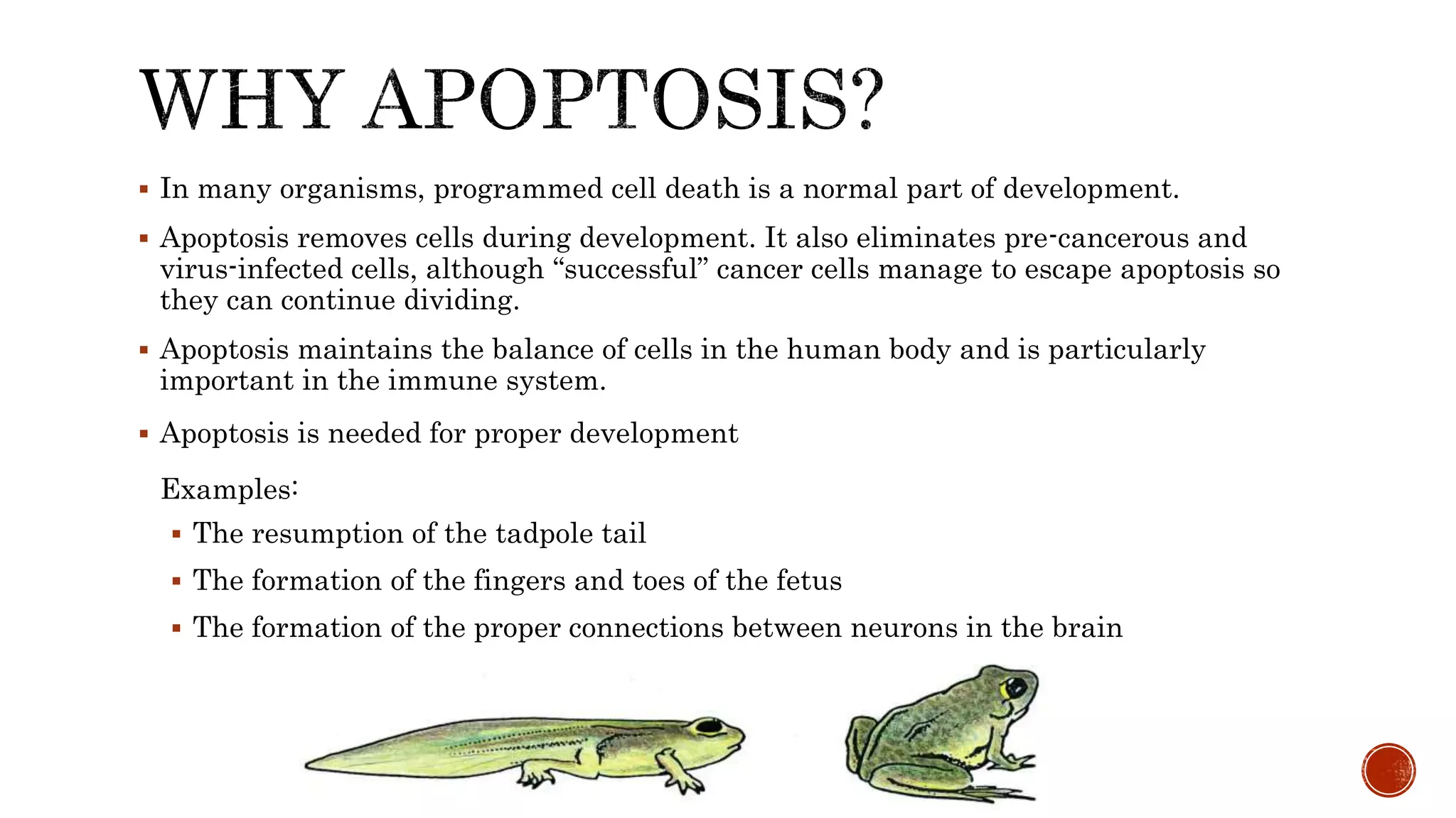
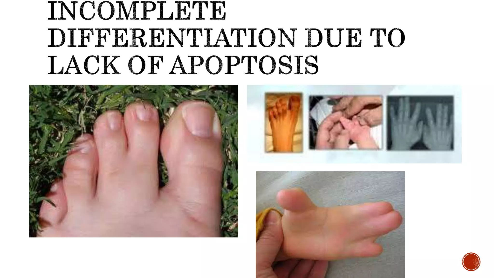
The resumption of the tadpole tail
The formation of the fingers and toes of the fetus
The formation of the proper connections between neurons in the brain
4.
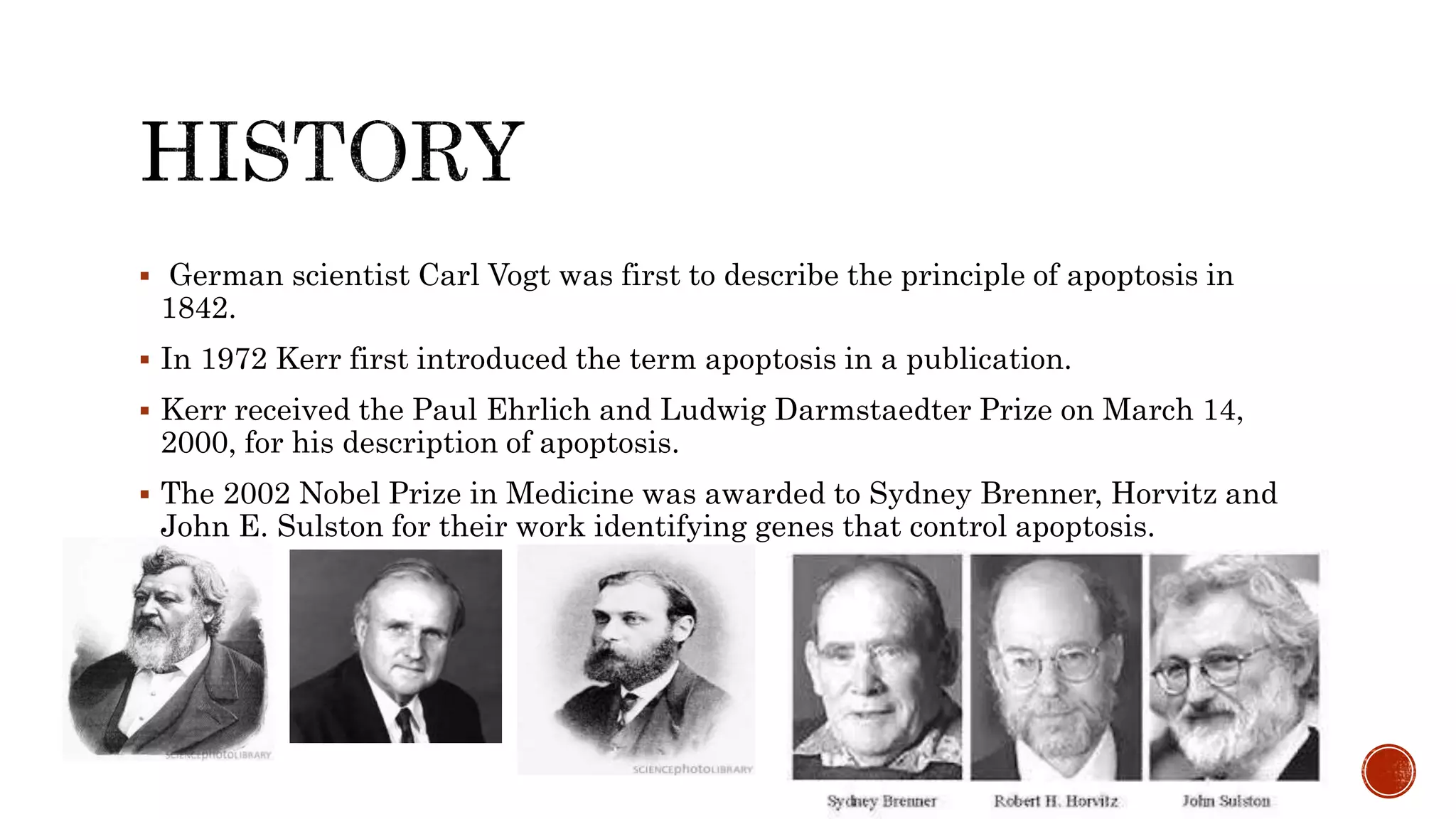
German scientistCarl Vogt was first to describe the principle of apoptosis in
1842.
In 1972 Kerr first introduced the term apoptosis in a publication.
Kerr received the Paul Ehrlich and Ludwig Darmstaedter Prize on March 14,
2000, for his description of apoptosis.
The 2002 Nobel Prize in Medicine was awarded to Sydney Brenner, Horvitz and
John E. Sulston for their work identifying genes that control apoptosis.
5.
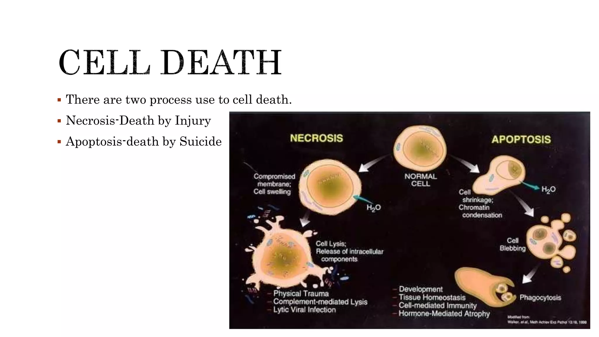
There aretwo process use to cell death.
Necrosis-Death by Injury
Apoptosis-death by Suicide
7.
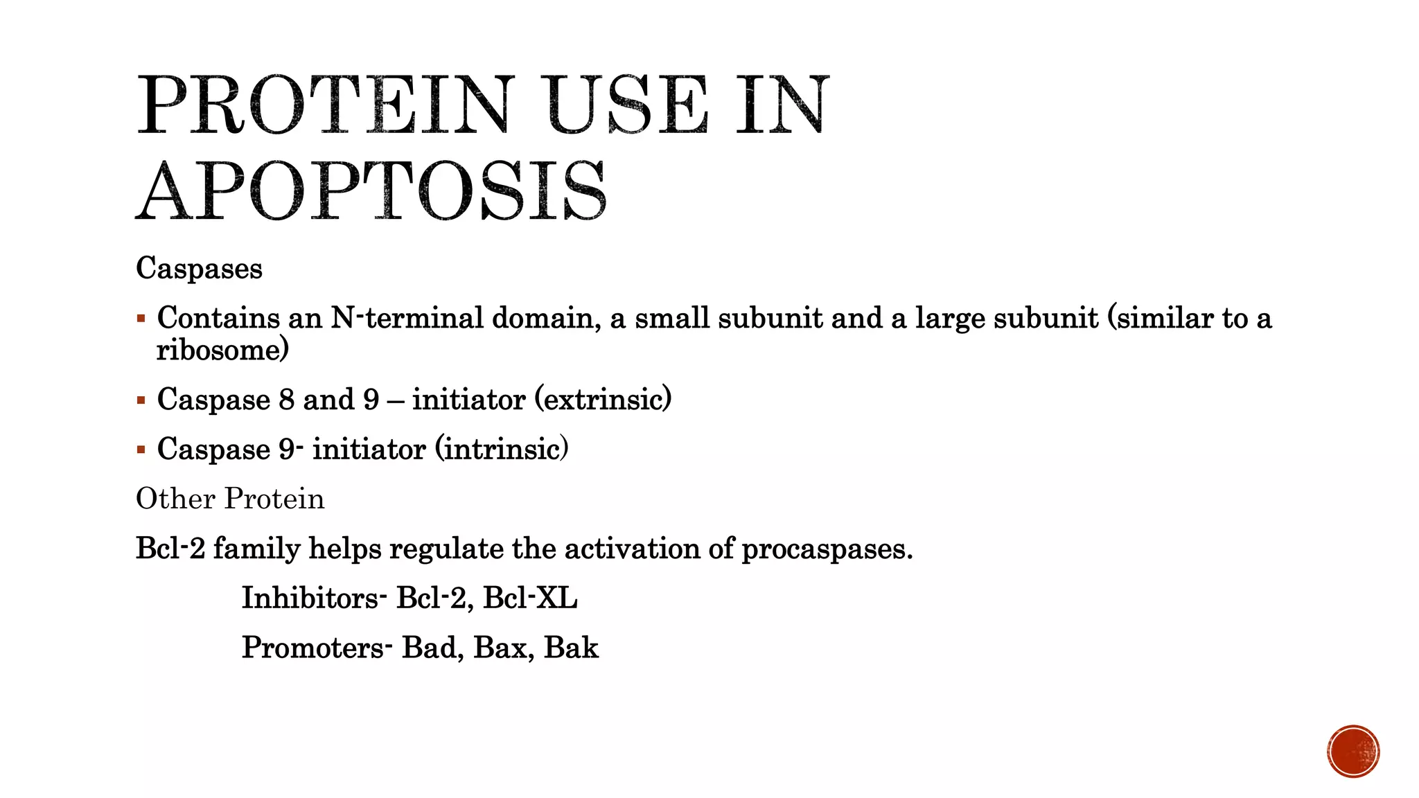
Caspases
Contains anN-terminal domain, a small subunit and a large subunit (similar to a
ribosome)
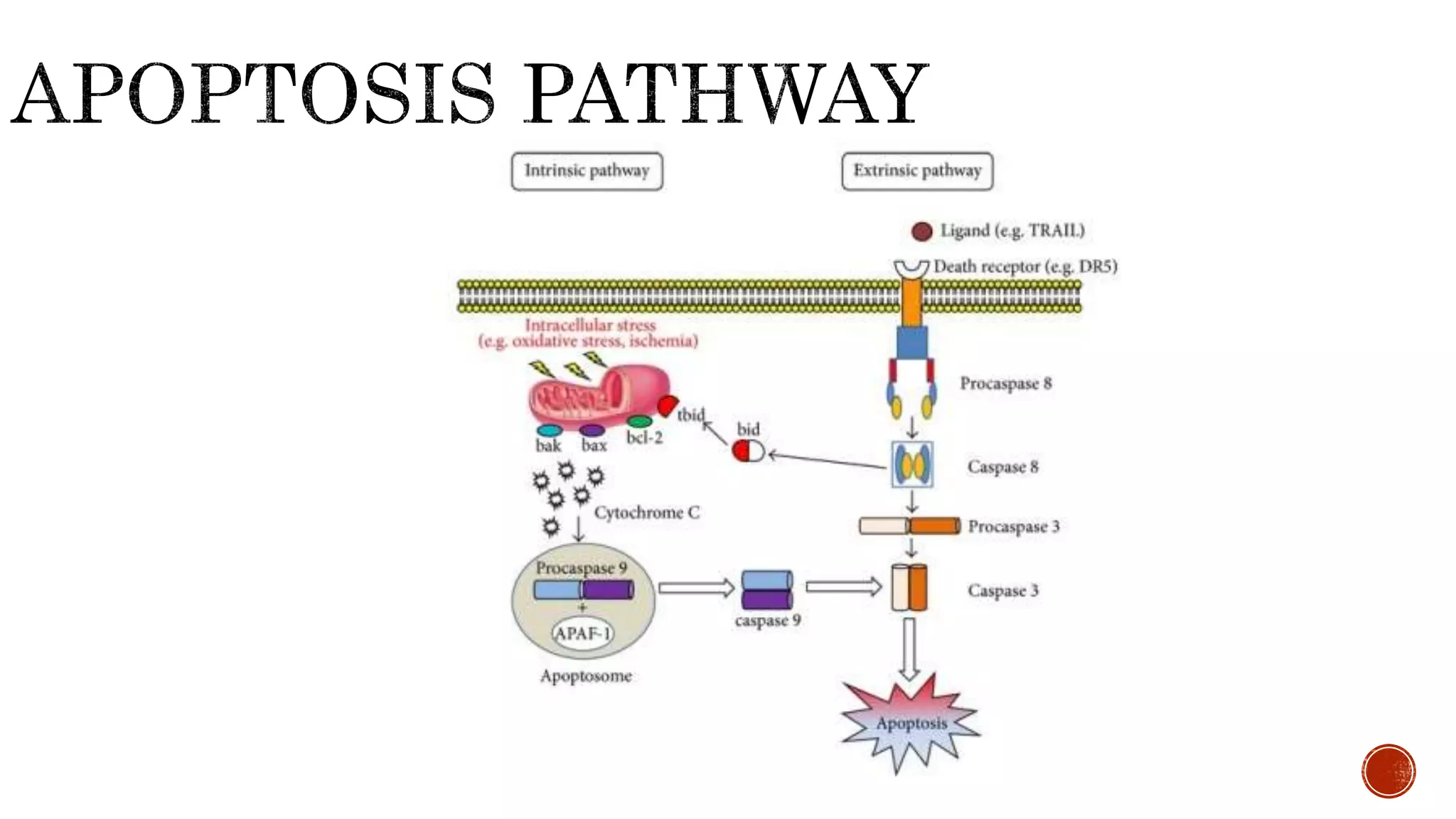
Caspase 8 and 9 – initiator (extrinsic)
Caspase 9- initiator (intrinsic)
Other Protein
Bcl-2 family helps regulate the activation of procaspases.
Inhibitors- Bcl-2, Bcl-XL
Promoters- Bad, Bax, Bak
9.
EXTRINSIC OR DEATH
RECEPTORPATHWAY
Extrinsic Pathway
Death Ligand
Death Receptors
Caspases
Cell Death
10.
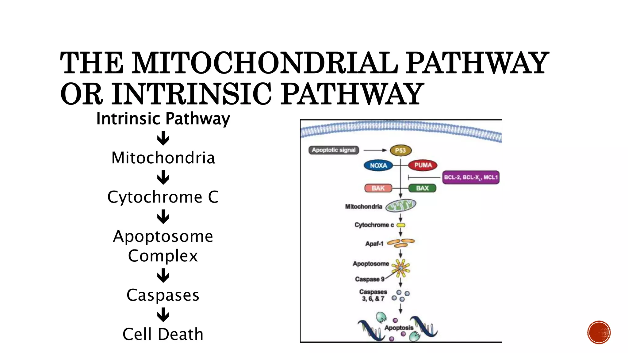
THE MITOCHONDRIAL PATHWAY
ORINTRINSIC PATHWAY
Intrinsic Pathway
Mitochondria
Cytochrome C
Apoptosome
Complex
Caspases
Cell Death
12.
• Important innormal physiology / development
– Development: Immune systems maturation, Morphogenesis.
– Adult: Immune privilege, DNA Damage and wound repair.
• Deficient apoptosis
– Cancer
– Autoimmunity