2
Learning objectives
At theend of the session the student should be
able to
1. Define apoptosis
2. Describe the features of apoptosis
3. Differentiate apoptosis from necrosis
4. Enumerate the steps involved in apoptosis
– intrinsic and extrinsic pathway
5. Discuss the physiological and applied importance
of apoptosis
3.
3
Introduction
• Total numberof cells is regulated by controlling
– rate of cell division and
– controlling rate of cell death
• Cell death:
1. When cell are no longer needed or become a threat they
undergo an orderly sequence of events called apoptosis. The
term apoptosis was coined by John Kerr, Andrew Wyllie and
A.R. Currie.
2. Cells that die as a result of acute injury undergo necrosis
4.
4
Physiological significance ofapoptosis
(Why apoptosis occurs?)
• Embryogenesis and fetal development
– In the central nervous system, large numbers of neurons are produced and
then die during the remodeling that occurs during development and
synapse formation.
– Removal of the webs between the fingers in fetal life
– Regression of duct systems in the course of sexual development in the fetus
• Hormone dependent involution
– Prostate glandular epithelium after castration
– Regression of lactating breast after weaning
• Cell loss in proliferating cell populations
– Immature lymphocytes
– Epithelial cells in the GI tract
– Elimination of self-reactive lymphocytes.
• Death of cells that have served their function
– Neutrophils, Lymphocytes
5.
5
Definition
• Apoptosis (Greekapo "away" + ptosis "fall") is a
pathway of cell death induced by a tightly regulated
suicide program controlled by specific genes.
• It is programmed sequence of molecular events, in
which the cell systematically destroys itself from
within and is then eaten by other cells, leaving no
trace.
• Hence it is a type of programmed cell death
• It can be called "cell suicide" in the sense that the cell's
own genes play an active role in its demise.
6.
6
Features of aapoptotic cell
• Characterized by the overall shrinkage in volume of the cell
• Collapse of cytoskeletal system
• Shrinkage of nucleus
• Loss of adhesion to neighboring cells
• Disintegration of the chromatin into small fragments
• Cell contents do not spill
• If cell is large it breaks into membrane enclosed fragments
called apoptotic bodies
• Engulfment of the “corpse” by macrophages or
neighboring cells
7.
7
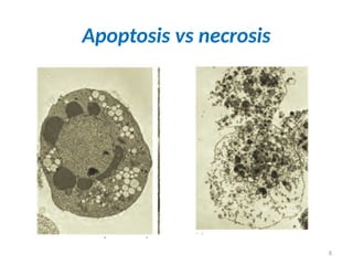
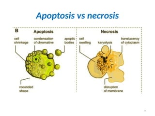
Apoptosis vs Necrosis
Apoptosis
•Death of individual or small group of cells
evoked by physiological stimuli.
• Morphological features:
- membrane blebbing without loss of
membrane integrity,
- condensation of chromatin,
- cell shrinkage,
- formation of apoptotic bodies.
• Biochemical changes:
- Genetically controlled activation of
enzymes
- ATP dependent process
- Generation of non random DNA
oligonucleosomes.
Necrosis
• Death of large contiguous groups of
cells or organ segments evoked by
pathological stimuli.
• Morphological features:
- loss of membrane integrity,
- random fragmentation of
chromatin,
- cellular swelling,
- cell lysis, swelling and disintegration
of organelles.
• Biochemical changes:
- Loss of ion homeostasis
- Passive process, no energy required
- Random DNA digestion
10
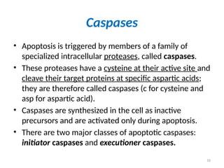
Caspases
• Apoptosis istriggered by members of a family of
specialized intracellular proteases, called caspases.
• These proteases have a cysteine at their active site and
cleave their target proteins at specific aspartic acids;
they are therefore called caspases (c for cysteine and
asp for aspartic acid).
• Caspases are synthesized in the cell as inactive
precursors and are activated only during apoptosis.
• There are two major classes of apoptotic caspases:
initiator caspases and executioner caspases.
11.
11
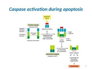
Caspases
• Initiator caspasesbegin the apoptotic process.
• Apoptotic signals cause their activation.
• Initiator caspases then activate executioner caspases.
• One initiator caspase complex can activate many
executioner caspases, resulting in an amplifying
proteolytic cascade.
• Once activated, executioner caspases catalyze the
widespread protein cleavage events that kills the cell.
13
Caspases
• Targets ofcaspases are the following:
– Focal adhesion kinase (FAK ): this disrupts cell adhesion,
leading to detachment of the apoptotic cell from its
neighbors.
– Lamins: make up the inner lining of the nuclear
envelope, cleavage of lamins leads to the disassembly of
the nuclear lamina and shrinkage of the nucleus.
– Proteins of the cytoskeleton: cleavage and consequent
inactivation of these proteins leads to change in cell
shape
– An endonuclease called caspase activated DNase (CAD):
once activated, CAD goes to the nucleus and attacks
DNA, severing it into fragments.
14.
14
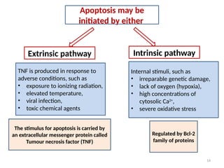
Apoptosis may be
initiatedby either
Extrinsic pathway Intrinsic pathway
TNF is produced in response to
adverse conditions, such as
• exposure to ionizing radiation,
• elevated temperature,
• viral infection,
• toxic chemical agents
The stimulus for apoptosis is carried by
an extracellular messenger protein called
Tumour necrosis factor (TNF)
Internal stimuli, such as
• irreparable genetic damage,
• lack of oxygen (hypoxia),
• high concentrations of
cytosolic Ca2+
,
• severe oxidative stress
Regulated by Bcl-2
family of proteins
15.
15
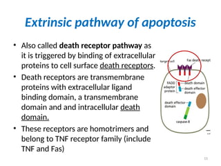
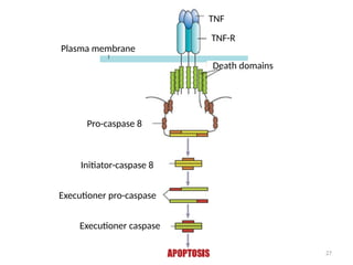
Extrinsic pathway ofapoptosis
• Also called death receptor pathway as
it is triggered by binding of extracellular
proteins to cell surface death receptors.
• Death receptors are transmembrane
proteins with extracellular ligand
binding domain, a transmembrane
domain and and intracellular death
domain.
• These receptors are homotrimers and
belong to TNF receptor family (include
TNF and Fas)
17
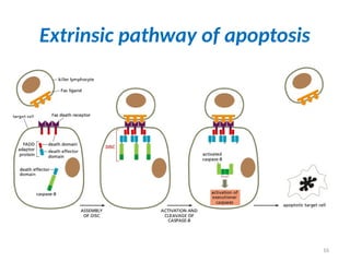
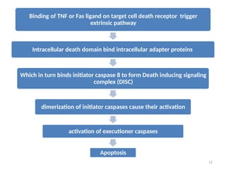
Binding of TNFor Fas ligand on target cell death receptor trigger
extrinsic pathway
Intracellular death domain bind intracellular adapter proteins
Which in turn binds initiator caspase 8 to form Death inducing signaling
complex (DISC)
dimerization of initiator caspases cause their activation
activation of executioner caspases
Apoptosis
18.
18
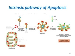
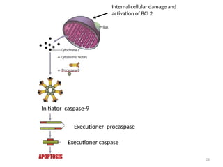
Intrinsic pathway ofapoptosis
• Pathway activated due to signals generated inside the cell often
due to stresses such as DNA damage or in response to
developmental signals.
• Also called mitochondrial pathway of apoptosis as it depends on
release of mitochondrial proteins into cytosol which activate
caspases.
• Key protein in this pathway is cytochrome c, a component of
electron transport chain, which when released into cytosol binds
an adapter protein Apaf1 (apoptotic protease activating factor-1)
causing Apaf1 to oligomerize into a heptamer called Apoptosome.
• Apoptosome recruits initiator caspase-9 which further activates
executioner caspases to induce apoptosis.
20
Regulation the Intrinsicpathway of
Apoptosis
• The intrinsic pathway of apoptosis is tightly
regulated to ensure that cells kill themselves
only when it is appropriate.
• Intracellular regulators of the intrinsic
pathway is the Bcl2 family of proteins.
• In mammals it regulates release of
cytochrome c and other mitochondrial
proteins into cytosol.
21.
21
Phagocytes remove apoptoticcells
• Phagocytosis of apoptotic cell by neighboring
cells or macrophage is facilitated by chemical
changes on surface of apoptotic cell.
• There occurs distribution of negatively
charged phospholipid ‘phosphatidylserine’ on
the cell surface along with loss of expression
of certain signal proteins on surface of
apoptotic cells seen in normal healthy cells.
22.
22
Excessive or InsufficientApoptosis
• Excessive apoptosis contribute to tissue damage.
• For example – heart attack and stroke. Many cells die due to
necrosis due to loss of blood supply, but in addition less
affected cells die by apoptosis.
• Less apoptosis causes:
o Autoimmune disease- eg. mutation of Fas ligand prevents
normal death of lymphocytes, leading to their accumulation in
spleen and lymph and development of autoimmune diseases.
o Cancer-
- Bcl2 gene mutation causing its excess production leads to
lymphoma.
- Mutation of p53, a tumor suppressor gene supresses apoptosis
causing cancer.
23.
23
• Abnormal apoptosismay occur in
autoimmune diseases, neurodegenerative
diseases, and cancer
• Therefore, selective manipulation of apoptotic
pathways may be an important approach for
treating cancer in the future.
24.
24
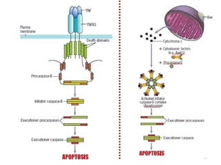
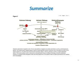
Summary: Main stepsof Apoptosis
• Death receptor (Extrinsic) pathway
• Mitochrondrial (Intrinsic) pathway
– The intrinsic and extrinsic pathways converge to a
caspase activation cascade.
• Execution Phase: Caspases execute the process
• Removal of dead cells:
– Dying cells secrete factors the recruit phagocytes.
– This facilitates prompt clearance
– Dead cells disappear without a trace and do not produce
inflammation.
25.
25
• Define Apoptosis.
•Why apoptosis occur?
• Name the pathways of apoptosis.
• Briefly describe steps of each pathway.
• What are caspases? What is their role in
apoptosis?
• How is apoptosis different from necrosis?
30
What is common?
•The intrinsic and extrinsic pathways finally
converge by activating the same enzymes i.e,
caspases
• Caspases are a group of cysteine proteases
responsible for triggering most, of the
changes observed during apoptosis
31.
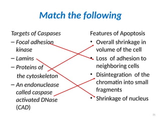
Match the following
Targetsof Caspases
– Focal adhesion
kinase
– Lamins
– Proteins of
the cytoskeleton
– An endonuclease
called caspase
activated DNase
(CAD)
Features of Apoptosis
• Overall shrinkage in
volume of the cell
• Loss of adhesion to
neighboring cells
• Disintegration of the
chromatin into small
fragments
• Shrinkage of nucleus
31
33
Learning objectives
At theend of the session the student must be
able to
1. Define apoptosis
2. Describe the features of apoptosis
3. Differentiate apoptosis from necrosis
4. Enumerate the steps involved in apoptosis
– intrinsic and extrinsic pathway
5. Discuss the physiological and applied
importance of apoptosis