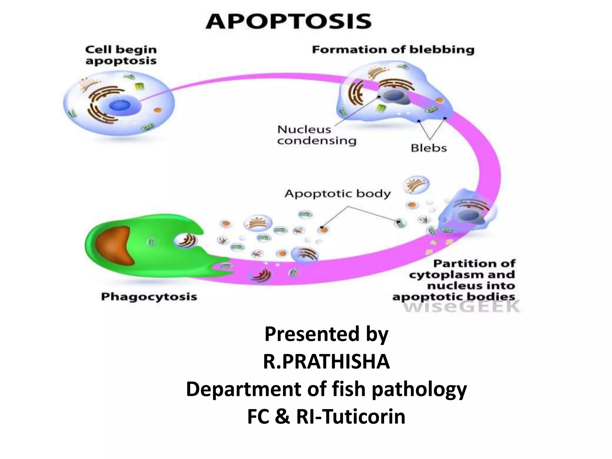
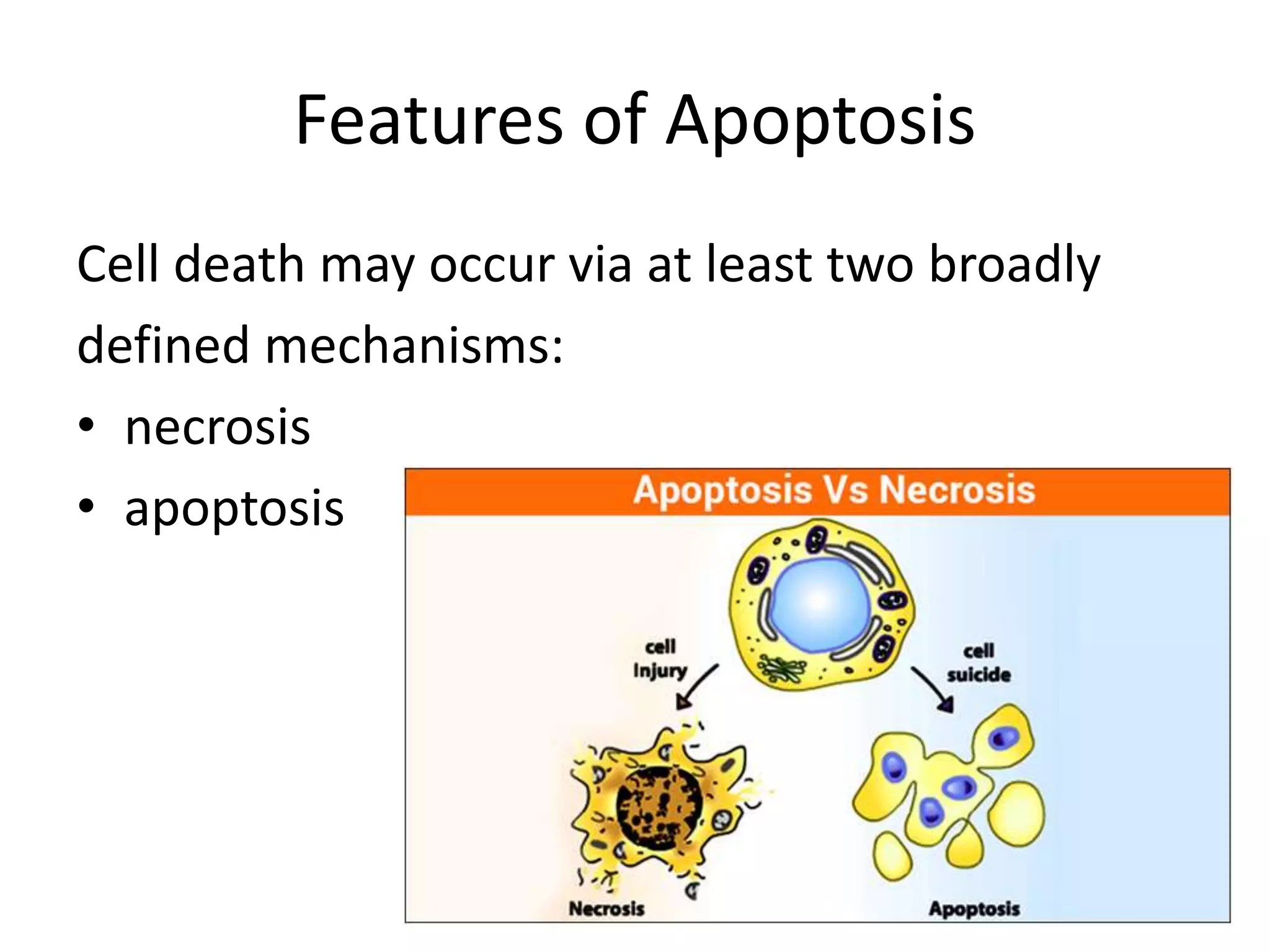
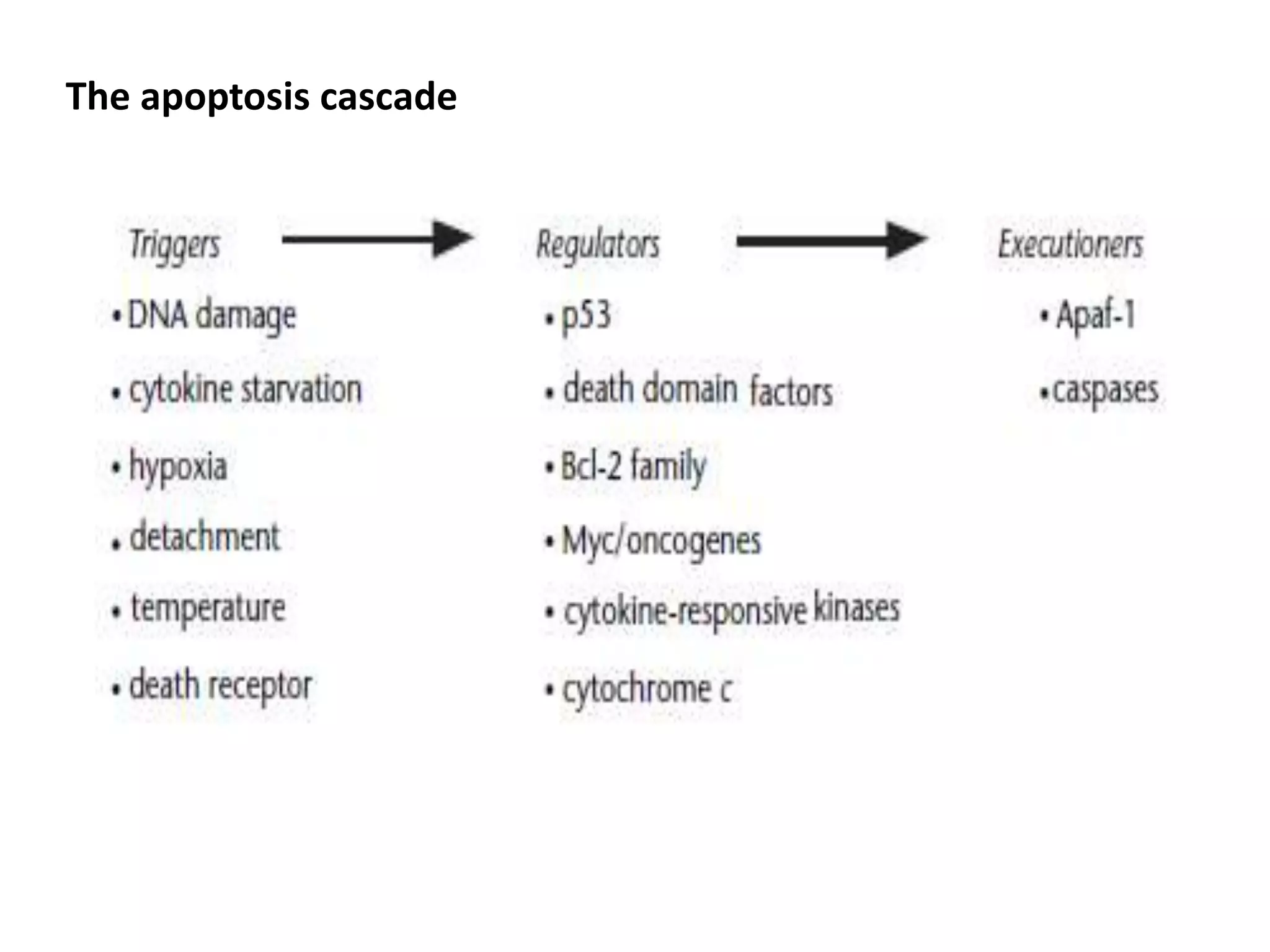
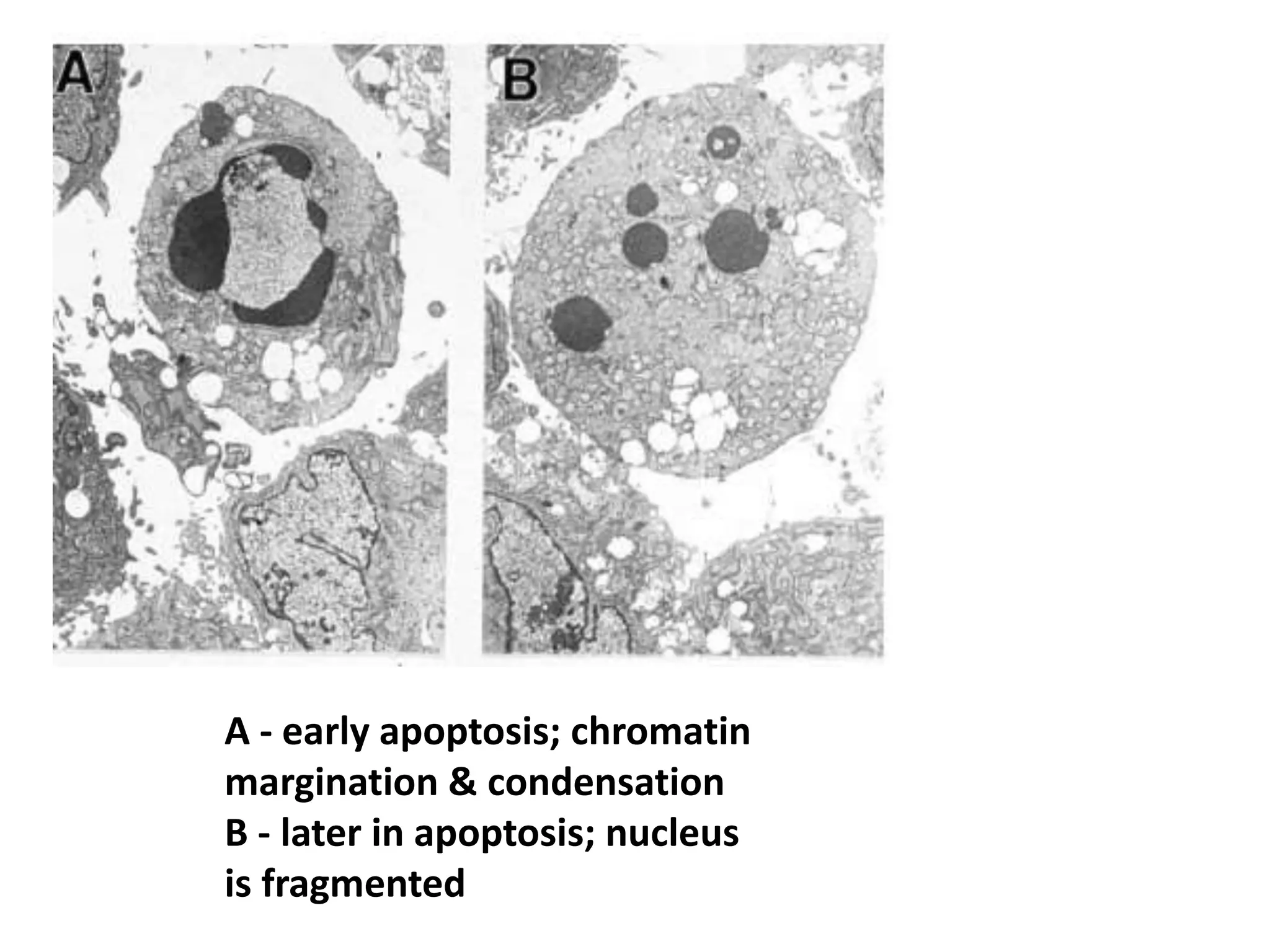
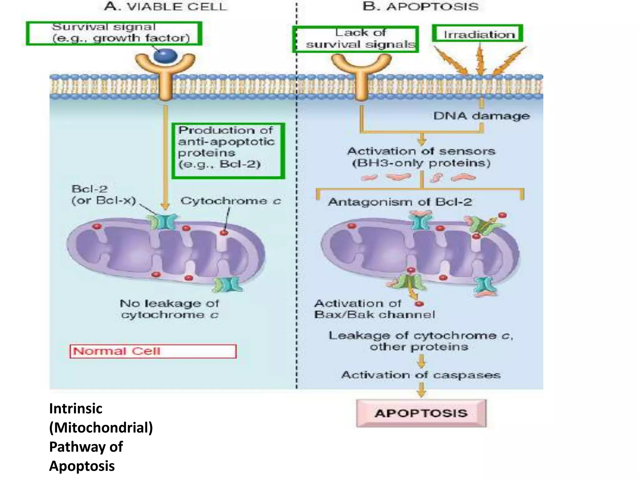
Presented by
R.PRATHISHA
Department of fish pathology
FC & RI-Tuticorin
Features of Apoptosis
Cell death may occur via at least two broadly
defined mechanisms:
• necrosis
• apoptosis
NECROSIS
• Usually affects large areas
of contiguous cells
• Control of intracellular
environment is lost early
• Cells swell and organelles
swell
APOPTOSIS
• Usually affects scattered
individual cells
• Control of intracellular
environment maintained in
early stages
• Cells contract
• Nuclear chromatin
marginates early,
• while injury is still
reversible
• When DNA is cleaved,which
is usually a late event,
fragments are random in
size (smear pattern in gels)
• Nuclear chromatin
marginates and chromatin
condenses,becoming very
compact
• Chromatin condensation
and DNA fragmentation
occur together; DNA
cleaved into multiples of
200 base pair units (ladder
pattern in gels)
• Cell membrane ruptures as
terminal event and cell
contents are released,which
are chemotactic
• Chemotactic factors lead to
neutrophil infiltration to
degrade dead cells
• Blebs form and apoptotic
bodies containing nuclear
Fragments are shed
• Phagocytosis of intact
apoptotic bodies, no
chemotactic factors are
generated
Apoptosis
• Pathway of cell death induced by a tightly
regulated suicide program.
• Controlled by specific genes.
• Fragmentation of DNA.
• Fragmentation of nucleus.
• Blebs form and apoptotic bodies are released.
• Apoptotic bodies are phagocytized.
 As many as 1011 cells die in an adult human per day to ensure
tissue homeostasis, and it is estimated that within a typical year,
the mass of cells a person loses through cell death is almost
equivalent to their entire body weight.
Apoptosis is also a protective mechanism, directing lysis of
virus-infected cells, foreign cells or incipient neoplasm.
Excess cell death can contribute to the acquired immune
deficiency syndrome (AIDS) and neurodegenerative diseases like
Alzheimer and Parkinson syndromes, and ischaemic injury such as
myocardial infarction.
Too little cell death could lead to cancer, persistent viral
infection, or autoimmune disorders.
Apoptosis plays a central role in the immune system
•Immature lymphocytes that bind to autoantigens are eliminated
by apoptosis
•Apoptosis in thymocytes is associated with the elimination of
self-reactive clones of developing T cells following their
interaction with antigens in the thymus
•B cells are subject to death by apoptosis throughout most
stages of their maturation and 60–70% cell loss has been
calculated during the pre-B to B cell transition in bone marrow
Inducers of Apoptosis and
Apoptotic Signalling
• Activators of apoptosis include
• Tumour necrosis factor α(TNFα),
• Fas ligand (FasL),
• Transforming growth factor β (TGFβ),
• Bax (and other proapoptotic Bcl-2 familymembers),and
• glucocorticoids
• In addition, aberrant oncogene expression (e.g. c-
myc), or normal tumour
• suppressor gene function (such as p53) may trigger
apoptosis under specific conditions.
Causes of Apoptosis
• Physiologic
• Pathologic
PHYSIOLOGIC APOPTOSIS
•Embryogenesis and fetal development.
• Hormone dependent involution.
• Cell loss in proliferating cell populations.
• Immature lymphocytes
• Epithelial cells in the GI tract
• Elimination of self-reactive lymphocytes.
• Death of cells that have served their function.
• Neutrophils, Lymphocytes
Pathologic
apoptosis:
• DNA damage due ‘to
radiation, chemotherapy.
• Accumulation of misfolded
proteins leads to ER stress
which ends with apoptosis.
• Cell death in viral
infections that induce
• apoptosis such as HIV and
Adenovirus or by the host
immune response such as
hepatitis.
• Organ atrophy after duct
obstruction
Genes that Regulate Apoptosis
• The ‘point of no return’ in apoptosis is
reached when caspases become
enzymatically active in cleaving target
proteins
The apoptosis cascade
• The Bcl-2 family of factors regulate caspase
activation either negatively (e.g. Bcl-2itself)or
positively(e.g. Bax)
• Among these upstream modulators are
oncogenes such as c-myc which activates
apoptosis in a manner which may be important
in tumorigenesis or cancer therapy.
• The tumour suppressor p53 induces apoptosis
under certain conditions, thereby accounting for
at least a portion of its tumour suppressive
activity.
cleavage of chromosomal DNA to a size
of several 100 kb,
then to 50 kb and
eventually to 200 bp
These oligosomal DNA fragments result in a distinct
laddering pattern on an ethidium bromide-stained
agarose gel that represents a hallmark of apoptosis
A - early apoptosis; chromatin
margination & condensation
B - later in apoptosis; nucleus
is fragmented
C - phagocytosis of apoptotic
cellular remnants by
adjacent cell
D - swollen, necrotic cell for
comparison
Mechanisms of Apoptosis
• Death receptor (Extrinsic)
pathway
• Mitochrondrial (Intrinsic)
pathway
• Execution Phase
• Removal of dead cells
Intrinsic -Mitochrondrial pathway
• Increased mitochondrial permeability with release of pro-
apoptotic molecules into the cytoplasm (cytochrome c).
• Synthesis of anti-apoptotic molecules (Bcl-2) promoted by
Growth factors.
• When cells are deprived of growth factors or subjected to
stress antiapoptotic molecules (Bcl-2) are lost.
• Mitochondrial membrane becomes permeable and proteins
that activate caspase leak out.
Intrinsic
(Mitochondrial)
Pathway of
Apoptosis
Extrinsic (Death receptor initiated) pathway
• Death receptors are members of the tumor necrosis factor
receptor family and a related protein called Fas (CD95).
• These molecules contain a death domain.
Fas and TNFa Receptor-mediated
Apoptotic Signalling
• Fas receptor (CD95) belongs to a family of receptors
including the tumour necrosis factor (TNF) and nerve
growth factor (NGF) receptors that utilize related
signalling pathways to regulate cell proliferation,
differentiation or death.
• Fas serves as a receptor on the cell surface for a ligand
(FasL), and the
• crosslinking of FasL to Fas receptor triggers
apoptosis on the target cells.
• The Fas apoptotic pathway is implicated in eliminating
unwanted activated lymphocytes or virus-infected
cells
Extrinsic (Death
Receptor-initiated)
Pathway of
Apoptosis
Executioners of Apoptosis: Caspases
• Caspases (cysteinyl aspartate-specific
proteinases) are a family of proteases containing
cysteine at their active sites.
• They are related to mammalian interleukin 1b-
converting enzyme (ICE/caspase1) and to the
nematode apoptotic gene product Ced-3
• That these proteases play critical roles in
executing programmed cell death
Eg: caspase-3 dominates in regulating neuronal
apoptosis
• The inhibition of caspases is also one of the major
strategies adopted by viruses to elude their
destruction through suicide of the infected cell
• Cytokine response modifier A (CrmA) -cowpox
virus- caspase-1 and caspase-8
• P35- baculovirus- all caspases
• Leakage of cytochrome c from mitochondria
might serve as a key signal to activate
procaspases and initiate the biochemical events
of death.
• Infectious pancreatic necrosis virus (IPNV), the
causative agent of a highly infectious disease
in salmonid fish, encodes a small
nonstructural protein designated VP5.
• This protein contains Bcl-2 homologous
domains and inhibits apoptosis
Execution Phase
• The intrinsic and extrinsic pathways converge to a caspase
activation cascade
• Caspases (cysteine-aspartic-acid-proteases) are conserved
across species
• Synthesized as inactive precursors; activated by proteolytic
cleavage
• Family of at least 12 proteases, a few of which are involved in
inflammation, and many of which are involved in apoptosis
How Caspases Disassemble a Cell
• Cleave structural proteins leading
to nuclear breakdown.
• Converts cytoplasmic DNase to
active form.
• DNase causes characteristic
internucleosomal cleavage of DNA.
Removal of Dead Cells
• Dying cells secrete factors the recruit phagocytes.
• This facilitates prompt clearance before they undergo
secondary necrosis.
• Dead cells disappear without a trace and do not
produce inflammation.
Antiapoptotic genes
Heavily studied antiapoptotic genes include
• Bcl-2 and
• Bcl-XL in mammalian cells,
• Ced-9 in C. elegans,
• p35 in baculovirus,
• and E1B 19K protein in adenovirus.
• Overexpression of Bcl-2 is capable of
antagonizing apoptosis triggered by c-myc or
p53
• Bcl-XL acts to inhibit apoptosis similarly to Bcl-
2,(Recent study shows that Bcl-X can be embedded into
either synthetic lipid vesicles or planar lipid bilayers Bcl-X may
sustain cell survival by maintaining mitochondrial membrane
potential)
Proapoptotic genes that prevent
apoptosis
Bcl-2 family
Bax
Bad Bid
• Bax expression has been shown to be
transcriptionally upregulated by p53 in certain
contexts
• Bad, is directly phosphorylated by the
phosphatidylinositol 3-kinase target Akt, a
potentially important regulator of survival
signals emanating from the cell membrane.
• Bid direct target of the Fas signalling pathway
Refrences
• Apoptosis:Molecular Mechanisms
Min Wu, Harvard Medical School, Boston,
Massachusetts, USA
Han-Fei Ding, Harvard Medical School, Boston,
Massachusetts, USA
David E Fisher, Harvard Medical School, Boston
Massachusetts, USA
• The Cellular Basis of Disease Cell Injury 3
Apoptosis and Necrosis Cellular Aging
Christine Hulette MD
• Infectious pancreatic necrosis virus induces apoptosis in vitro
and in vivo independent of VP5 expression
Nina Santia, Ane Sandtrøb, Hilde Sindrec, Haichen Songd, Jiann-
Ruey Honge, Beate Thub,
Jen-Leih Wuf, Vikram N. Vakhariad, Øystein Evensenb,*
VIROLOGY ELSIVIER
• ApoReview Introduction to Apoptosis
• Extrinsic and Intrinsic Apoptosis Signal Pathway Review
Zhao Hongmei
Additional information is available at the end of the chapter
http://dx.doi.org/10.5772/50129
• Fish & Shell fish Immunology Apoptosis in thymus of teleost fish
Nicla Romano a,*, Giuseppina Ceccarelli a, Cecilia Capreraa,
Elisabetta Caccia a, Maria Rosaria Baldassini a, Giovanna Marino ba
Department of Ecology and Biology, Tuscia University, Viterbo, Italy
b National Institute for Environmental Protection and Research
(ISPRA), Rome, Italy
• APOPTOSIS: PROGRAMMED CELL DEATH AND ITS CLINICAL
IMPLICATIONS
Goran Bjelaković1, Aleksandar Nagorni1, Marija Bjelaković2, Ivanka
Stamenković1, Rade Arsić1, Vuka Katić3
1Department of Internal Medicine-Gastroenterology and Hepatology,
2Institute of Anatomy, 3Institute of Pathology, Medical Faculty,
Niš, Serbia and Montenegro E-mail: goranb@junis.ni.ac.yu
Apoptosis

Apoptosis

  • 1.
    Presented by R.PRATHISHA Department offish pathology FC & RI-Tuticorin
  • 2.
    Features of Apoptosis Celldeath may occur via at least two broadly defined mechanisms: • necrosis • apoptosis
  • 3.
    NECROSIS • Usually affectslarge areas of contiguous cells • Control of intracellular environment is lost early • Cells swell and organelles swell APOPTOSIS • Usually affects scattered individual cells • Control of intracellular environment maintained in early stages • Cells contract
  • 4.
    • Nuclear chromatin marginatesearly, • while injury is still reversible • When DNA is cleaved,which is usually a late event, fragments are random in size (smear pattern in gels) • Nuclear chromatin marginates and chromatin condenses,becoming very compact • Chromatin condensation and DNA fragmentation occur together; DNA cleaved into multiples of 200 base pair units (ladder pattern in gels)
  • 5.
    • Cell membraneruptures as terminal event and cell contents are released,which are chemotactic • Chemotactic factors lead to neutrophil infiltration to degrade dead cells • Blebs form and apoptotic bodies containing nuclear Fragments are shed • Phagocytosis of intact apoptotic bodies, no chemotactic factors are generated
  • 6.
    Apoptosis • Pathway ofcell death induced by a tightly regulated suicide program. • Controlled by specific genes. • Fragmentation of DNA. • Fragmentation of nucleus. • Blebs form and apoptotic bodies are released. • Apoptotic bodies are phagocytized.
  • 8.
     As manyas 1011 cells die in an adult human per day to ensure tissue homeostasis, and it is estimated that within a typical year, the mass of cells a person loses through cell death is almost equivalent to their entire body weight. Apoptosis is also a protective mechanism, directing lysis of virus-infected cells, foreign cells or incipient neoplasm. Excess cell death can contribute to the acquired immune deficiency syndrome (AIDS) and neurodegenerative diseases like Alzheimer and Parkinson syndromes, and ischaemic injury such as myocardial infarction. Too little cell death could lead to cancer, persistent viral infection, or autoimmune disorders.
  • 9.
    Apoptosis plays acentral role in the immune system •Immature lymphocytes that bind to autoantigens are eliminated by apoptosis •Apoptosis in thymocytes is associated with the elimination of self-reactive clones of developing T cells following their interaction with antigens in the thymus •B cells are subject to death by apoptosis throughout most stages of their maturation and 60–70% cell loss has been calculated during the pre-B to B cell transition in bone marrow
  • 10.
    Inducers of Apoptosisand Apoptotic Signalling • Activators of apoptosis include • Tumour necrosis factor α(TNFα), • Fas ligand (FasL), • Transforming growth factor β (TGFβ), • Bax (and other proapoptotic Bcl-2 familymembers),and • glucocorticoids • In addition, aberrant oncogene expression (e.g. c- myc), or normal tumour • suppressor gene function (such as p53) may trigger apoptosis under specific conditions.
  • 11.
    Causes of Apoptosis •Physiologic • Pathologic
  • 12.
    PHYSIOLOGIC APOPTOSIS •Embryogenesis andfetal development. • Hormone dependent involution. • Cell loss in proliferating cell populations. • Immature lymphocytes • Epithelial cells in the GI tract • Elimination of self-reactive lymphocytes. • Death of cells that have served their function. • Neutrophils, Lymphocytes
  • 14.
    Pathologic apoptosis: • DNA damagedue ‘to radiation, chemotherapy. • Accumulation of misfolded proteins leads to ER stress which ends with apoptosis. • Cell death in viral infections that induce • apoptosis such as HIV and Adenovirus or by the host immune response such as hepatitis. • Organ atrophy after duct obstruction
  • 15.
    Genes that RegulateApoptosis • The ‘point of no return’ in apoptosis is reached when caspases become enzymatically active in cleaving target proteins
  • 16.
  • 17.
    • The Bcl-2family of factors regulate caspase activation either negatively (e.g. Bcl-2itself)or positively(e.g. Bax) • Among these upstream modulators are oncogenes such as c-myc which activates apoptosis in a manner which may be important in tumorigenesis or cancer therapy. • The tumour suppressor p53 induces apoptosis under certain conditions, thereby accounting for at least a portion of its tumour suppressive activity.
  • 18.
    cleavage of chromosomalDNA to a size of several 100 kb, then to 50 kb and eventually to 200 bp These oligosomal DNA fragments result in a distinct laddering pattern on an ethidium bromide-stained agarose gel that represents a hallmark of apoptosis
  • 20.
    A - earlyapoptosis; chromatin margination & condensation B - later in apoptosis; nucleus is fragmented
  • 21.
    C - phagocytosisof apoptotic cellular remnants by adjacent cell D - swollen, necrotic cell for comparison
  • 22.
    Mechanisms of Apoptosis •Death receptor (Extrinsic) pathway • Mitochrondrial (Intrinsic) pathway • Execution Phase • Removal of dead cells
  • 24.
    Intrinsic -Mitochrondrial pathway •Increased mitochondrial permeability with release of pro- apoptotic molecules into the cytoplasm (cytochrome c). • Synthesis of anti-apoptotic molecules (Bcl-2) promoted by Growth factors. • When cells are deprived of growth factors or subjected to stress antiapoptotic molecules (Bcl-2) are lost. • Mitochondrial membrane becomes permeable and proteins that activate caspase leak out.
  • 25.
  • 26.
    Extrinsic (Death receptorinitiated) pathway • Death receptors are members of the tumor necrosis factor receptor family and a related protein called Fas (CD95). • These molecules contain a death domain.
  • 27.
    Fas and TNFaReceptor-mediated Apoptotic Signalling • Fas receptor (CD95) belongs to a family of receptors including the tumour necrosis factor (TNF) and nerve growth factor (NGF) receptors that utilize related signalling pathways to regulate cell proliferation, differentiation or death. • Fas serves as a receptor on the cell surface for a ligand (FasL), and the • crosslinking of FasL to Fas receptor triggers apoptosis on the target cells. • The Fas apoptotic pathway is implicated in eliminating unwanted activated lymphocytes or virus-infected cells
  • 29.
  • 30.
    Executioners of Apoptosis:Caspases • Caspases (cysteinyl aspartate-specific proteinases) are a family of proteases containing cysteine at their active sites. • They are related to mammalian interleukin 1b- converting enzyme (ICE/caspase1) and to the nematode apoptotic gene product Ced-3 • That these proteases play critical roles in executing programmed cell death Eg: caspase-3 dominates in regulating neuronal apoptosis
  • 31.
    • The inhibitionof caspases is also one of the major strategies adopted by viruses to elude their destruction through suicide of the infected cell • Cytokine response modifier A (CrmA) -cowpox virus- caspase-1 and caspase-8 • P35- baculovirus- all caspases • Leakage of cytochrome c from mitochondria might serve as a key signal to activate procaspases and initiate the biochemical events of death.
  • 32.
    • Infectious pancreaticnecrosis virus (IPNV), the causative agent of a highly infectious disease in salmonid fish, encodes a small nonstructural protein designated VP5. • This protein contains Bcl-2 homologous domains and inhibits apoptosis
  • 33.
    Execution Phase • Theintrinsic and extrinsic pathways converge to a caspase activation cascade • Caspases (cysteine-aspartic-acid-proteases) are conserved across species • Synthesized as inactive precursors; activated by proteolytic cleavage • Family of at least 12 proteases, a few of which are involved in inflammation, and many of which are involved in apoptosis
  • 34.
    How Caspases Disassemblea Cell • Cleave structural proteins leading to nuclear breakdown. • Converts cytoplasmic DNase to active form. • DNase causes characteristic internucleosomal cleavage of DNA.
  • 35.
    Removal of DeadCells • Dying cells secrete factors the recruit phagocytes. • This facilitates prompt clearance before they undergo secondary necrosis. • Dead cells disappear without a trace and do not produce inflammation.
  • 37.
    Antiapoptotic genes Heavily studiedantiapoptotic genes include • Bcl-2 and • Bcl-XL in mammalian cells, • Ced-9 in C. elegans, • p35 in baculovirus, • and E1B 19K protein in adenovirus.
  • 38.
    • Overexpression ofBcl-2 is capable of antagonizing apoptosis triggered by c-myc or p53 • Bcl-XL acts to inhibit apoptosis similarly to Bcl- 2,(Recent study shows that Bcl-X can be embedded into either synthetic lipid vesicles or planar lipid bilayers Bcl-X may sustain cell survival by maintaining mitochondrial membrane potential)
  • 39.
    Proapoptotic genes thatprevent apoptosis Bcl-2 family Bax Bad Bid
  • 40.
    • Bax expressionhas been shown to be transcriptionally upregulated by p53 in certain contexts • Bad, is directly phosphorylated by the phosphatidylinositol 3-kinase target Akt, a potentially important regulator of survival signals emanating from the cell membrane.
  • 41.
    • Bid directtarget of the Fas signalling pathway
  • 42.
    Refrences • Apoptosis:Molecular Mechanisms MinWu, Harvard Medical School, Boston, Massachusetts, USA Han-Fei Ding, Harvard Medical School, Boston, Massachusetts, USA David E Fisher, Harvard Medical School, Boston Massachusetts, USA • The Cellular Basis of Disease Cell Injury 3 Apoptosis and Necrosis Cellular Aging Christine Hulette MD
  • 43.
    • Infectious pancreaticnecrosis virus induces apoptosis in vitro and in vivo independent of VP5 expression Nina Santia, Ane Sandtrøb, Hilde Sindrec, Haichen Songd, Jiann- Ruey Honge, Beate Thub, Jen-Leih Wuf, Vikram N. Vakhariad, Øystein Evensenb,* VIROLOGY ELSIVIER • ApoReview Introduction to Apoptosis • Extrinsic and Intrinsic Apoptosis Signal Pathway Review Zhao Hongmei Additional information is available at the end of the chapter http://dx.doi.org/10.5772/50129
  • 44.
    • Fish &Shell fish Immunology Apoptosis in thymus of teleost fish Nicla Romano a,*, Giuseppina Ceccarelli a, Cecilia Capreraa, Elisabetta Caccia a, Maria Rosaria Baldassini a, Giovanna Marino ba Department of Ecology and Biology, Tuscia University, Viterbo, Italy b National Institute for Environmental Protection and Research (ISPRA), Rome, Italy • APOPTOSIS: PROGRAMMED CELL DEATH AND ITS CLINICAL IMPLICATIONS Goran Bjelaković1, Aleksandar Nagorni1, Marija Bjelaković2, Ivanka Stamenković1, Rade Arsić1, Vuka Katić3 1Department of Internal Medicine-Gastroenterology and Hepatology, 2Institute of Anatomy, 3Institute of Pathology, Medical Faculty, Niš, Serbia and Montenegro E-mail: goranb@junis.ni.ac.yu

Editor's Notes

  • #18 The apoptosis cascade: triggers, regulators and effectors (executioners). A variety of triggers both pathological and physiological (e.g. during normal development) can activate apoptosis. Numerous regulators include factors which can dampen or amplify the apoptotic signal, as well as intermediates which are essential participants in a specific apoptotic pathway (e.g. p53). Executioners are activated as downstream effectors. Their activation represents a point of no return in the life or death of a cell.
  • #31  Fas receptor signalling pathways employ a cytoplasmic protein motif known as the death domain (DD) in the receptors and certain adaptor proteins. The death domain is a conserved sequence of 80 amino acids. This DD motif is also found in adaptor proteins MORT1/FADD, TRADD and RIP. The death domain of Fas binds to MORT1/FADD. In addition, MORT1/FADD interacts with caspase-8, a member of the ICE/Ced-3 protease family, through another motif designated the death effector domain (DED). Recent evidence suggests that caspase-8 in turn cleaves Bid, a Bcl-2 family protein which may regulate mitochondrial integrity in a manner which further activates the apoptotic cascade.