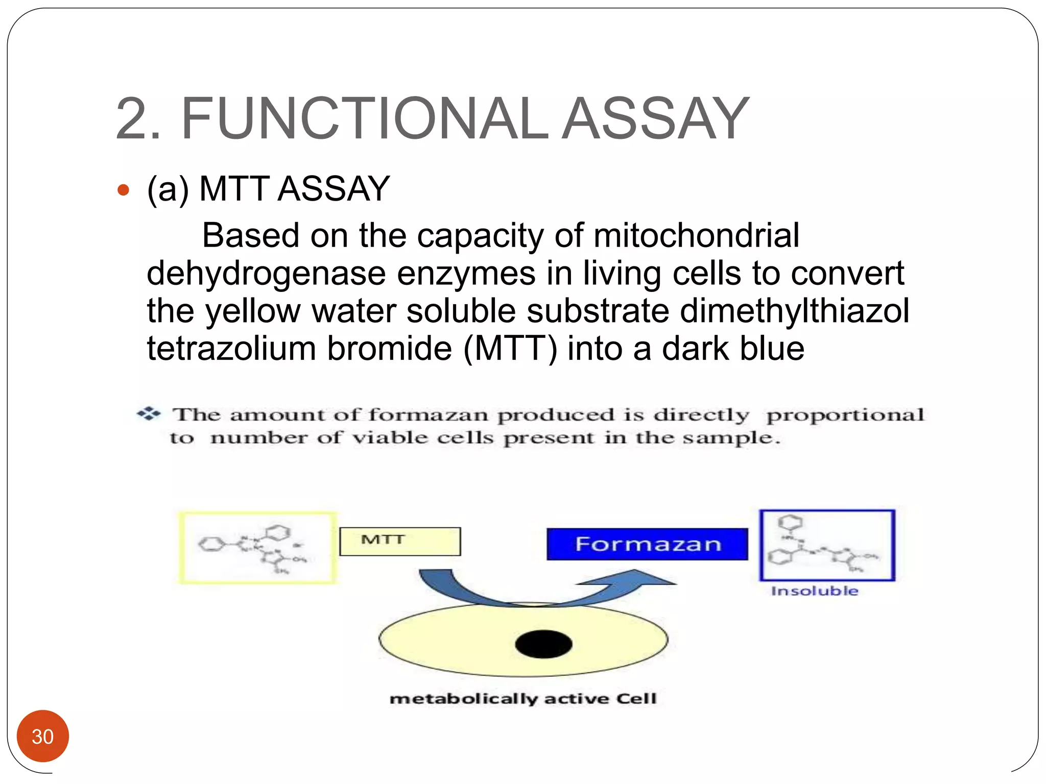
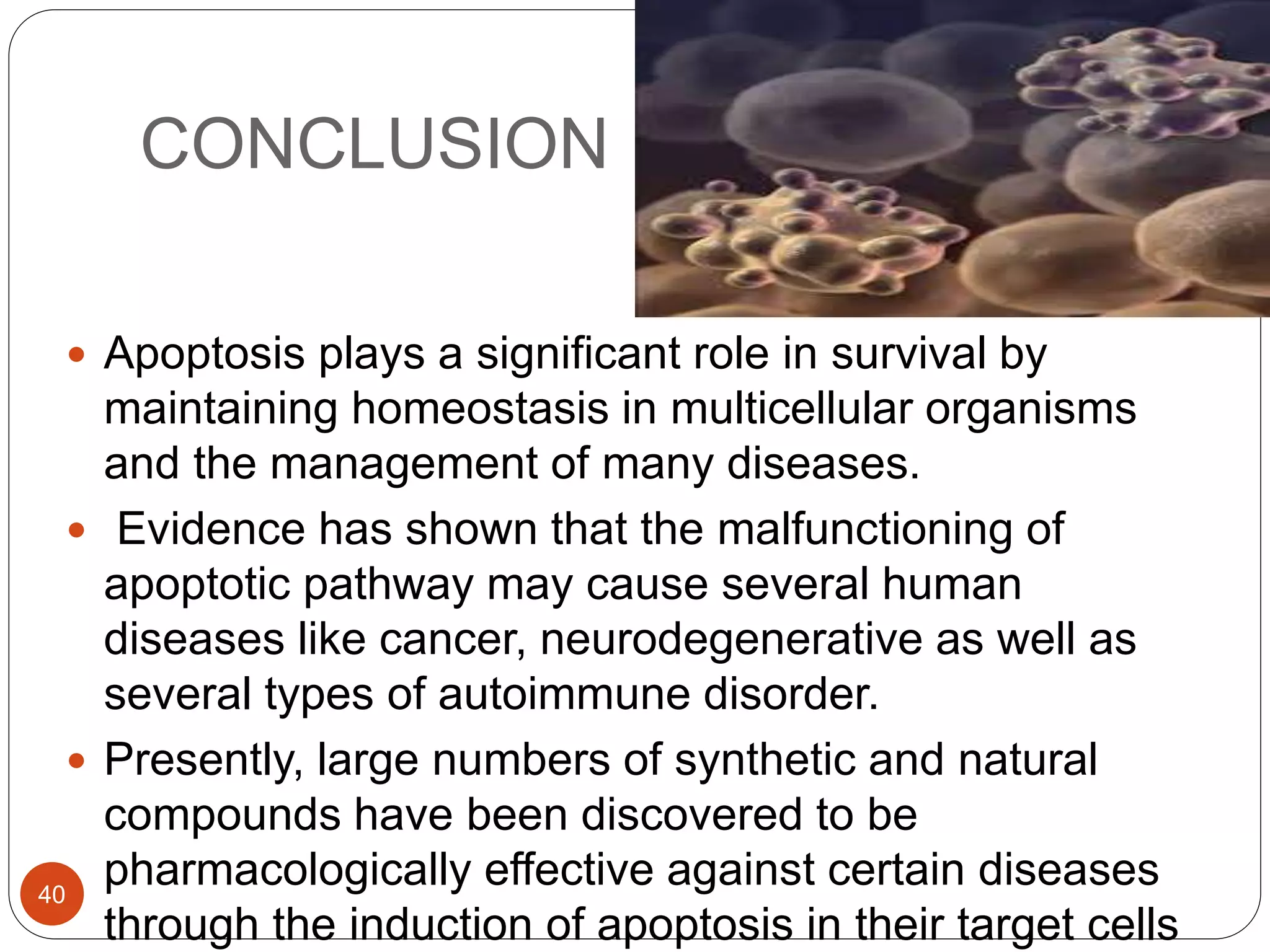
1
PRESENTED BY- AMEENA MEHABOOB
Ist YEAR MPHARM2
INTRODUCTION
3
4
5
6
1. APOPTOSIS DURING
EMBRYOGENESIS
FORMATION OF FREE AND
INDEPENDENT DIGITS
•
•
DEVELOPMENT OF THE BRAIN
DEVELOPMENT OF
REPRODUCTIVE ORGANS
7
8
9
3. CELL LOSS IN PROLIFERATING
CELL POPULATIONS
Immature lymphocytes in bone marrow
and thymus that fail to express useful
antigen receptors
B lymphocytes in germinal centers
Epithelial cells in intestinal crypts
10
4. ELIMINATION OF POTENTIALLY HARMFUL SELF-
REACTIVE LYMPHOCYTES
5. DEATH OF HOST CELLS THAT HAVE
SERVED THEIR USEFUL PURPOSE
11
12
DNA
DAMAGE
ACCUMULATIO
N OF MIS-
FOLDED
PROTEINS
CELL INJURY
IN CERTAIN
INFECTIONS
PATHOLOGICA
L ATROPHY IN
PARENCHYMA
L ORGANS
AFTER DUCT
OBSTRUCTION
13
14
15
16
 Fundamental event: activation of
CASPASES
 They are central initiators and executioners
of apoptosis
 Synthesized as inactive zymogens
(procaspases)
• Cysteine
proteases
• Cysteine-
dependent
ASPartate-specific
proteASES
CASPASES
17
18
TYPES OF CASPASES
Inflammatory
caspases:
1,4,5,13
Initiator
caspases:
2,8,9,10
Interact with
effector
caspases
Effector
caspases:
3,6,7,14
Initiate cell
death
19
20
21
ASSAYS TO DETECT
APOPTOSIS
22
MEMBRANE
INTEGRITY
ASSAY
EXCLUSION
DYES
FLOURESCENT
DYES
ANNEXIN V
LDH ASSAY
FUNCTIONAL
ASSAY
MTT, XTT
ASSAY
CRYSTAL
VIOLET/ACID
PHOSPHATASE
ASSAY
NEUTRAL RED
ASSAY
DNA
LABELLING
ASSAY
FLOURESCENT
CONJUGATES
TUNNEL ASSAY
COMET ASSAY
23
MORPHOLOGICAL
AND MECHANISM
BASED
MICROSCOPIC
OBSERVATION
CASPASE
3DETECTION
REPRODUCTIVE
ASSAY
COLONY
FORMATION
ASSAY
IMMUNOHISTOCHE
MISTRY
ANNEXIN V
P53
24
1. MEMBRANE INTEGRITY
ASSAY
 (a) EXCLUSION
DYES
25
 (b) FLOURESCENT
DYES
26
(c) ANNEXIN V ASSAY
•In apoptosis, earliest indication is translocation of phosphatidyl
serine from inner to outer leaflet of plasma membrane
•Once exposed, binding sites become available for Annexin V
(Ca2+ dependent, phospholipid binding protein)
•Annexin V can be conjugated to biotin or to a flourochrome for
detection of apoptotic cells.
• PS translocation also occurs during necrosis, Annexin V is not
an absolute marker for apoptosis
•So it is used in conjunction with vital dyes such as 7-amino
actinomysis or propidium iodide which bind to nucleic acids, but
can only penetrate the plasma membrane integrity is breached,
as occurs in later stages of apoptosis
27
28
(d) LDH LEAKAGE ASSAY
 PRINCIPLE:
Tumour cells possess high concentration of
intracellular LDH and cleavage of a tetrazolium
salt occurs when LDH is present in culture
supernatent.
29
2. FUNCTIONAL ASSAY
 (a) MTT ASSAY
Based on the capacity of mitochondrial
dehydrogenase enzymes in living cells to convert
the yellow water soluble substrate dimethylthiazol
tetrazolium bromide (MTT) into a dark blue
formazan product which is insoluble in water
30
31
32
COMET ASSAY
 Simple procedure using a microgel and
electrophoresis to detect DNA damage
 Damage in DNA looks like a comet, so its
called comet assay
 Technique is further developed and introduced
the use of high alkaline conditions. Thus not
only double strand breaks but also single
strand breaks can be detected
33
34
ENDONUCLEASE ASSAY
 During apoptosis endonuclease enzymes are
activated to cleave genomic DNA into many
smaller fragments.
35
36
PHYSIOLOGICAL IMPLICATIONS
OF APOPTOSIS
CNS
• During embryonic development of the nervous system, surplus of cells
are produced. Oxidative stress and calcium influx can induce
apoptosis in the mature central nervous system
VIRAL INFECTIONS
• Many viruses inhibit apoptosis in their target cells thereby prolonging
host cell life and in order to permit viral replication
IMMUNE SYSTEM
• Usually, any dysfunction of the apoptotic pathway causes autoimmune
disease, immunodeficiencies and lymphoid malignancies.
RENAL SYSTEM
• Embryological development of the kidney involves periods of growth
and apoptosis which are reflected by the levels of Bcl-2 present
37
GIT
• Gastrointestinal diseases may be associated with
excessive or defective apoptosis. Shigella dysenteriae
causes excessive apoptosis of macrophages in the lamina
propria of the intestine. Progressive inhibition of apoptosis
appears to be involved in the pathogenesis of
gastrointestinal neoplasia, in particular colorectal cancer
CANCER
• Evidence shows that failure to initiate apoptosis following
DNA damage may cause cancer
REPRODUCTIVE SYSTEM
• Apoptosis is continually inhibited in many tissues of the
reproductive system owing to the presence of trophic
hormones from the pituitary, gonads(testes and ovary)
and uterus38
THERAPEUTIC POTENTALS OF
APOPTOSIS
• Corticosteroids induce eosinophil apoptosis but
inhibit neutrophil apoptosis
• The treatment of asthmatic patients using
corticosteroids causes eosinophil death and
macrophage engulfment
Inflammatory
Disease
• Cytotoxic drugs induce apoptosis in human
gastrointestinal cancer cells
• Chronic ingestion of nonsteroidal anti-inflammatory
drugs may be useful in preventing colonic cancer,
possibly by induction of apoptosis
Gastrointestinal
Tract
• Anti cancer drugs induce apoptosis of
cancer cells
Cancer
39
CONCLUSION
 Apoptosis plays a significant role in survival by
maintaining homeostasis in multicellular organisms
and the management of many diseases.
 Evidence has shown that the malfunctioning of
apoptotic pathway may cause several human
diseases like cancer, neurodegenerative as well as
several types of autoimmune disorder.
 Presently, large numbers of synthetic and natural
compounds have been discovered to be
pharmacologically effective against certain diseases
through the induction of apoptosis in their target cells
40
REFERENCES
 Apoptosis: Its Physiological Implication And
Therapeutic Possibilities, Chidinma Adanna Levi,
Vincent C. Ejere et alIOSR Journal of Pharmacy
and Biological Sciences Volume 9, Issue 1 Ver. V
(Feb. 2014)
 The cell: A molecular approach, Geoffrey M
Cooper, Robert E Hausmann, 4th edition, pg
1010-1025
 Robbins Basic pathology, 9th edition, pg 10-35
41
42

Apoptosis

  • 1.
  • 2.
    PRESENTED BY- AMEENAMEHABOOB Ist YEAR MPHARM2
  • 3.
  • 4.
  • 5.
  • 6.
  • 7.
    1. APOPTOSIS DURING EMBRYOGENESIS FORMATIONOF FREE AND INDEPENDENT DIGITS • • DEVELOPMENT OF THE BRAIN DEVELOPMENT OF REPRODUCTIVE ORGANS 7
  • 8.
  • 9.
  • 10.
    3. CELL LOSSIN PROLIFERATING CELL POPULATIONS Immature lymphocytes in bone marrow and thymus that fail to express useful antigen receptors B lymphocytes in germinal centers Epithelial cells in intestinal crypts 10
  • 11.
    4. ELIMINATION OFPOTENTIALLY HARMFUL SELF- REACTIVE LYMPHOCYTES 5. DEATH OF HOST CELLS THAT HAVE SERVED THEIR USEFUL PURPOSE 11
  • 12.
  • 13.
    DNA DAMAGE ACCUMULATIO N OF MIS- FOLDED PROTEINS CELLINJURY IN CERTAIN INFECTIONS PATHOLOGICA L ATROPHY IN PARENCHYMA L ORGANS AFTER DUCT OBSTRUCTION 13
  • 14.
  • 15.
  • 16.
  • 17.
     Fundamental event:activation of CASPASES  They are central initiators and executioners of apoptosis  Synthesized as inactive zymogens (procaspases) • Cysteine proteases • Cysteine- dependent ASPartate-specific proteASES CASPASES 17
  • 18.
  • 19.
    TYPES OF CASPASES Inflammatory caspases: 1,4,5,13 Initiator caspases: 2,8,9,10 Interactwith effector caspases Effector caspases: 3,6,7,14 Initiate cell death 19
  • 20.
  • 21.
  • 22.
  • 23.
    MEMBRANE INTEGRITY ASSAY EXCLUSION DYES FLOURESCENT DYES ANNEXIN V LDH ASSAY FUNCTIONAL ASSAY MTT,XTT ASSAY CRYSTAL VIOLET/ACID PHOSPHATASE ASSAY NEUTRAL RED ASSAY DNA LABELLING ASSAY FLOURESCENT CONJUGATES TUNNEL ASSAY COMET ASSAY 23
  • 24.
  • 25.
    1. MEMBRANE INTEGRITY ASSAY (a) EXCLUSION DYES 25
  • 26.
  • 27.
    (c) ANNEXIN VASSAY •In apoptosis, earliest indication is translocation of phosphatidyl serine from inner to outer leaflet of plasma membrane •Once exposed, binding sites become available for Annexin V (Ca2+ dependent, phospholipid binding protein) •Annexin V can be conjugated to biotin or to a flourochrome for detection of apoptotic cells. • PS translocation also occurs during necrosis, Annexin V is not an absolute marker for apoptosis •So it is used in conjunction with vital dyes such as 7-amino actinomysis or propidium iodide which bind to nucleic acids, but can only penetrate the plasma membrane integrity is breached, as occurs in later stages of apoptosis 27
  • 28.
  • 29.
    (d) LDH LEAKAGEASSAY  PRINCIPLE: Tumour cells possess high concentration of intracellular LDH and cleavage of a tetrazolium salt occurs when LDH is present in culture supernatent. 29
  • 30.
    2. FUNCTIONAL ASSAY (a) MTT ASSAY Based on the capacity of mitochondrial dehydrogenase enzymes in living cells to convert the yellow water soluble substrate dimethylthiazol tetrazolium bromide (MTT) into a dark blue formazan product which is insoluble in water 30
  • 31.
  • 32.
  • 33.
    COMET ASSAY  Simpleprocedure using a microgel and electrophoresis to detect DNA damage  Damage in DNA looks like a comet, so its called comet assay  Technique is further developed and introduced the use of high alkaline conditions. Thus not only double strand breaks but also single strand breaks can be detected 33
  • 34.
  • 35.
    ENDONUCLEASE ASSAY  Duringapoptosis endonuclease enzymes are activated to cleave genomic DNA into many smaller fragments. 35
  • 36.
  • 37.
    PHYSIOLOGICAL IMPLICATIONS OF APOPTOSIS CNS •During embryonic development of the nervous system, surplus of cells are produced. Oxidative stress and calcium influx can induce apoptosis in the mature central nervous system VIRAL INFECTIONS • Many viruses inhibit apoptosis in their target cells thereby prolonging host cell life and in order to permit viral replication IMMUNE SYSTEM • Usually, any dysfunction of the apoptotic pathway causes autoimmune disease, immunodeficiencies and lymphoid malignancies. RENAL SYSTEM • Embryological development of the kidney involves periods of growth and apoptosis which are reflected by the levels of Bcl-2 present 37
  • 38.
    GIT • Gastrointestinal diseasesmay be associated with excessive or defective apoptosis. Shigella dysenteriae causes excessive apoptosis of macrophages in the lamina propria of the intestine. Progressive inhibition of apoptosis appears to be involved in the pathogenesis of gastrointestinal neoplasia, in particular colorectal cancer CANCER • Evidence shows that failure to initiate apoptosis following DNA damage may cause cancer REPRODUCTIVE SYSTEM • Apoptosis is continually inhibited in many tissues of the reproductive system owing to the presence of trophic hormones from the pituitary, gonads(testes and ovary) and uterus38
  • 39.
    THERAPEUTIC POTENTALS OF APOPTOSIS •Corticosteroids induce eosinophil apoptosis but inhibit neutrophil apoptosis • The treatment of asthmatic patients using corticosteroids causes eosinophil death and macrophage engulfment Inflammatory Disease • Cytotoxic drugs induce apoptosis in human gastrointestinal cancer cells • Chronic ingestion of nonsteroidal anti-inflammatory drugs may be useful in preventing colonic cancer, possibly by induction of apoptosis Gastrointestinal Tract • Anti cancer drugs induce apoptosis of cancer cells Cancer 39
  • 40.
    CONCLUSION  Apoptosis playsa significant role in survival by maintaining homeostasis in multicellular organisms and the management of many diseases.  Evidence has shown that the malfunctioning of apoptotic pathway may cause several human diseases like cancer, neurodegenerative as well as several types of autoimmune disorder.  Presently, large numbers of synthetic and natural compounds have been discovered to be pharmacologically effective against certain diseases through the induction of apoptosis in their target cells 40
  • 41.
    REFERENCES  Apoptosis: ItsPhysiological Implication And Therapeutic Possibilities, Chidinma Adanna Levi, Vincent C. Ejere et alIOSR Journal of Pharmacy and Biological Sciences Volume 9, Issue 1 Ver. V (Feb. 2014)  The cell: A molecular approach, Geoffrey M Cooper, Robert E Hausmann, 4th edition, pg 1010-1025  Robbins Basic pathology, 9th edition, pg 10-35 41
  • 42.