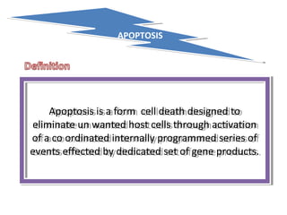
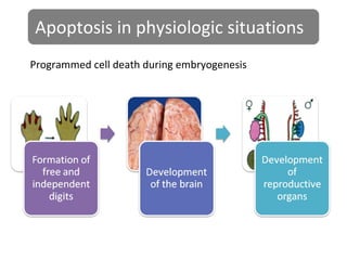
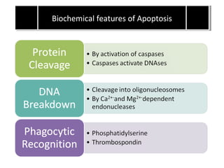
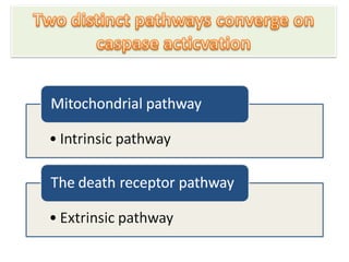
Cell death, particularly apoptosis, is probably one of the
most widely-studied subjects among cell biologists.
Understanding apoptosis in disease conditions is very
important as it not only gives insights into the pathogenesis
of a disease but may also leaves clues on how
the disease can be treated. In cancer, there is a loss of
balance between cell division and cell death and cells
that should have died did not receive the signals to do
so. The problem can arise in any one step along the way
of apoptosis.Apoptosis is an ordered and orchestrated cellular process that occurs in physiological and pathological conditions.
It is also one of the most studied topics among cell biologists. An understanding of the underlying mechanism of
apoptosis is important as it plays a pivotal role in the pathogenesis of many diseases. In some, the problem is due
to too much apoptosis, such as in the case of degenerative diseases while in others, too little apoptosis is the
culprit. Cancer is one of the scenarios where too little apoptosis occurs, resulting in malignant cells that will not
die. The mechanism of apoptosis is complex and involves many pathways. Defects can occur at any point along
these pathways, leading to malignant transformation of the affected cells, tumour metastasis and resistance to
anticancer drugs. Despite being the cause of problem, apoptosis plays an important role in the treatment of
cancer as it is a popular target of many treatment strategies. The abundance of literature suggests that targeting
apoptosis in cancer is feasible. However, many troubling questions arise with the use of new drugs or treatment
strategies that are designed to enhance apoptosis and critical tests must be passed before they can be used safely
in human subjects.. It is used,
in contrast to necrosis, to describe the situation in
which a cell actively pursues a course toward death
upon receiving certain stimule
Apoptosis is a
-pathway of cell death that is
-induced by an internally regulated program
-in which cells destined to die activate intrinsic enzymes that --degrade the cells’ own nuclear DNA and also nuclear and cytoplasmic proteins
-With minimal host reaction.
Seminar about apoptosis a type of programmed cell death in Soran University by Mahmood Khaleel Pirani (MSc Student) for Medical Physiology lecture on 24 November 2018
Apoptosis is the programmed cell death. Aim of cancer therapy is to destroy the invading cells. Cancerous cells can be destroyed by increasing apoptosis.
it can occur in both physiological and pathological conditions. It is different from necrosis. In necrosis, the cell contents leak out and lead to inflammation. But in apoptosis there is no cellular leakage, only apoptotic bodies are formed. They are then engulfed by macrophages.
this is a series of notes on general pathology, useful for undergraduate and post graduate pathology students. Notes have been prepared from standard textbooks and are in a format easy to reproduce in exams.
Hi! I am Komal Sankaran, M.Sc. Biotechnology (Pune University Gold Medalist, 2013), CSIR-NET SPM fellow (Jun- 2014, 4th rank), CSIR-NET- LS (Dec 2013, 2nd rank), DBT JRF category- I. Please contact if anyone is interested in Life Sciences CSIR-NET coaching in Pune (Khadki area).
Email- komalsan91@gmail.com
Knee anatomy and clinical tests 2024.pdfvimalpl1234
This includes all relevant anatomy and clinical tests compiled from standard textbooks, Campbell,netter etc..It is comprehensive and best suited for orthopaedicians and orthopaedic residents.
micro teaching on communication m.sc nursing.pdfAnurag Sharma
Microteaching is a unique model of practice teaching. It is a viable instrument for the. desired change in the teaching behavior or the behavior potential which, in specified types of real. classroom situations, tends to facilitate the achievement of specified types of objectives.
Basavarajeeyam is a Sreshta Sangraha grantha (Compiled book ), written by Neelkanta kotturu Basavaraja Virachita. It contains 25 Prakaranas, First 24 Chapters related to Rogas& 25th to Rasadravyas.
Title: Sense of Smell
Presenter: Dr. Faiza, Assistant Professor of Physiology
Qualifications:
MBBS (Best Graduate, AIMC Lahore)
FCPS Physiology
ICMT, CHPE, DHPE (STMU)
MPH (GC University, Faisalabad)
MBA (Virtual University of Pakistan)
Learning Objectives:
Describe the primary categories of smells and the concept of odor blindness.
Explain the structure and location of the olfactory membrane and mucosa, including the types and roles of cells involved in olfaction.
Describe the pathway and mechanisms of olfactory signal transmission from the olfactory receptors to the brain.
Illustrate the biochemical cascade triggered by odorant binding to olfactory receptors, including the role of G-proteins and second messengers in generating an action potential.
Identify different types of olfactory disorders such as anosmia, hyposmia, hyperosmia, and dysosmia, including their potential causes.
Key Topics:
Olfactory Genes:
3% of the human genome accounts for olfactory genes.
400 genes for odorant receptors.
Olfactory Membrane:
Located in the superior part of the nasal cavity.
Medially: Folds downward along the superior septum.
Laterally: Folds over the superior turbinate and upper surface of the middle turbinate.
Total surface area: 5-10 square centimeters.
Olfactory Mucosa:
Olfactory Cells: Bipolar nerve cells derived from the CNS (100 million), with 4-25 olfactory cilia per cell.
Sustentacular Cells: Produce mucus and maintain ionic and molecular environment.
Basal Cells: Replace worn-out olfactory cells with an average lifespan of 1-2 months.
Bowman’s Gland: Secretes mucus.
Stimulation of Olfactory Cells:
Odorant dissolves in mucus and attaches to receptors on olfactory cilia.
Involves a cascade effect through G-proteins and second messengers, leading to depolarization and action potential generation in the olfactory nerve.
Quality of a Good Odorant:
Small (3-20 Carbon atoms), volatile, water-soluble, and lipid-soluble.
Facilitated by odorant-binding proteins in mucus.
Membrane Potential and Action Potential:
Resting membrane potential: -55mV.
Action potential frequency in the olfactory nerve increases with odorant strength.
Adaptation Towards the Sense of Smell:
Rapid adaptation within the first second, with further slow adaptation.
Psychological adaptation greater than receptor adaptation, involving feedback inhibition from the central nervous system.
Primary Sensations of Smell:
Camphoraceous, Musky, Floral, Pepperminty, Ethereal, Pungent, Putrid.
Odor Detection Threshold:
Examples: Hydrogen sulfide (0.0005 ppm), Methyl-mercaptan (0.002 ppm).
Some toxic substances are odorless at lethal concentrations.
Characteristics of Smell:
Odor blindness for single substances due to lack of appropriate receptor protein.
Behavioral and emotional influences of smell.
Transmission of Olfactory Signals:
From olfactory cells to glomeruli in the olfactory bulb, involving lateral inhibition.
Primitive, less old, and new olfactory systems with different path
Integrating Ayurveda into Parkinson’s Management: A Holistic ApproachAyurveda ForAll
Explore the benefits of combining Ayurveda with conventional Parkinson's treatments. Learn how a holistic approach can manage symptoms, enhance well-being, and balance body energies. Discover the steps to safely integrate Ayurvedic practices into your Parkinson’s care plan, including expert guidance on diet, herbal remedies, and lifestyle modifications.
3. APOPTOSISAPOPTOSIS
Apoptosis is a form cell death designed to
eliminate un wanted host cells through activation
of a co ordinated internally programmed series of
events effected by dedicated set of gene products.
Apoptosis is a form cell death designed to
eliminate un wanted host cells through activation
of a co ordinated internally programmed series of
events effected by dedicated set of gene products.
9. Apoptosis in pathologic situations
Apoptosis eliminates cells that are genetically altered or
injured beyond repair without eliciting a severe host reaction,
thus keeping the damage as contained as possible.
13. • Activation of several members of cystine
family called caspases.can be divided into two
groups initiator and executioner.
• Initiator include caspases 8 and 9.
• Executioners including caspases 3 and 6.
• DNA breakdown by calcium and magnesium
dependent endonucleases into fragments
whose sizes are multiples of 180 units to 200
base pairs.fragments may be visualized by
electrophoresis. As DNA ladder.
• Dead cells are recognized by phogocytes by
the help of phospholipid such as
phosphatidylserine from inner leaflet to outer
leaflet of membrane.
14.
15. • Intrinsic (mitochondrial)pathway of apoptosis
• This pathway is result of increased
mitochondrial permeability and release of pro
apoptotic molecules into cytoplasm.
• Release of mitochondrial proteins is controlled
by pro and anti apoptotic members of Bcl
family of proteins.
• Bcl-2,Bcl-x and Mcl-1 are anti apoptotic
proteins they inhibit apoptosis.
16. • When cells are deprived of survival singals or
their DNA is damaged or misfolded proteins
induces ER stress,sensors of damage or stress
are activated.
• These sensors are also members of Bcl
family.they include proteins called Bim,Bid,and
Bad.
• The sensors activate two critical effectors
Bax,Bak
17. EXTRINSIC PATHWAY
• Death receptors are members of TNF receptor
family .
• Best known death receptors are type 1 TNF
receptor and related protein called Fas
(CD95).
• The machenism of apoptosis induced by these
death receptor expressed on many cell types.
• Fas is expressed on T cells that recognize self
antigens and on some cytotoxic T
lymphocytes.
18. • When FasL binds to Fas,three or more
molecules of Fas are brought togather and
there cytoplasmic death domains form a
binding site for a adapter protein that also
contains a death domain and is called FADD.
• FADD than activated the inactive form of pro
caspase-8 to active form of caspase-8.
• This pathway of apoptosis can be inhibited by
a protein called FLIP.
19. • After intiating pathways the executioner
phase is activated,executioner caspases
iclude caspase 3 and 6.
• These caspases once activated,cleave an
inhibitor of cytoplasmic DNase,thus DNase
enzyme activated,this Dnase enzyme break
down of DNA into nucleosome.
• Caspaes degraded structural component of
nuclear matrix & thus promote fragmentation
of nuclei.
20. REMOVAL OF DEAD CELLS
• In healthy cells phosphatidylserine is present
on inner leaflet of plasma membrane but in
apoptotic cells this phospholipid flips out and
is expressed on outer layer where it is
recognized by several macrophage
receptors.which help in removal of death
cells.
During development many cells are produced in excess which eventually undergo programmed cell death
A particularly instructive example for the implication of programmed cell death in animal development is the formation of free and independent digits by massive cell death in the interdigital mesenchymal tissue [Zuzarte-Luis, 2002]. Other examples are the development of the brain, during which half of the neurons that are initially created will die in later stages when the adult brain is formed [Hutchins, 1998] and the development of the reproductive organs [Meier, 2000]. Also cells of an adult organism constantly undergo physiological cell death which must be balanced with proliferation in order to maintain homeostasis in terms of constant cell numbers. The majority of the developing lymphocytes die either during genetic rearrangement events in the formation of the antigen receptor, during negative selection or in the periphery, thereby tightly controlling the pool of highly efficient and functional but not self-reactive immune cells and at the same time keeping lymphocyte numbers relatively constant [Rathmell, 2002].
Taken together, apoptotic processes are of widespread biological significance, being involved in e.g. development, differentiation, proliferation/homoeostasis, regulation and function of the immune system and in the removal of defect and therefore harmful cells. Thus, dysfunction or dysregulation of the apoptotic program is implicated in a variety of pathological conditions. Defects in apoptosis can result in cancer, autoimmune diseases and spreading of viral infections, while neurodegenerative disorders, AIDS and ischaemic diseases are caused or enhanced by excessive apoptosis [Fadeel, 1999a].
Role of mitochondria in apoptosis
Mitochondria play an important role in the regulation of cell death. They contain many pro-apoptotic proteins such as Apoptosis Inducing Factor (AIF), Smac/DIABLO and cytochrome C. These factors are released from the mitochondria following the formation of a pore in the mitochondrial membrane called the Permeability Transition pore, or PT pore. These pores are thought to form through the action of the pro-apoptotic members of the bcl-2 family of proteins, which in turn are activated by apoptotic signals such as cell stress, free radical damage or growth factor deprivation. Mitochondria also play an important role in amplifying the apoptotic signalling from the death receptors, with receptor recruited caspase 8 activating the pro-apoptotic bcl-2 protein, Bid.
Role of Bcl-2 proteins
The bcl-2 proteins are a family of proteins involved in the response to apoptosis. Some of these proteins (such as bcl-2 and bcl-XL) are anti-apoptotic, while others (such as Bad, Bax or Bid) are pro-apoptotic. The sensitivity of cells to apoptotic stimuli can depend on the balance of pro- and anti-apoptotic bcl-2 proteins. When there is an excess of pro-apoptotic proteins the cells are more sensitive to apoptosis, when there is an excess of anti-apoptotic proteins the cells will tend to be more resistant. An excess of pro-apoptotic bcl-2 proteins at the surface of the mitochondria is thought to be important in the formation of the PT pore.
An animation illustrating the general principles is shown below.
The pro-apoptotic bcl-2 proteins are often found in the cytosol where they act as sensors of cellular damage or stress. Following cellular stress they relocate to the surface of the mitochondria where the anti-apoptotic proteins are located. This interaction between pro- and anti-apoptotic proteins disrupts the normal function of the anti-apoptotic bcl-2 proteins and can lead to the formation of pores in the mitochondria and the release of cytochrome C and other pro-apoptotic molecules from the intermembrane space. This in turn leads to the formation of the apoptosome and the activation of the caspase cascade.
The release of cytochrome C from the mitochondria is a particularly important event in the induction of apoptosis. Once cytochrome C has been released into the cytosol it is able to interact with a protein called Apaf-1. This leads to the recruitment of pro-caspase 9 into a multi-protein complex with cytochrome C and Apaf-1 called the apoptosome. Formation of the apoptosome leads to activation of caspase 9 and the induction of apoptosis.
The role of mitochondria in the induction of apoptosis is summarised in the figure below.
Apoptosis can be triggered by various stimuli from outside or inside the cell, e.g. by ligation of cell surface receptors, by DNA damage as a cause of defects in DNA repair mechanisms, treatment with cytotoxic drugs or irradiation, by a lack of survival signals, contradictory cell cycle signalling or by developmental death signals. Death signals of such diverse origin nevertheless appear to eventually activate a common cell death machinery leading to the characteristic features of apoptotic cell death.