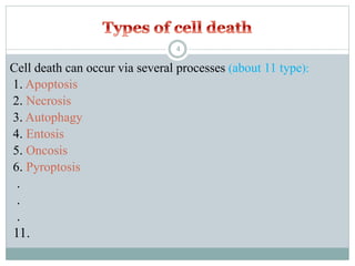
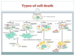
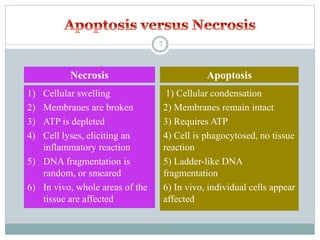
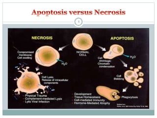
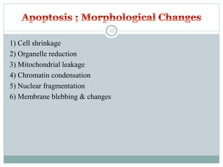
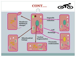
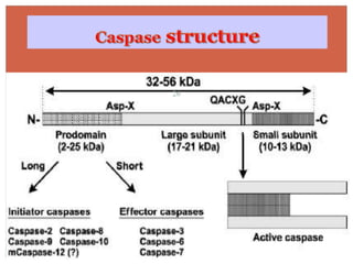
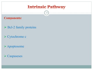
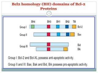
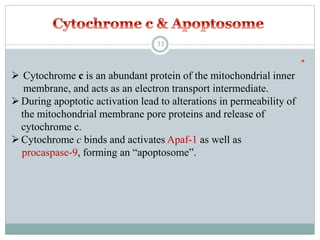
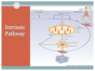
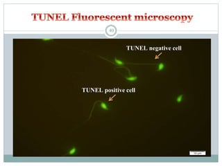
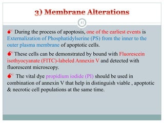
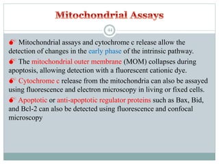
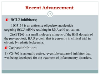
Apoptosis
SUBMITED BY
AMIT KUMAR
MPHARM (I Year)
1
 In humans, the rate of cell growth and cell death is balanced to
maintain the weight of the body.
2
Life cannot exist without cellular death
3
Cell death can occur via several processes (about 11 type):
1. Apoptosis
2. Necrosis
3. Autophagy
4. Entosis
5. Oncosis
6. Pyroptosis
.
.
.
11.
4
5
Apoptosis = “normal” or “programmed” cell death
Apoptosis is the physiological cell death which
unwanted or useless cells are eliminated during
development and other normal biological processes.
Necrosis = “accidental” or “ordinary” cell death
Necrosis is the pathological cell death which occurs
when cells are exposed to a serious physical or chemical
insult (hypoxia, hyperthermia, ischemia).
6
1) Cellular condensation
2) Membranes remain intact
3) Requires ATP
4) Cell is phagocytosed, no tissue
reaction
5) Ladder-like DNA
fragmentation
6) In vivo, individual cells appear
affected
1) Cellular swelling
2) Membranes are broken
3) ATP is depleted
4) Cell lyses, eliciting an
inflammatory reaction
5) DNA fragmentation is
random, or smeared
6) In vivo, whole areas of the
tissue are affected
Necrosis Apoptosis
7
8
The word ‘‘apoptosis’’ comes from the
ancient Greek, meaning the:
‘‘falling of petals from a flower’’ or
‘‘of leaves from a tree in autumn’’
In 1964 Lockshin, study on programmed cell death.
The term apoptosis (a-po-toe-sis) was first used in a
now-classic paper by Kerr et al 1972 to describe a
morphologically distinct form of cell death.
9
 Apoptosis or programmed cell death (PCD) is a mode of cell
death that occurs under normal physiological conditions and
the cell is an active participant in its own demise (“cellular
suicide”).
 It is important for the development of multicellular organism
(embryonic development) and homeostasis of their tissues
(adult).
10
11
1) Cell shrinkage
2) Organelle reduction
3) Mitochondrial leakage
4) Chromatin condensation
5) Nuclear fragmentation
6) Membrane blebbing & changes
12
Mitochondrial
leakage
Organelle
reduction
Cell
shrinkage
Nuclear
fragmentation
Chromatin
condensation
Membrane
blebbing &
changes
13
Characteristic biochemical changes in cells undergoing
apoptosis
1)Chromosomal DNA cleaved into fragments
2)Change in the plasma membrane – phosphatidylserine
in the outer leaflet
3)Loss of electrical potential across the inner membrane of the
mitochondria
4)Relocation of cytochrome c from the intermembrane space of
the mitochondria to the cytosol
14
Apoptosis is a beneficial and important phenomenon:
 In embryo
1. During embryonic development, help to digit formation.
 Lack of apoptosis in humans
can lead to webbed fingers
called “syndactyly ”.
15
2. Normal event in development of the nervous system
16
 In adult
 Normal cell turn over
 Tissue homeostasis
 Induction and maintenance of immune tolerance
 Development of the nervous system
 Endocrine-dependent tissue atrophy
 Elimination of activated, damaged and abnormal cells
17
 In animals
Embryonic Chicken Foot
Embryonic Mouse Paw
Tail absorption of the tadpole
18
APOPTOSIS
19
Extrinsic pathway (death receptor- mediated events)
Intrinsic pathway (mitochondria- mediated events)
20
 Death Receptors
 Death Ligands
 Adaptor Proteins
 Caspaseses
21
 “Death receptors” that are members of the tumor necrosis factor
(TNF) receptor superfamily.
 Death receptors have a cytoplasmic domain of about 80
amino acids called the “death domain”.
 This death domain plays a critical role in transmitting the death
signal from the cell surface to the intracellular signaling
pathways.
22
 The best characterized receptors & ligands corresponding
death receptors include:
FasR (CD95/APO1) FasL
DR3 Apo3L
DR4 (TRAIL-R1) Apo2L
DR5 (TRAIL-R2) Apo2L
TNFR1 TNF-α
TNFR2 TNF-ß
LigandsReceptors
23
 Apoptotic adaptor proteins play a critical role in
regulating pro- and anti-apoptotic signalling
pathways
Adaptor proteins;
1)FADD (Fas-associated death domain)
2) TRADD (TNF receptor-associated death domain), are
recruited to ligand-activated, oligomerized death receptors
to mediate apoptotic signalling pathways.
24
Caspases= Cysteinyl aspartate specific proteases
 A family of intracellular cysteine proteases that play a pivotal
role in the initiation and execution of apoptosis.
 At least 14 different members of caspases in mammalian cells
have been identified
 All are synthesized as inactive proenzymes (zymogen) with
32-56 kDa
25
26
 To date, ten major caspases have been identified and broadly
categorized into:
 Signaling or Initiator caspases
 Effector or Executioner caspases
 Inflammatory caspases
 The other caspases that have been identified include:
Caspases
 Central role in cascade of apoptotic events is played by
caspase 3 (CPP32)
27
28
 Bcl-2 family proteins
 Cytochrome c
 Apoptosome
 Caspaseses
29
 The control & regulation of apoptotic mitochondrial events occurs
through members of the Bcl-2 family of proteins
 Anti-apoptotic proteins include Bcl-2, Bcl-x, Bcl-XL, Bcl-w
 Pro-apoptotic proteins include Bax, Bak, Bid, Bad, Bim, Bik
 The main mechanism of action of the Bcl-2 family of proteins
is the regulation of cytochrome c release from the
mitochondria via alteration of mitochondrial membrane
permeability.
30
31
32

 Cytochrome c is an abundant protein of the mitochondrial inner
membrane, and acts as an electron transport intermediate.
During apoptotic activation lead to alterations in permeability of
the mitochondrial membrane pore proteins and release of
cytochrome c.
Cytochrome c binds and activates Apaf-1 as well as
procaspase-9, forming an “apoptosome”.
33
 The stimuli that initiate the intrinsic pathway produce intracellular
signals such as radiation (DNA damage), absence of certain growth
factors, hormones and cytokines.
 All of these stimuli cause changes in the mitochondrial outer
membrane permeabilization (MOMP)
 Release of pro-apoptotic proteins such as cytochrome c,
Smac/DIABLO, AIF, endonuclease G and CAD from the inter-
membrane space into the cytosol.
 Cytochrome c binds and activates Apaf-1 as well as
procaspase-9, forming an “apoptosome”.
 Caspase-9 activation, subsequent caspase-3 activation and
cell death.
34
35
Assay for apoptosis
36
 Apoptosis assays, based on
methodology, can be classified into six
major groups:
1. Cytomorphological alterations
2. DNA fragmentation
3. Detection of caspases, cleaved
substrates, regulators and inhibitors
4. Membrane alterations
5. Detection of apoptosis in whole mounts
6. Mitochondrial assays
37
 The evaluation of hematoxylin and eosin-stained tissue sections
with light microscopy does allow the visualization of apoptotic
cells.
 This method detects the later events of apoptosis
 TEM is considered the gold standard to confirm apoptosis:
1) Electron-dense nucleus
2) Nuclear fragmentation
3) Disorganized cytoplasmic organelles
4) large clear Vacuoles
5) Intact cell membrane
6) Blebs at the cell surface
38
 DNA fragmentation occurs in the later phase of apoptosis.
 TUNEL (Terminal dUTP Nick End-Labeling) assay quantifies
the incorporation of deoxyuridine triphosphate (dUTP) at single and
double stranded DNA breaks in a reaction catalyzed by the template
independent enzyme, terminal deoxynucleotidyl transferase (TdT).
 Incorporated dUTP is labeled such that breaks can be quantified
either by flowcytometry, fluorescent microscopy, or light
microscopy.
39
TUNEL positive cell
TUNEL negative cell
40
 During the process of apoptosis, one of the earliest events is
Externalization of Phosphatidylserine (PS) from the inner to the
outer plasma membrane of apoptotic cells.
These cells can be demonstrated by bound with Fluorescein
isothyocyanate (FITC)-labeled Annexin V and detected with
fluorescent microscopy.
 The vital dye propidium iodide (PI) should be used in
combination of annexin V that help in distinguish viable , apoptotic
& necrotic cell populations at the same time.
41
42
 Apoptosis can also be visualized in whole mounts of embryos
or tissues using dyes such as acridine orange (AO), Nile blue
sulfate (NBS), and neutral red (NR).
 Since these dyes are acidophilic, they are concentrated in areas
of high lysosomal and phagocytotic activity.
43
 Mitochondrial assays and cytochrome c release allow the
detection of changes in the early phase of the intrinsic pathway.
 The mitochondrial outer membrane (MOM) collapses during
apoptosis, allowing detection with a fluorescent cationic dye.
 Cytochrome c release from the mitochondria can also be assayed
using fluorescence and electron microscopy in living or fixed cells.
 Apoptotic or anti-apoptotic regulator proteins such as Bax, Bid,
and Bcl-2 can also be detected using fluorescence and confocal
microscopy
44
45
 BCL2 inhibitors;
1)G3139 is an antisense oligodeoxynucleotide
targeting BCL2 mRNA resulting in RNAse H activation.
2)ABT263 is a small molecule mimetic of the BH3 domain of
the pro-apoptotic BAD protein that is currently in clinical trial in
chronic lymphatic leukaemia.
Caspaseinhibitors;
1) VX-765 is an orally active, reversible caspase-1 inhibitor that
was being developed for the treatment of inflammatory disorders.
46
2) Emricasan is a novel, irreversible, orally active pan-caspase
inhibitor that has been investigated for the treatment of chronic
HCV infection and liver transplantation rejection.
3) NCX-1000, a small-molecule inhibitor that selectively inhibits
caspase-3, -8 and -9 in the micromolar range, was in phase II
clinical trials for the treatment of chronic liver disease.
4) PAC-1, a small molecule that induces both procaspase-3
activation in vitro and apoptosis in several cancer cell lines.
47
‫تشکر‬ ‫با‬
Thanks for your attention
48

Apoptosis

  • 1.
  • 2.
     In humans,the rate of cell growth and cell death is balanced to maintain the weight of the body. 2
  • 3.
    Life cannot existwithout cellular death 3
  • 4.
    Cell death canoccur via several processes (about 11 type): 1. Apoptosis 2. Necrosis 3. Autophagy 4. Entosis 5. Oncosis 6. Pyroptosis . . . 11. 4
  • 5.
  • 6.
    Apoptosis = “normal”or “programmed” cell death Apoptosis is the physiological cell death which unwanted or useless cells are eliminated during development and other normal biological processes. Necrosis = “accidental” or “ordinary” cell death Necrosis is the pathological cell death which occurs when cells are exposed to a serious physical or chemical insult (hypoxia, hyperthermia, ischemia). 6
  • 7.
    1) Cellular condensation 2)Membranes remain intact 3) Requires ATP 4) Cell is phagocytosed, no tissue reaction 5) Ladder-like DNA fragmentation 6) In vivo, individual cells appear affected 1) Cellular swelling 2) Membranes are broken 3) ATP is depleted 4) Cell lyses, eliciting an inflammatory reaction 5) DNA fragmentation is random, or smeared 6) In vivo, whole areas of the tissue are affected Necrosis Apoptosis 7
  • 8.
  • 9.
    The word ‘‘apoptosis’’comes from the ancient Greek, meaning the: ‘‘falling of petals from a flower’’ or ‘‘of leaves from a tree in autumn’’ In 1964 Lockshin, study on programmed cell death. The term apoptosis (a-po-toe-sis) was first used in a now-classic paper by Kerr et al 1972 to describe a morphologically distinct form of cell death. 9
  • 10.
     Apoptosis orprogrammed cell death (PCD) is a mode of cell death that occurs under normal physiological conditions and the cell is an active participant in its own demise (“cellular suicide”).  It is important for the development of multicellular organism (embryonic development) and homeostasis of their tissues (adult). 10
  • 11.
  • 12.
    1) Cell shrinkage 2)Organelle reduction 3) Mitochondrial leakage 4) Chromatin condensation 5) Nuclear fragmentation 6) Membrane blebbing & changes 12
  • 13.
  • 14.
    Characteristic biochemical changesin cells undergoing apoptosis 1)Chromosomal DNA cleaved into fragments 2)Change in the plasma membrane – phosphatidylserine in the outer leaflet 3)Loss of electrical potential across the inner membrane of the mitochondria 4)Relocation of cytochrome c from the intermembrane space of the mitochondria to the cytosol 14
  • 15.
    Apoptosis is abeneficial and important phenomenon:  In embryo 1. During embryonic development, help to digit formation.  Lack of apoptosis in humans can lead to webbed fingers called “syndactyly ”. 15
  • 16.
    2. Normal eventin development of the nervous system 16
  • 17.
     In adult Normal cell turn over  Tissue homeostasis  Induction and maintenance of immune tolerance  Development of the nervous system  Endocrine-dependent tissue atrophy  Elimination of activated, damaged and abnormal cells 17
  • 18.
     In animals EmbryonicChicken Foot Embryonic Mouse Paw Tail absorption of the tadpole 18
  • 19.
  • 20.
    Extrinsic pathway (deathreceptor- mediated events) Intrinsic pathway (mitochondria- mediated events) 20
  • 21.
     Death Receptors Death Ligands  Adaptor Proteins  Caspaseses 21
  • 22.
     “Death receptors”that are members of the tumor necrosis factor (TNF) receptor superfamily.  Death receptors have a cytoplasmic domain of about 80 amino acids called the “death domain”.  This death domain plays a critical role in transmitting the death signal from the cell surface to the intracellular signaling pathways. 22
  • 23.
     The bestcharacterized receptors & ligands corresponding death receptors include: FasR (CD95/APO1) FasL DR3 Apo3L DR4 (TRAIL-R1) Apo2L DR5 (TRAIL-R2) Apo2L TNFR1 TNF-α TNFR2 TNF-ß LigandsReceptors 23
  • 24.
     Apoptotic adaptorproteins play a critical role in regulating pro- and anti-apoptotic signalling pathways Adaptor proteins; 1)FADD (Fas-associated death domain) 2) TRADD (TNF receptor-associated death domain), are recruited to ligand-activated, oligomerized death receptors to mediate apoptotic signalling pathways. 24
  • 25.
    Caspases= Cysteinyl aspartatespecific proteases  A family of intracellular cysteine proteases that play a pivotal role in the initiation and execution of apoptosis.  At least 14 different members of caspases in mammalian cells have been identified  All are synthesized as inactive proenzymes (zymogen) with 32-56 kDa 25
  • 26.
  • 27.
     To date,ten major caspases have been identified and broadly categorized into:  Signaling or Initiator caspases  Effector or Executioner caspases  Inflammatory caspases  The other caspases that have been identified include: Caspases  Central role in cascade of apoptotic events is played by caspase 3 (CPP32) 27
  • 28.
  • 29.
     Bcl-2 familyproteins  Cytochrome c  Apoptosome  Caspaseses 29
  • 30.
     The control& regulation of apoptotic mitochondrial events occurs through members of the Bcl-2 family of proteins  Anti-apoptotic proteins include Bcl-2, Bcl-x, Bcl-XL, Bcl-w  Pro-apoptotic proteins include Bax, Bak, Bid, Bad, Bim, Bik  The main mechanism of action of the Bcl-2 family of proteins is the regulation of cytochrome c release from the mitochondria via alteration of mitochondrial membrane permeability. 30
  • 31.
  • 32.
  • 33.
      Cytochrome cis an abundant protein of the mitochondrial inner membrane, and acts as an electron transport intermediate. During apoptotic activation lead to alterations in permeability of the mitochondrial membrane pore proteins and release of cytochrome c. Cytochrome c binds and activates Apaf-1 as well as procaspase-9, forming an “apoptosome”. 33
  • 34.
     The stimulithat initiate the intrinsic pathway produce intracellular signals such as radiation (DNA damage), absence of certain growth factors, hormones and cytokines.  All of these stimuli cause changes in the mitochondrial outer membrane permeabilization (MOMP)  Release of pro-apoptotic proteins such as cytochrome c, Smac/DIABLO, AIF, endonuclease G and CAD from the inter- membrane space into the cytosol.  Cytochrome c binds and activates Apaf-1 as well as procaspase-9, forming an “apoptosome”.  Caspase-9 activation, subsequent caspase-3 activation and cell death. 34
  • 35.
  • 36.
  • 37.
     Apoptosis assays,based on methodology, can be classified into six major groups: 1. Cytomorphological alterations 2. DNA fragmentation 3. Detection of caspases, cleaved substrates, regulators and inhibitors 4. Membrane alterations 5. Detection of apoptosis in whole mounts 6. Mitochondrial assays 37
  • 38.
     The evaluationof hematoxylin and eosin-stained tissue sections with light microscopy does allow the visualization of apoptotic cells.  This method detects the later events of apoptosis  TEM is considered the gold standard to confirm apoptosis: 1) Electron-dense nucleus 2) Nuclear fragmentation 3) Disorganized cytoplasmic organelles 4) large clear Vacuoles 5) Intact cell membrane 6) Blebs at the cell surface 38
  • 39.
     DNA fragmentationoccurs in the later phase of apoptosis.  TUNEL (Terminal dUTP Nick End-Labeling) assay quantifies the incorporation of deoxyuridine triphosphate (dUTP) at single and double stranded DNA breaks in a reaction catalyzed by the template independent enzyme, terminal deoxynucleotidyl transferase (TdT).  Incorporated dUTP is labeled such that breaks can be quantified either by flowcytometry, fluorescent microscopy, or light microscopy. 39
  • 40.
    TUNEL positive cell TUNELnegative cell 40
  • 41.
     During theprocess of apoptosis, one of the earliest events is Externalization of Phosphatidylserine (PS) from the inner to the outer plasma membrane of apoptotic cells. These cells can be demonstrated by bound with Fluorescein isothyocyanate (FITC)-labeled Annexin V and detected with fluorescent microscopy.  The vital dye propidium iodide (PI) should be used in combination of annexin V that help in distinguish viable , apoptotic & necrotic cell populations at the same time. 41
  • 42.
  • 43.
     Apoptosis canalso be visualized in whole mounts of embryos or tissues using dyes such as acridine orange (AO), Nile blue sulfate (NBS), and neutral red (NR).  Since these dyes are acidophilic, they are concentrated in areas of high lysosomal and phagocytotic activity. 43
  • 44.
     Mitochondrial assaysand cytochrome c release allow the detection of changes in the early phase of the intrinsic pathway.  The mitochondrial outer membrane (MOM) collapses during apoptosis, allowing detection with a fluorescent cationic dye.  Cytochrome c release from the mitochondria can also be assayed using fluorescence and electron microscopy in living or fixed cells.  Apoptotic or anti-apoptotic regulator proteins such as Bax, Bid, and Bcl-2 can also be detected using fluorescence and confocal microscopy 44
  • 45.
  • 46.
     BCL2 inhibitors; 1)G3139is an antisense oligodeoxynucleotide targeting BCL2 mRNA resulting in RNAse H activation. 2)ABT263 is a small molecule mimetic of the BH3 domain of the pro-apoptotic BAD protein that is currently in clinical trial in chronic lymphatic leukaemia. Caspaseinhibitors; 1) VX-765 is an orally active, reversible caspase-1 inhibitor that was being developed for the treatment of inflammatory disorders. 46
  • 47.
    2) Emricasan isa novel, irreversible, orally active pan-caspase inhibitor that has been investigated for the treatment of chronic HCV infection and liver transplantation rejection. 3) NCX-1000, a small-molecule inhibitor that selectively inhibits caspase-3, -8 and -9 in the micromolar range, was in phase II clinical trials for the treatment of chronic liver disease. 4) PAC-1, a small molecule that induces both procaspase-3 activation in vitro and apoptosis in several cancer cell lines. 47
  • 48.