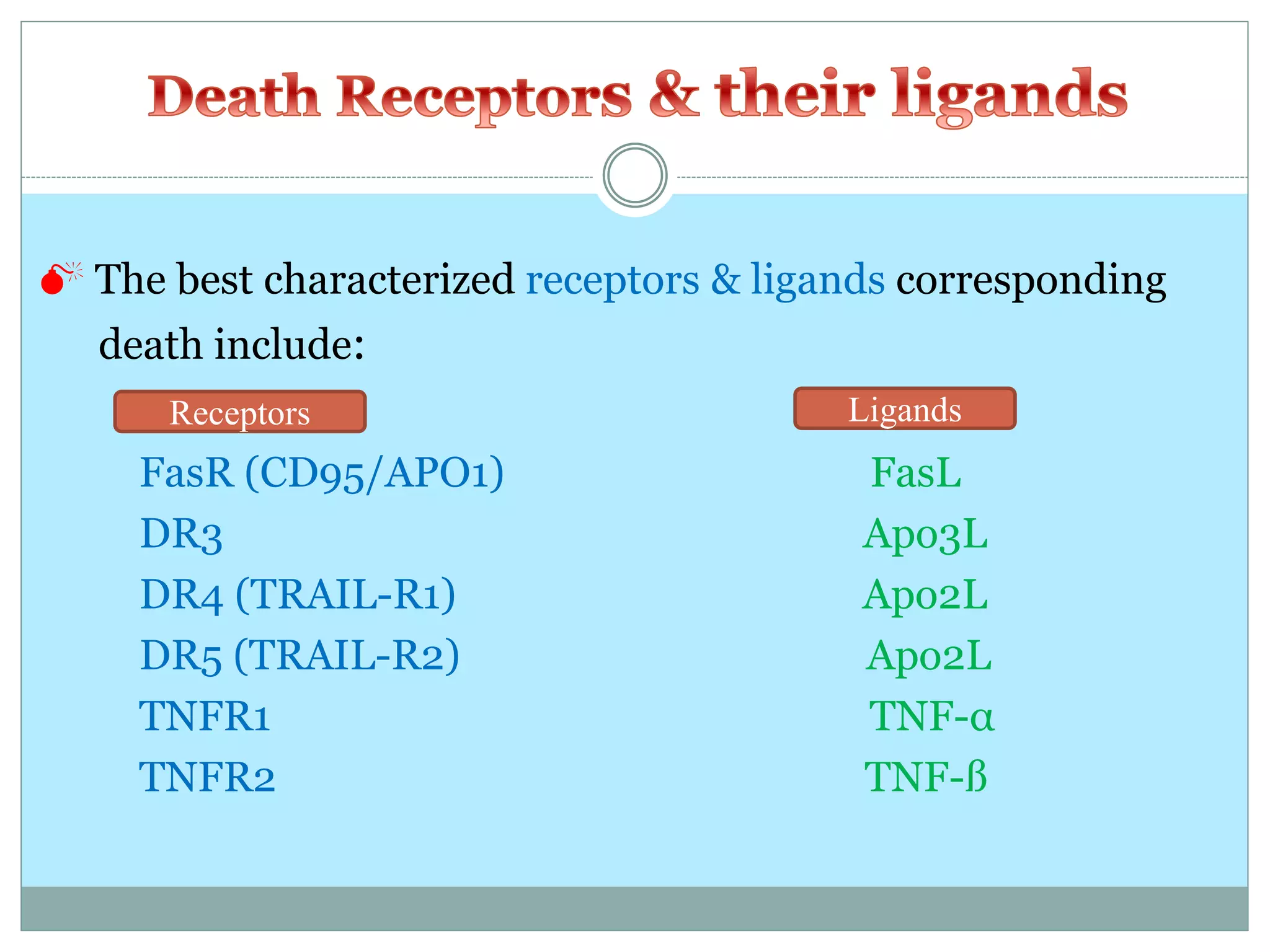
Apoptosis
By
Mrs Sanchita Choubey
(M.Sc., PGDCR, Pursuing Ph. D)
Assistant Professor of Microbiology
Dr. D Y Patil Arts Commerce and Science College Pimpri, Pune
 In humans, the rate of cell growth and cell death is
balanced to maintain the weight of the body.
 Apoptosis = “normal” or “programmed” cell death
 Necrosis = “accidental” or “ordinary” cell death
 The word ‘‘apoptosis’’ comes from the
ancient Greek, meaning the:
‘‘falling of petals from a flower’’
 The term apoptosis (a-po-toe-sis) was first used in a
now-classic paper by Kerr et al 1972 to describe a
morphologically distinct form of cell death.
 Apoptosis or programmed cell death (PCD) is a mode of
cell death that occurs under normal physiological
conditions and the cell is an active participant in its own
demise (“cellular suicide”).
 It is important for the development of multicellular
organism (embryonic development) and homeostasis of
their tissues (adult).
Apoptosis is a beneficial and important phenomenon:
 In embryo
1. During embryonic development, help to digit formation.
 Lack of apoptosis in humans
can lead to webbed fingers
called “ syndactyly ”.
2. Normal event in development of the nervous system
Apoptosis is a beneficial and important phenomenon:
 In adult
 Normal cell turn over
 Tissue homeostasis
 Induction and maintenance of immune tolerance
 Development of the nervous system
 Endocrine-dependent tissue atrophy
 Elimination of activated, damaged and abnormal cells
Apoptosis is a beneficial and important phenomenon:
 In animals
Embryonic Chicken Foot
Embryonic Mouse Paw
Tail absorption of the tadpole
 Apoptosis is the physiological cell death which unwanted or
useless cells are eliminated during development and other
normal biological processes.
 Necrosis is the pathological cell death which occurs when
cells are exposed to a serious physical or chemical insult
(hypoxia, hyperthermia, ischemia).
 There are many observable morphological and biochemical
differences between necrosis and apoptosis:
 Morphological features
Necrotic cells Apoptotic cells
 Volume enlargement  Volume reduction
 Swelling of cytoplasm  Shrinking of cytoplasm
& mitochondria  No loss of membrane integrity
 Loss of membrane integrity
 No vesicle formation  Formation of apoptotic bodies
 Condensation of chromatin &
DNA fragmentation
 Biochemical features
Necrotic cells
 Loss of regulation of ion homeostasis
 No energy requirement (passive process, also occurs at 4 °C)
Apoptotic cells
 Tightly regulated process
 Energy(ATP)- dependent (active process, doesn’t occur at 4 °C)
 Release of various factors into cytosol by mitochondria
 Activation of caspase cascade
 Takes about 30 - 60 min.
 In the tissue only about 5% cells is affected by the PCD.
 Physiological changes and phagocytosis is very fast.
Extrinsic pathway (death receptor- mediated events)
Intrinsic pathway (mitochondria- mediated events)
 Death Receptors
 Death Ligands
 Adaptor Proteins
 Caspaseses
Caspases= Cysteinyl aspartate specific proteases
 A family of intracellular cysteine proteases that play a
pivotal role in the initiation and execution of apoptosis.
 At least 14 different members of caspases in mammalian
cells have been identified
 All are synthesized as inactive proenzymes (zymogen)
with 32-56 kDa
 To date, ten major caspases have been identified and broadly
categorized into:
 Signaling or Initiator caspases
 Effector or Executioner caspases
 Inflammatory caspases
 The other caspases that have been identified include:
Caspases
 Central role in cascade of apoptotic events is played by
caspase 3 (CPP32)
 “Death receptors” that are members of the tumor necrosis
factor (TNF) receptor superfamily.
 Death receptors have a cytoplasmic domain of about 80
amino acids called the “death domain”.
 This death domain plays a critical role in transmitting the
death signal from the cell surface to the intracellular
signaling pathways.
 The best characterized receptors & ligands corresponding
death include:
FasR (CD95/APO1) FasL
DR3 Apo3L
DR4 (TRAIL-R1) Apo2L
DR5 (TRAIL-R2) Apo2L
TNFR1 TNF-α
TNFR2 TNF-ß
Ligands
Receptors
and
 Binding of trimeric FasL to Fas
 Trimerization and clustering of Fas
 Recruitment of Fas-associated death
domain (FADD) to Fas
 Recruitment of caspase-8 to FADD
 Formation of Death-Inducing Signaling
Complex (DISC )
 Activation of caspase-8 (autoactivation)
 Activation of effector caspases
 Apoptosis
 Bcl-2 family proteins
 Cytochrom c
 Adaptor proteins
 Caspaseses
 The stimuli that initiate the intrinsic pathway produce
intracellular signals such as radiation (DNA damage),
absence of certain growth factors, hormones and cytokines.
 All of these stimuli cause changes in the mitochondrial outer
membrane permeabilization (MOMP)
 Release of pro-apoptotic proteins such as cytochrome c,
Smac/DIABLO, AIF, endonuclease G and CAD from the
inter-membrane space into the cytosol.
 Cytochrome c binds and activates Apaf-1 as well as
procaspase-9, forming an “apoptosome”.
 Caspase-9 activation, subsequent caspase-3 activation and
cell death.
 The control & regulation of apoptotic mitochondrial events
occurs through members of the Bcl-2 family of proteins
 Anti-apoptotic proteins include Bcl-2, Bcl-x, Bcl-XL, Bcl-w
 Pro-apoptotic proteins include Bax, Bak, Bid, Bad, Bim, Bik
 The main mechanism of action of the Bcl-2 family of proteins
is the regulation of cytochrome c release from the
mitochondria via alteration of mitochondrial membrane
permeability.
Aberrant cell death can lead to many human diseases:
 Decreased apoptosis Cancer, Autoimmune disorders
 Excessive apoptosis Neurodegenerative and
immunodeficiency (AIDS) disorders , Ischemia
NOTE: Properties of carcinogenic agents (chemical agents as
well as radiations) are the growth-inhibition power and the
ability to induce cell death. These properties are widely used in
anticancer chemo- and radiotherapies
‫تشکر‬ ‫با‬
Thanks for your attention

Apoptosis

  • 1.
    Apoptosis By Mrs Sanchita Choubey (M.Sc.,PGDCR, Pursuing Ph. D) Assistant Professor of Microbiology Dr. D Y Patil Arts Commerce and Science College Pimpri, Pune
  • 2.
     In humans,the rate of cell growth and cell death is balanced to maintain the weight of the body.
  • 3.
     Apoptosis =“normal” or “programmed” cell death  Necrosis = “accidental” or “ordinary” cell death
  • 4.
     The word‘‘apoptosis’’ comes from the ancient Greek, meaning the: ‘‘falling of petals from a flower’’  The term apoptosis (a-po-toe-sis) was first used in a now-classic paper by Kerr et al 1972 to describe a morphologically distinct form of cell death.
  • 5.
     Apoptosis orprogrammed cell death (PCD) is a mode of cell death that occurs under normal physiological conditions and the cell is an active participant in its own demise (“cellular suicide”).  It is important for the development of multicellular organism (embryonic development) and homeostasis of their tissues (adult).
  • 6.
    Apoptosis is abeneficial and important phenomenon:  In embryo 1. During embryonic development, help to digit formation.  Lack of apoptosis in humans can lead to webbed fingers called “ syndactyly ”.
  • 7.
    2. Normal eventin development of the nervous system
  • 8.
    Apoptosis is abeneficial and important phenomenon:  In adult  Normal cell turn over  Tissue homeostasis  Induction and maintenance of immune tolerance  Development of the nervous system  Endocrine-dependent tissue atrophy  Elimination of activated, damaged and abnormal cells
  • 9.
    Apoptosis is abeneficial and important phenomenon:  In animals Embryonic Chicken Foot Embryonic Mouse Paw Tail absorption of the tadpole
  • 10.
     Apoptosis isthe physiological cell death which unwanted or useless cells are eliminated during development and other normal biological processes.  Necrosis is the pathological cell death which occurs when cells are exposed to a serious physical or chemical insult (hypoxia, hyperthermia, ischemia).
  • 11.
     There aremany observable morphological and biochemical differences between necrosis and apoptosis:  Morphological features Necrotic cells Apoptotic cells  Volume enlargement  Volume reduction  Swelling of cytoplasm  Shrinking of cytoplasm & mitochondria  No loss of membrane integrity  Loss of membrane integrity  No vesicle formation  Formation of apoptotic bodies  Condensation of chromatin & DNA fragmentation
  • 12.
     Biochemical features Necroticcells  Loss of regulation of ion homeostasis  No energy requirement (passive process, also occurs at 4 °C) Apoptotic cells  Tightly regulated process  Energy(ATP)- dependent (active process, doesn’t occur at 4 °C)  Release of various factors into cytosol by mitochondria  Activation of caspase cascade
  • 13.
     Takes about30 - 60 min.  In the tissue only about 5% cells is affected by the PCD.  Physiological changes and phagocytosis is very fast.
  • 14.
    Extrinsic pathway (deathreceptor- mediated events) Intrinsic pathway (mitochondria- mediated events)
  • 15.
     Death Receptors Death Ligands  Adaptor Proteins  Caspaseses
  • 16.
    Caspases= Cysteinyl aspartatespecific proteases  A family of intracellular cysteine proteases that play a pivotal role in the initiation and execution of apoptosis.  At least 14 different members of caspases in mammalian cells have been identified  All are synthesized as inactive proenzymes (zymogen) with 32-56 kDa
  • 18.
     To date,ten major caspases have been identified and broadly categorized into:  Signaling or Initiator caspases  Effector or Executioner caspases  Inflammatory caspases  The other caspases that have been identified include: Caspases  Central role in cascade of apoptotic events is played by caspase 3 (CPP32)
  • 19.
     “Death receptors”that are members of the tumor necrosis factor (TNF) receptor superfamily.  Death receptors have a cytoplasmic domain of about 80 amino acids called the “death domain”.  This death domain plays a critical role in transmitting the death signal from the cell surface to the intracellular signaling pathways.
  • 20.
     The bestcharacterized receptors & ligands corresponding death include: FasR (CD95/APO1) FasL DR3 Apo3L DR4 (TRAIL-R1) Apo2L DR5 (TRAIL-R2) Apo2L TNFR1 TNF-α TNFR2 TNF-ß Ligands Receptors
  • 21.
  • 22.
     Binding oftrimeric FasL to Fas  Trimerization and clustering of Fas  Recruitment of Fas-associated death domain (FADD) to Fas  Recruitment of caspase-8 to FADD  Formation of Death-Inducing Signaling Complex (DISC )  Activation of caspase-8 (autoactivation)  Activation of effector caspases  Apoptosis
  • 23.
     Bcl-2 familyproteins  Cytochrom c  Adaptor proteins  Caspaseses
  • 24.
     The stimulithat initiate the intrinsic pathway produce intracellular signals such as radiation (DNA damage), absence of certain growth factors, hormones and cytokines.  All of these stimuli cause changes in the mitochondrial outer membrane permeabilization (MOMP)  Release of pro-apoptotic proteins such as cytochrome c, Smac/DIABLO, AIF, endonuclease G and CAD from the inter-membrane space into the cytosol.  Cytochrome c binds and activates Apaf-1 as well as procaspase-9, forming an “apoptosome”.  Caspase-9 activation, subsequent caspase-3 activation and cell death.
  • 26.
     The control& regulation of apoptotic mitochondrial events occurs through members of the Bcl-2 family of proteins  Anti-apoptotic proteins include Bcl-2, Bcl-x, Bcl-XL, Bcl-w  Pro-apoptotic proteins include Bax, Bak, Bid, Bad, Bim, Bik  The main mechanism of action of the Bcl-2 family of proteins is the regulation of cytochrome c release from the mitochondria via alteration of mitochondrial membrane permeability.
  • 27.
    Aberrant cell deathcan lead to many human diseases:  Decreased apoptosis Cancer, Autoimmune disorders  Excessive apoptosis Neurodegenerative and immunodeficiency (AIDS) disorders , Ischemia NOTE: Properties of carcinogenic agents (chemical agents as well as radiations) are the growth-inhibition power and the ability to induce cell death. These properties are widely used in anticancer chemo- and radiotherapies
  • 28.